. 1999 May 17;145(4):865-76.
doi: 10.1083/jcb.145.4.865.

Neurotractin, a novel neurite outgrowth-promoting Ig-like protein that interacts with CEPU-1 and LAMP

Affiliations

Neurotractin, a novel neurite outgrowth-promoting Ig-like protein that interacts with CEPU-1 and LAMP

A Marg et al. J Cell Biol. .

Abstract

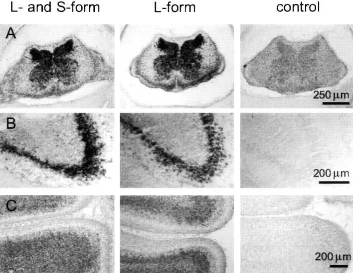
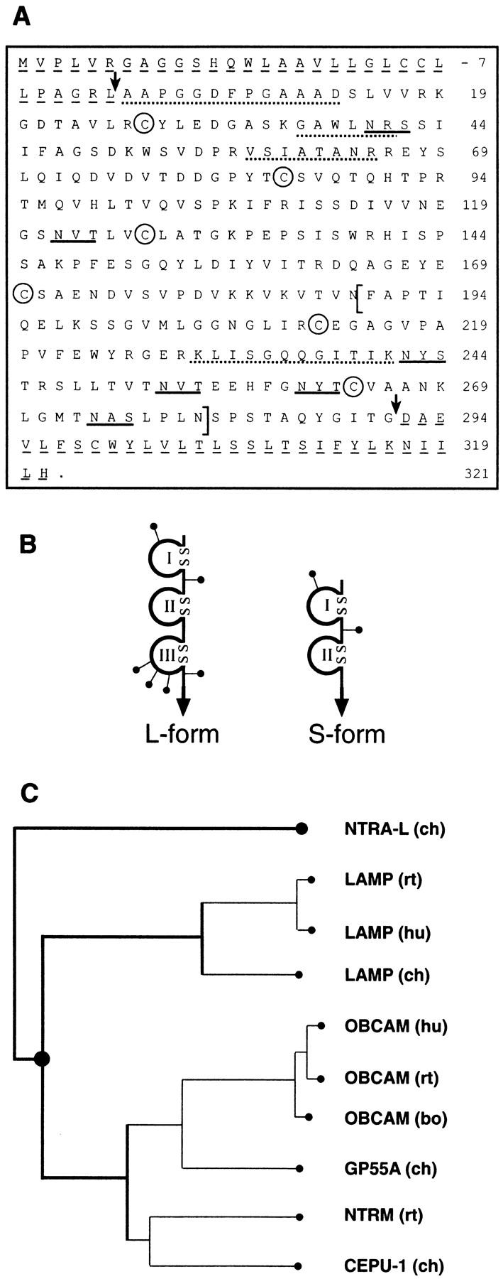
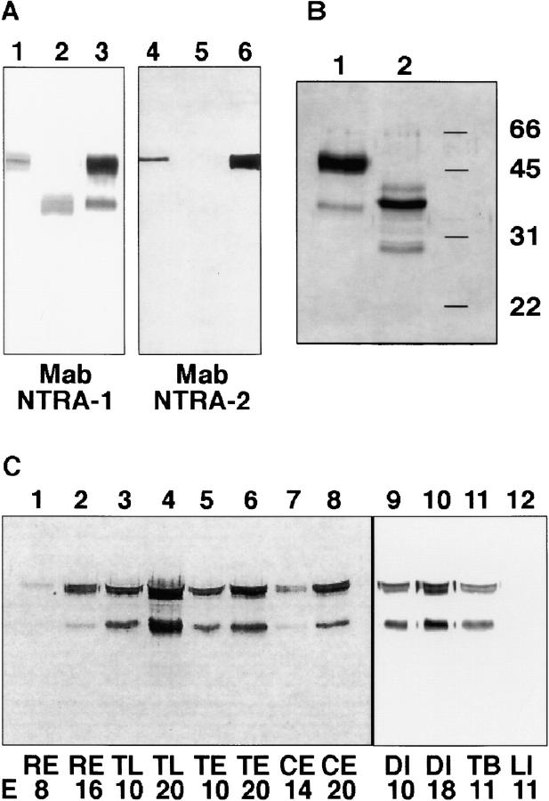
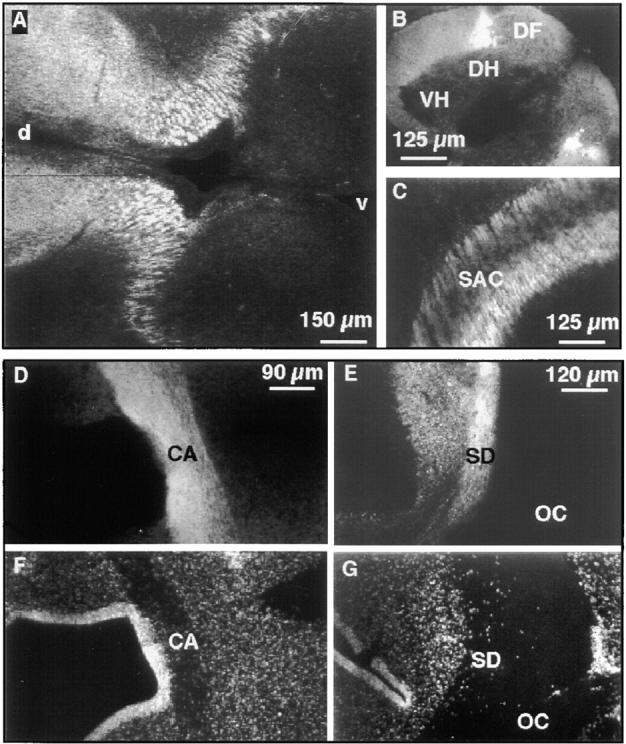
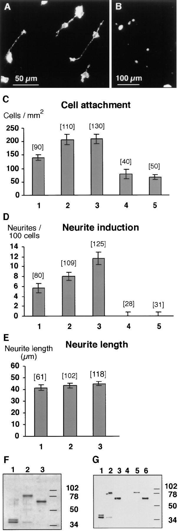
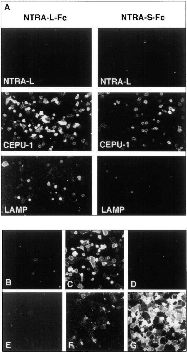
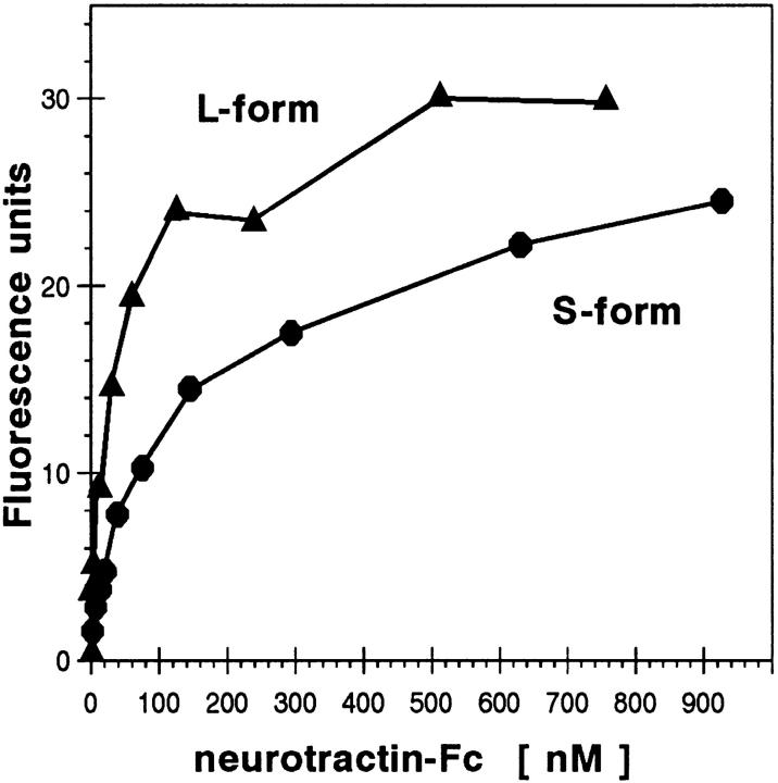
The formation of axon tracts in nervous system histogenesis is the result of selective axon fasciculation and specific growth cone guidance in embryonic development. One group of proteins implicated in neurite outgrowth, fasciculation, and guidance is the neural members of the Ig superfamily (IgSF). In an attempt to identify and characterize new proteins of this superfamily in the developing nervous system, we used a PCR-based strategy with degenerated primers that represent conserved sequences around the characteristic cysteine residues of Ig-like domains. Using this approach, we identified a novel neural IgSF member, termed neurotractin. This GPI-linked cell surface glycoprotein is composed of three Ig-like domains and belongs to the IgLON subgroup of neural IgSF members. It is expressed in two isoforms with apparent molecular masses of 50 and 37 kD, termed L-form and S-form, respectively. Monoclonal antibodies were used to analyze its biochemical features and histological distribution. Neurotractin is restricted to subsets of developing commissural and longitudinal axon tracts in the chick central nervous system. Recombinant neurotractin promotes neurite outgrowth of telencephalic neurons and interacts with the IgSF members CEPU-1 (KD = 3 x 10(-8) M) and LAMP. Our data suggest that neurotractin participates in the regulation of neurite outgrowth in the developing brain.


Figures

Figure 4
Figure 4
Localization of neurotractin mRNA in the developing nervous system. In situ hybridizations with two different neurotractin antisense probes: one derived from the second Ig-like domain which detects both isoforms (first column) and one derived from the third Ig-like domain which is L-form specific (second row). Both probes gave essentially the same staining patterns in chick E12 cervical spinal cord (A), in a subpopulation of neurons in E12 ventral telencephalon (B), and in the internal granular layer of E16 cerebellum (C). Control hybridizations with sense probes did not give significant signals (third column).
Figure 1
Figure 1
Primary structure, domain models of neurotractin and sequence relationship to other IgLON members. (A) Primary structure of neurotractin. The predicted NH2-terminal signal peptide and the COOH-terminal hydrophobic segment are underlined by dashed lines and arrows indicate the mature NH2 terminus and COOH terminus. Putative N-linked glycosylation sites are underlined and characteristic cysteine residues of Ig-like domains are labeled by circles. To obtain independent evidence that the protein which had been isolated by immunoaffinity chromatography (see Fig. 2 B, lane 1) is identical with that predicted by the cDNA clones, a sample of it as well as peptides derived from a tryptic digest were subjected to Edman degradation. Evaluation of the partial internal as well as the NH2-terminal sequence (indicated by dotted lines) confirms that they match this cDNA sequence. The alternatively spliced and L-form–specific third Ig-like domain is indicated by brackets. The sequences of S-form and L-form are available from GenBank/EMBL/DDBJ under accession numbers AJ132998 and AJ132999, respectively. (B) Domain models of neurotractin-L (large) and -S (small). Ig-like domains are drawn as loops that are closed by disulfide bridges, putative N-linked glycosylation sites are shown as lines ending with dots and the GPI-anchor is represented by an arrow. (C) Neurotractin is a novel protein belonging to the IgLON subgroup. Sequence relationship of neurotractin-L to other members of the IgLON subgroup was examined with the PILEUP program from the GCG package (University of Wisconsin, Madison, WI) and sequences have been taken from Schofield et al., 1989; Lippman et al., 1992; Shark and Lee, 1995; Struyk et al., 1995; Pimenta et al., 1995, ; Spaltmann and Brümmendorf, 1996; Brümmendorf et al., 1997; Hancox et al., 1997. GP55-A is a partial sequence lacking most likely a short stretch at the NH2 terminus (Wilson et al., 1996). ch, chicken; hu, human; bo, bovine; rt, rat.
Figure 2
Figure 2
Neurotractin occurs in two isoforms and is upregulated in development. (A) Lysates of COS cells that had been transfected with the L-form (lanes 1 and 4) or S-form (lanes 2 and 5) of neurotractin were compared with mAb NTRA-1 immunoaffinity isolate (lanes 3 and 6) by SDS-PAGE. In Western blots mAb NTRA-1 stains both isoforms as well as both bands in the immunoaffinity isolate (lanes 1–3) whereas mAb NTRA-2 detects only the L-form and the larger band of the immunoaffinity isolate (lanes 4–6). (B) Neurotractin that was isolated by immunoaffinity chromatography from adult chicken brains using mAb NTRA-1 was subjected to SDS-PAGE and detected by silver staining. Neurotractin resolves in two bands, one of 50 kD and one of 37 kD (lane 1). Deglycosylation by endoglycosidase F/peptide-N-glycosidase F leads to a reduction of the molecular mass to 38 and 30 kD, respectively (lane 2). The 40-kD component (lane 2) represents a deglycosylation intermediate. (C) Samples of different regions of embryonic chick brain from early and late developmental stages were solubilized in SDS-PAGE sample buffer, resolved by SDS-PAGE, and probed with mAb NTRA-1 directed to neurotractin (each lane represents 30 μl of 1% brain homogenate). Comparison of samples from early stages with those from late stages shows that in each analyzed brain region neurotractin expression increases during development (lanes 1–10). Neurotractin can be detected in total brain but not in liver (lanes 11 and 12). E, embryonic day; RE, retina; TL, telencephalon; TE, tectum; CE, cerebellum; DI, diencephalon; TB, total brain; LI, liver.
Figure 3
Figure 3
Neurotractin expression on subsets of embryonic axon tracts. Immunohistochemical analysis of E7 diencephalon with mAb NTRA-1 shows crosscut longitudinal neurotractin positive axon tracts (A). In the E9 spinal cord dorsolateral longitudinal axons show strong neurotractin expression (B). In tectum of embryonic day 8 neurotractin is found on axons of the stratum album centrale (C). Staining of a horizontal section of E9 ventral forebrain with mAb NTRA-1 shows strong expression of neurotractin on axons of the anterior commissure (D). This axon tract is also discernible as a nuclei-poor zone as revealed by DNA staining (F). A horizontal section at the level of the E9 optic chiasm reveals neurotractin expression on axons of the supraoptic decussation (E). Bisbenzimide staining of cellular nuclei in the same section outlines the axons of the supraoptic decussation as well as those of retinal ganglion cells in the optic chiasm as an extended nuclei-poor region (G). Note that neurotractin is restricted to the supraoptic decussation and is lacking on the retinal ganglion cell axons. At E9, the supraoptic decussation is not yet subdivided into the dorsal, ventral and subventral regions which can be distinguished later in development (Ehrlich et al., 1988). VH, ventral horn; DH, dorsal horn; DF, dorsal funiculus; SAC, stratum album centrale; d, dorsal; v, ventral; CA, commissura anterior; SD, supraoptic decussation; OC, optic chiasm.
Figure 5
Figure 5
Neurotractin-L promotes neurite outgrowth and cell attachment of telencephalic neurons. (A) Cells isolated from E8 telencephalon attach to a neurotractin-L substratum which promotes neurite outgrowth. (B) Cell attachment to the control substrate consisting of Fc protein without neurotractin domains is shown for comparison. (C) Density of telencephalic neurons adhering to different substrates. Fc fusion proteins were coated as follows: neurotractin L-form with 25 μg/ml (1), 50 μg/ml (2), and 100 μg/ml (3), Fc control protein (4), and neurotractin S-form (5) with 100 μg/ml. Data were pooled from independent experiments, histogram bars show median values, error bars represent SEM, and the number of analyzed images (450 μm × 450 μm) is given in parentheses. The difference between Fc control protein and 25 μg/ml neurotractin-L is statistically significant (P = 0.003, Mann Whitney U test). (D) Neurotractin-induced telencephalic neurite outgrowth after 40 h of incubation on Fc fusion proteins coated as in C. The differences between Fc control protein and 25 μg/ml neurotractin-L (P < 0.001), between 25 μg/ml and 50 μg/ ml (P < 0.05), and between 50 μg/ml and 100 μg/ml (P < 0.05) are statistically significant (Mann Whitney U test). Lower neurotractin-L concentrations in the coating solution (12.5 μg/ml) did not result in significant neurite extension above background values (data not shown). (E) Increasing amounts of neurotractin do not influence the average length of telencephalic neurites. Fc fusion protein of neurotractin L-form was coated as indicated in C. (F) SDS-PAGE (10%) of fusion proteins used in neurite outgrowth experiments and binding studies followed by silver staining. Purified Fc fusion proteins of L-form (lane 2) and S-form (lane 3) neurotractin reveal the expected molecular masses of 80 and 65 kD, respectively. The Fc domains are resolved in a 39-kD component and a 36-kD degradation product (lane 1). (G) SDS-PAGE (10%) and Western blot analyses show that neurotractin-L (lane 2), neurotractin-S (lane 3), and the Fc domains (lane 1) can be detected with polyclonal Fc domain-specific antibodies. The 36-kD component (lanes 2 in F and G) which can also be observed in the Fc control protein (lanes 1 in F and G) is a degradation product. Neurotractin-specific polyclonal antibodies identify both neurotractin isoforms (lanes 5 and 6) but do not react with the Fc domains (lane 4).
Figure 6
Figure 6
Molecular interaction of neurotractin with CEPU-1 and LAMP. (A) Neurotractin-L (first row), CEPU-1 (middle row), and LAMP (bottom row) were expressed on the surface of CHO cells by transient transfection. Confluent layers of cells were incubated with 10 μg/ml of purified Fc fusion proteins of neurotractin L-form (left column) and S-form (right column). After washing, Fc domains of fusion proteins were detected by Cy3-conjugated secondary antibody directed to human IgG. Control experiments showed that the percentage of transfected cells and their surface expression levels were indistinguishable for NTRA-L, CEPU-1, and LAMP. CHO cells expressing CEPU-1 on their surface that had been preincubated with PI-PLC (B) or control buffer (C) were tested with COS cell supernatants containing neurotractin-L Fc fusion protein. CHO cells expressing F11 (D) or GPI-linked Fc domains (E) do not bind neurotractin-L fusion protein. Furthermore, COS cells which express CEPU-1 (G) or LAMP (F) on their surface were also found to bind soluble recombinant neurotractin-L.
Figure 7
Figure 7
Equilibrium binding of recombinant neurotractin. CHO cell transfectants expressing CEPU-1 were incubated with increasing concentrations of neurotractin fusion proteins, followed by a constant saturating amount of fluorochrome-conjugated Fc domain–specific antibody. Average fluorescence intensity that was measured in at least three independent experiments is given. Soluble Fc control protein gave a signal of <5 fluorescence units if applied to CEPU-1 transfectants at a concentration of 6.2 μM under the same conditions.

Similar articles

Cited by

References

    1. Beckmann MP, Cerretti DP, Baum P, Vanden T, Bos, James L, Farrah T, Kozlosky C, Hollingsworth T, Shilling H, Maraskovsky E, et al. Molecular characterization of a family of ligands for Eph-related tyrosine kinase receptors. EMBO (Eur Mol Biol Organ) J. 1994;13:3757–3762. - PMC - PubMed
    1. Bisswanger, H. 1979. Theorie und Methoden der Enzymkinetik. Verlag Chemie, Weinheim.
    1. Brümmendorf T, Spaltmann F, Treubert U. Cloning and characterization of a neural cell recognition molecule on axons of the retinotectal system and spinal cord. Eur J Neurosci. 1997;9:1105–1116. - PubMed
    1. Brümmendorf T, Kenwrick S, Rathjen FG. Neural cell recognition molecule L1: from cell biology to human hereditary brain malformations. Curr Opin Neurobiol. 1998;8:87–97. - PubMed
    1. Brümmendorf T, Rathjen FG. Cell adhesion molecules 1: immunoglobulin superfamily. Prot Profile. 1995;2:963–1108. - PubMed

Publication types

MeSH terms

Associated data