. 1999 May 15;103(10):1409-18.
doi: 10.1172/JCI4682.

Estrogen's bone-protective effects may involve differential IL-1 receptor regulation in human osteoclast-like cells

Affiliations

Estrogen's bone-protective effects may involve differential IL-1 receptor regulation in human osteoclast-like cells

T Sunyer et al. J Clin Invest. .

Abstract

Declining estrogen levels during the first postmenopausal decade lead to rapid bone loss and increased fracture risk that can be reversed by estrogen replacement therapy. The bone-protective effects of estrogen may involve suppression of inflammatory cytokines that promote osteoclastogenesis and bone resorption, such as IL-1, TNF-alpha, and IL-6. We investigated whether estrogen modulates IL-1 actions on human osteoclasts (OCs) and other bone cell types. Isolated human OCs and primary bone marrow-derived OC-like cells expressed both the signaling (IL-1RI) and decoy (IL-1RII) IL-1 receptors, whereas only IL-1RI was detected in osteoblasts. IL-1RII/IL-1RI mRNA ratios and release of soluble IL-1RII (sIL-1RII) were lower in OC-like cells derived from women in the late postmenopausal period compared with younger women, but were unrelated to male donor age, suggesting that estrogen might play a role in regulating IL-1 receptor levels in vivo. Estrogen directly reduced in vitro OC-like cell IL-1RI mRNA levels while increasing IL-1RII mRNA levels and sIL-1RII release. These estrogenic events were associated with inhibited IL-1-mediated cytokine (IL-8) mRNA induction and cell survival, i.e., increased apoptosis. In contrast, estrogen did not alter IL-1R levels or IL-1 responsiveness in primary human osteoblasts or bone marrow stromal cells. We conclude that one novel mechanism by which estrogen exerts bone-protective effects may include a selective modulation of IL-1R isoform levels in OC or OC-like cells, thereby reducing their IL-1 responsiveness and cell survival. Conversely, this restraint on IL-1 actions may be lost as estrogen levels decline in aging women, contributing to an enhanced OC-mediated postmenopausal bone loss.


Figures

Figure 1
Figure 1
hOC and hOCL cells express mRNA for both the type I and type II IL-1 receptors. (a) Schematic representation of the human IL-1 receptor genes. Hatched bars represent the extracellular regions of the proteins; filled bars represent the transmembrane domains; and open bars represent the intracellular tails. Arrowheads indicate the location of primers used in RT-PCR. (b) RT-PCR of IL-1RI (I) and IL-1RII (II) from hOCs directly isolated from a femoral head of a 76-year-old woman undergoing hip replacement surgery, and from hOCL and hOBL cells generated from a single bone marrow mononuclear preparation derived from a 58-year-old woman. Amplicons were separated in 1.5% agarose gels and viewed with ethidium bromide. Similar results were obtained by RT-PCR analysis for IL-1RI and IL-1RII in hOCs directly isolated from osteopenic bone specimens obtained from 6 patients (4 women of 71, 76, 78, and 78 years of age; and 2 men of 56 and 78 years of age) undergoing hip replacement surgery, and a patient with implant osteolysis (a 71-year-old man). St., molecular weight standards.
Figure 2
Figure 2
IL-1 induction of IL-8 in hOCL cells is mediated by the signaling IL-1RI. Bone marrow–derived hOCL cells were preincubated for 1 hour in the absence (0) or presence of neutralizing antibodies to the signaling IL-1RI (5 or 50 μg/mL) or the decoy IL-1RII (1 or 5 μg/mL) at concentrations that represent approximately 1–10 times their ND50 (antibody concentration required for half-maximal inhibition of IL-1 activity as determined by the manufacturer). Cell cultures were then treated for 16 hours with 10–9 M IL-1β (+) or vehicle (–), and the cells were harvested for protein determination. IL-8 cytokine release was measured in the conditioned medium, normalized by the total cellular protein in each culture dish, and expressed as the mean ± SEM of nanograms of IL-8 released per milligrams of cell protein. Significant differences from control cultures: **P < 0.01. Significant differences from IL-1–treated cultures: +P < 0.05. Determined by Bonferroni post-ANOVA test for multiple comparisons.
Figure 3
Figure 3
Steady-state mRNA levels for IL-1RI and IL-1RII vary among hOCL cells generated from different donors. (a) Northern analysis of poly(A)-selected RNA from hOCL cells derived from 2 different individuals (designated 1 and 2) sequentially hybridized with the indicated probes. Transcripts were identified of the following approximate sizes: 5.6-kb IL-1RI, 1.4-kb IL-1RII, 1.4-kb GAPDH, and 2.0-kb β-actin. Amounts of each radioactive probe used and times of gel exposure for autoradiography were as follows: IL-1RI, 3 × 10⁶ cpm/mL, 7 days; IL-1RII, 3 × 10⁶ cpm/mL, 14 days; GAPDH, 2 × 10⁶ cpm/mL, 20 hours; and β-actin, 1 × 10⁶ cpm/mL, 60 hours. (b) Representative RPA for IL-1RI and IL-1RII using total RNA from hOCL and hOBL cells that were obtained from the same bone marrow mononuclear cultures of 2 different individuals (designated 3 and 4). Each sample was probed simultaneously with IL-1RI (I) and GAPDH, or in a parallel reaction with IL-1RII (II) and GAPDH. To attribute correctly the protected bands as signals for IL-1RI, IL-1RII, and GAPDH, reactions were first performed with each individual probe alone. Specific protected bands were of the expected sizes, which differed from one another, and were clearly discernible from full-length unprotected probes (which were included along with molecular size markers in the gels). The data shown in b were obtained from a single RPA and set of labeled probes. Additional RPAs performed with hOCL cells derived from 25 other donors yielded similar variations in their relative steady-state mRNA levels of the 2 IL-1Rs, a feature inherent to the hOCL cell preparations themselves (see Figure 4).
Figure 4
Figure 4
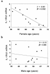
IL-1RII/IL-1RI steady-state mRNA ratios in hOCL cells decrease with increasing age of female, but not male, bone marrow donors. Steady-state mRNA levels for IL-1RI, IL-1RII, and the internal control GAPDH were measured in hOCL cells generated from 11 women ranging from 51 to 72 years of age (a), and from 11 men ranging from 40 to 68 years of age (b), as described in Figure 3b. No bone marrow samples were available from female donors of 40 to 50 years of age. The IL-1RII/IL-1RI mRNA ratios were calculated as follows: the signal intensity of each IL-1R protected band was divided by the signal intensity of the corresponding internal GAPDH control for that reaction, and the normalized value for IL-1RII was then divided by the normalized value for IL-1RI (Table 1). P, statistical significance as determined by ANOVA; r, correlation coefficient. Typically, as many RNA samples as possible were run in a single RPA (using the same gel and probes) to enable direct comparisons. In addition, RNA obtained from 2 different hOCL cell cultures each was reanalyzed in 3 separate RPA trials to calculate the variances associated with determining the IL-1RII/IL-1RI mRNA ratios across independent RPA experiments. Thus, independent assessments of the IL-1RII/IL-1RI ratio for hOCL cells derived from a 72-year-old woman yielded values of 0.28, 0.3, and 0.32 with a variance of 0.0004, and hOCL cells from a 51-year-old woman yielded ratios of 2.4, 1.8, and 2.0 with a variance of 0.093.
Figure 5
Figure 5
17β-estradiol differentially regulates IL-1RI and IL-1RII mRNA levels in hOCL. (a) hOC and hOCL cells express mRNA for ER-α. RT-PCR analysis of ER-α and GAPDH was performed using total RNA of immunomagnetically purified hOC directly isolated from a femoral head or from purified bone marrow–derived hOCL cells. Amplicons were separated in 1.5% agarose gels and stained with ethidium bromide. (be) 17β-estradiol decreased signaling IL-1RI and increased decoy IL-1RII mRNA levels in hOCL cells. hOCL cell cultures derived from 5 different bone marrow donors (2 women and 3 men, 1 to 3 replicate wells each) were incubated for 4 hours with vehicle (0), 10 or 100 nM 17β-estradiol (E2), or 100 nM of the inactive analogue 17α-estradiol. Total RNA was extracted, and steady-state mRNA levels for IL-1RI, IL-1RII, and GAPDH were measured by RPA. (b and c) Representative RPA for IL-1RI and GAPDH mRNA (b), and for IL-1RII and GAPDH mRNA (c). (d and e) Protected IL-1R mRNA bands were quantified and normalized to GAPDH. The data are expressed as a percentage of vehicle control and represent the mean ± SEM of the IL-1RI/GAPDH ratio (d) or the IL-1RII/GAPDH ratio (e). Significant differences from control cultures: *P < 0.05 as determined by Bonferroni post-ANOVA test for multiple comparisons. St., molecular weight standards.
Figure 6
Figure 6
17β-estradiol pretreatment of hOCL cells, but not hOBL cells, inhibits IL-1 induction of IL-8 mRNA. hOCL cells (a and b) or hOBL cells (c and d) were preincubated for 4 hours with 10–7 M 17β-estradiol (E2, +) or vehicle (–). After pretreatment, the same modulators were added to the cell cultures simultaneously with 10–9 M IL-1β (IL-1, +) or vehicle (–), and the cells were further incubated for another 4 hours. Total RNA was extracted, and IL-8 mRNA was analyzed by RPA as a functional measure of IL-1 biologic responsiveness. (a and c) Representative RPA for IL-8 mRNA levels in hOCL and hOBL, respectively. (b and d) IL-8 mRNA protected bands from 3 independent hOCL (b) or hOBL (d) cell cultures were quantified and normalized to GAPDH. Additional results obtained using a primary hOB cell culture were indistinguishable from those obtained with the hOBL cell cultures. Data are expressed as a percentage of maximal IL-8 mRNA levels induced by IL-1β and represent the mean ± SEM. Significant differences from control cultures: **P < 0.01, ***P < 0.001. Significant differences from IL-1β–treated cultures: ++P < 0.01 as determined by the Bonferroni post-ANOVA test.
Figure 7
Figure 7
17β-estradiol pretreatment of hOCL cells inhibits IL-1–promoted cell survival. hOCL cells cultured on glass coverslips were preincubated for 4 hours with 10–7 M 17β-estradiol (E2, +) or vehicle (–). After pretreatment, the same modulators were added to the cell cultures simultaneously with 10–9 M IL-1β (IL-1, +) or vehicle (–), and the cells were further incubated for another 18 hours. hOCL cells were then stained with annexin V-fluorescein to identify apoptotic cells, and a total of 2,400–3,700 hOCL cells for each experimental condition were viewed in 10–16 fields and counted by fluorescence microscopy. Data are expressed as the mean ± SEM percentage of annexin V–labeled apoptotic hOCL cells relative to total hOCL cells per field for each condition. No necrotic cells (stained with propidium iodide) were detected in these cultures. Significant differences from control cultures: **P < 0.001. Significant differences from IL-1–treated cultures: +P < 0.05.

Similar articles

Cited by

References

    1. Turner R, Riggs B, Spelsberg T. Skeletal effects of estrogen. Endocr Rev. 1994;15:275–300. - PubMed
    1. Smith E, et al. Estrogen resistance caused by a mutation in the estrogen-receptor gene in a man. N Engl J Med. 1994;331:1056–1061. - PubMed
    1. Korach K, et al. Estrogen receptor gene disruption: molecular characterization and experimental and clinical phenotypes. Recent Prog Horm Res. 1996;51:159–186. - PubMed
    1. Horowitz M. Cytokines and estrogen in bone: anti-osteoporotic effects. Science. 1993;260:626–627. - PubMed
    1. Pensler JM, Radosevich JA, Higbee R, Langman CB. Osteoclasts isolated from membranous bone in children exhibit nuclear estrogen and progesterone receptors. J Bone Miner Res. 1990;5:797–802. - PubMed

Publication types

MeSH terms