. 1999 May 15;103(10):1459-67.
doi: 10.1172/JCI6377.

Identification of a contractile deficit in adult cardiac myocytes expressing hypertrophic cardiomyopathy-associated mutant troponin T proteins

Affiliations

Identification of a contractile deficit in adult cardiac myocytes expressing hypertrophic cardiomyopathy-associated mutant troponin T proteins

E M Rust et al. J Clin Invest. .

Abstract

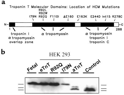
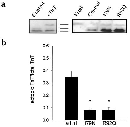
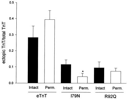
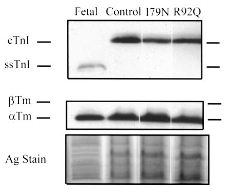
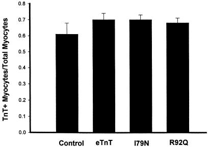
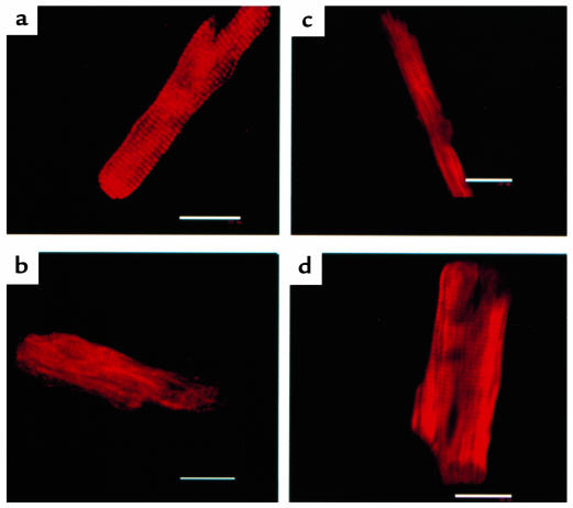
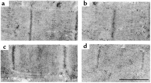
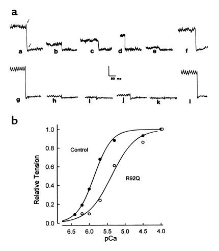
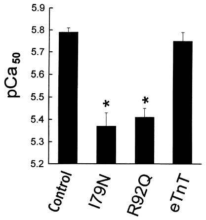
The direct effects of expressing hypertrophic cardiomyopathy-associated (HCM-associated) mutant troponin T (TnT) proteins on the force generation of single adult cardiac myocytes have not been established. Replication-defective recombinant adenovirus vectors were generated for gene transfer of HCM-associated I79N and R92Q mutant cardiac TnT cDNAs into fully differentiated adult cardiac myocytes in primary culture. We tested the hypothesis that the mutant TnT proteins would be expressed and incorporated into the cardiac sarcomere and would behave as dominant-negative proteins to directly alter calcium-activated force generation at the level of the single cardiac myocyte. Interestingly, under identical experimental conditions, the ectopic expression of the mutant TnTs was significantly less ( approximately 8% of total) than that obtained with expression of wild-type TnT ( approximately 35%) in the myocytes. Confocal imaging of immunolabeled TnT showed a regular periodic pattern of localization of ectopic mutant TnT that was not different than that in normal controls, suggesting that mutant TnT incorporation had no deleterious effects on sarcomeric architecture. Direct measurements of isometric force production in single cardiac myocytes demonstrated marked desensitization of submaximal calcium-activated tension, with unchanged maximum tension generation in mutant TnT-expressing myocytes compared with control myocytes. Collectively, these results demonstrate an impaired expression of the mutant protein and a disabling of cardiac contraction in the submaximal range of myoplasmic calcium concentrations. Our functional results suggest that development of new pharmacological, chemical, or genetic approaches to sensitize the thin-filament regulatory protein system could ameliorate force deficits associated with expression of I79N and R92Q in adult cardiac myocytes.


Figures

Figure 1
Figure 1
(a) Schematic of the functional domains of the TnT protein and the location of the I79N and R92Q mutations in the protein. (b) Western blot showing expression of the eTnT, R92Q, I79N, and aTnT protein products in HEK 293 cells infected with the recombinant adenoviral constructs AdCMVeTnT, AdCMVR92Q, AdCMVI79N, and AdCMVaTnT. The migration patterns (from top to bottom) of the embryonic isoform, a minor adult isoform, and the major adult isoform are shown next to the blot. Control is from adult rat heart.
Figure 2
Figure 2
Expression of wild-type and mutant TnT proteins in adult cardiac myocytes. (a) Western blots showing representative expression of control, eTnT-, I79N-,and R92Q-infected adult cardiac myocytes. The antibody used was either an anti-cTnT mAb (MAB 1695; left blot) or an anti–striated muscle TnT mAb (T6277, clone JLT-12; right blot). The migration pattern markers are shown between the 2 blots (from top to bottom: the embryonic isoform, a minor adult isoform, and the major adult isoform). (b) The fraction of the total TnT (endogenous aTnT + ectopic TnT) protein that is either eTnT (n = 19), I79N (n = 23), or R92Q (n = 20). Scanning densitometry was performed to determine the ectopic TnT/total TnT ratio. Bars represent the mean ± SE. *Significantly different than eTnT.
Figure 3
Figure 3
Comparison of ectopic protein expression in membrane-intact and membrane-permeabilized myocyte samples. Scanning densitometry was performed to determine the ectopic TnT/total TnT ratio. eTnT: n = 9 (intact), n = 11 (permeabilized); I79N: n = 11 (intact), n = 12 (permeabilized); R92Q: n = 7 (intact), n = 13 (permeabilized). Bars represent the mean ± SE. *Significant difference between intact and permeabilized. For each ectopic protein, membrane-intact and membrane-permeabilized data were compared using an unpaired t test. P = 0.23 for eTnT; P = 0.04 for I79N; and P = 0.57 for R92Q.
Figure 4
Figure 4
Tropomyosin and troponin I isoform expression pattern in adult myocytes following gene transfer. Western blots and silver-stained gel (to show loading) of control, AdCMVI79N-infected, and AdCMVR92Q-infected adult cardiac myocytes. The migration patterns of cardiac troponin I (cTnI), slow skeletal troponin I (ssTnI), β-tropomyosin (β-Tm), and α-tropomyosin (α-Tm) are shown by markers placed next to the figure.
Figure 5
Figure 5
Summary of scoring of immunofluorescence data for cardiac myocytes following gene transfer. The graph shows the fraction of total rod-shaped myocytes counted that showed a striated pattern of TnT staining (TnT+). Refer to Methods for details on scoring coverslips. Numbers of coverslips examined were as follows: control = 3; eTnT = 7; I79 = 11; and R92Q = 11. Bars represent the mean ± SE.
Figure 6
Figure 6
Representative confocal images of myocytes labeled by indirect immunofluorescence for TnT. (a) Control myocyte. (b) eTnT-expressing myocyte. (c) I79N-expressing myocyte. (d) R92Q-expressing myocyte. Scale bar: 20 μm.
Figure 7
Figure 7
Representative transmission electron micrographs comparing sarcomeric ultrastructure between control myocytes and myocytes after gene transfer. (a) Control. (b) eTnT. (c) R92Q. (d) I79N. Scale bar in d represents 1 μm for a, 0.8 μm for b, 1.1 μm for c, and 1 μm for d.
Figure 8
Figure 8
Direct determination of calcium-activated force generation in single adult cardiac myocytes. (a) Fast time-base records of isometric tension for a single control cardiac myocyte (records a–f) and a single cardiac myocyte after AdCMVR92Q gene transfer (records g–l). pCa values for records a–l are as follows: a and g = 4.0; b and h = 6.2; c and i = 6.0; d and j = 5.7; e and k = 9.0; f and l = 4.0. (b) Summary of the tension-pCa relationships for the control (filled circles) and R92Q (open circles) single cardiac myocytes shown in a. Tension values are normalized to the maximum tension value obtained in each myocyte at pCa 4.0.
Figure 9
Figure 9
Summary of the effects of wild-type and mutant TnT expression on the pCa50 of adult single cardiac myocytes. n = 14 (control); n = 6 (I79N); n = 7 (R92Q); and n = 13 (eTnT). *Significant difference of mutant-expressing myocytes compared with controls or wild-type (eTnT). Values are mean ± SE.

References

    1. Bonne G, Carrier L, Richard P, Hainque B, Schwartz K. Familial hypertrophic cardiomyopathy: from mutations to functional defects. Circ Res. 1998;83:580–593. - PubMed
    1. Maron BJ, Bonow RO, Cannon RO, III, Leon MB, Epstein SE. Hypertrophic cardiomyopathy. I. Interrelations of clinical manifestations, pathophysiology, and therapy. N Engl J Med. 1987;316:780–789. - PubMed
    1. Maron BJ, Bonow RO, Cannon RO, III, Leon MB, Epstein SE. Hypertrophic cardiomyopathy. II. Interrelations of clinical manifestations, pathophysiology, and therapy. N Engl J Med. 1987;316:844–852. - PubMed
    1. Marian AJ. Sudden cardiac death in patients with hypertrophic cardiomyopathy: from bench to bedside with an emphasis on genetic markers. Clin Cardiol. 1995;18:189–198. - PubMed
    1. Thierfelder L, et al. α-tropomyosin and cardiac troponin T mutations cause familial hypertrophic cardiomyopathy: a disease of the sarcomere. Cell. 1994;77:701–712. - PubMed

Publication types

MeSH terms