

Molecular correlates of the calcium-independent, depolarization-activated K+ currents in rat atrial myocytes
- PMID: 10332091
- PMCID: PMC2269341
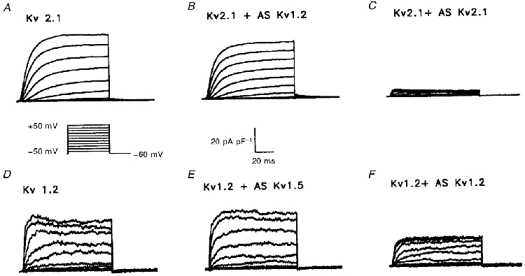
- DOI: 10.1111/j.1469-7793.1999.0407t.x
Molecular correlates of the calcium-independent, depolarization-activated K+ currents in rat atrial myocytes
Abstract
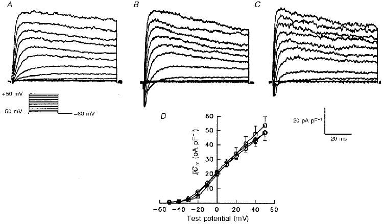
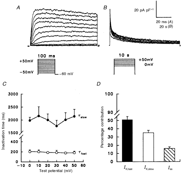
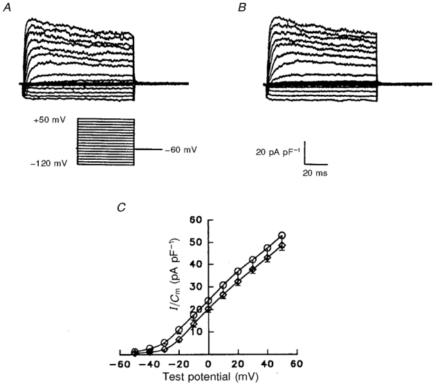
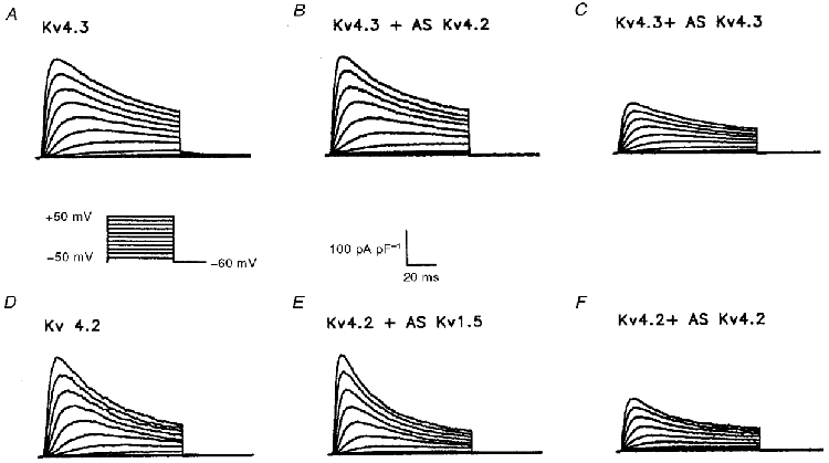
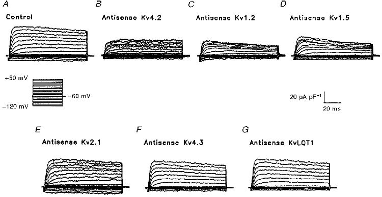
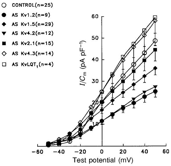
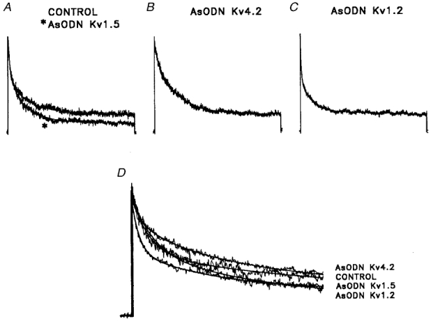
1. In adult rat atrial myocytes, three kinetically distinct Ca2+-independent depolarization-activated outward K+ currents, IK, fast, IK,slow and Iss, have been separated and characterized. 2. To test directly the hypothesis that different voltage-dependent K+ channel (Kv channel) alpha subunits underlie rat atrial IK,fast, IK, slow and Iss, the effects of antisense oligodeoxynucleotides (AsODNs) targeted against the translation start sites of the Kv alpha subunits Kv1.2, Kv1.5, Kv4.2, Kv4.3, Kv2.1 and KvLQT1 were examined. 3. Control experiments on heterologously expressed Kv alpha subunits revealed that each AsODN is selective for the subunit against which it was targeted. 4. Peak outward K+ currents were attenuated significantly in rat atrial myocytes exposed to AsODNs targeted against Kv4.2, Kv1.2 and Kv1.5, whereas AsODNs targeted against Kv2.1, Kv4.3 and KvLQT1 were without effects. 5. No measurable effects on inwardly rectifying K+ currents (IK1) were observed in atrial cells exposed to any of the Kv alpha subunit AsODNs. 6. Kinetic analysis of the currents evoked during long (10 s) depolarizing voltage steps revealed that AsODNs targeted against Kv4.2, Kv1.2 and Kv1.5 selectively attenuate rat atrial IK,fast, IK, slow and Iss, respectively, thus demonstrating that the molecular correlates of rat atrial IK,fast, IK,slow and Iss are distinct. 7. The lack of effect of the Kv4.3 AsODNs on peak outward K+ currents reveals that Kv4.2 and Kv4.3 do not heteromultimerize in rat atria in vivo. In addition, the finding that Kv1.2 and Kv1.5 contribute to distinct K+ currents in rat atrial myocytes demonstrates that Kv1.2 and Kv1.5 also do not associate in rat atria in vivo.
Figures
References
-
- Barhanin J, Lesage F, Guillemare E, Fink M, Lazdunski M, Romey G. K(v)LQT1 and IsK (minK) proteins associate to form the I-Ks cardiac potassium current. Nature. 1996;384:78–80. - PubMed
-
- Barry DM, Nerbonne JM. Myocardial potassium channels: Electrophysiological and molecular diversity. Annual Review of Physiology. 1996;58:363–394. - PubMed
-
- Barry DM, Trimmer JS, Merlie JP, Nerbonne JM. Differential expression of voltage-gated potassium channel subunits in adult rat heart: relation to functional potassium channels? Circulation Research. 1995;77:361–369. - PubMed
-
- Barry DM, Xu H, Schuessler RB, Nerbonne JM. Functional knockout of the transient outward current, Long QT syndrome and cardiac remodelling in mice expressing a dominant negative Kv4 α subunit. Circulation Research. 1998;83:560–567. - PubMed
Publication types
MeSH terms
Substances
Grants and funding
LinkOut - more resources
Full Text Sources
Miscellaneous