. 1999 Jun 1;517 ( Pt 2)(Pt 2):575-90.
doi: 10.1111/j.1469-7793.1999.0575t.x.

Quantitative analysis of peristalsis in the guinea-pig small intestine using spatio-temporal maps

Affiliations

Quantitative analysis of peristalsis in the guinea-pig small intestine using spatio-temporal maps

G W Hennig et al. J Physiol. .

Abstract

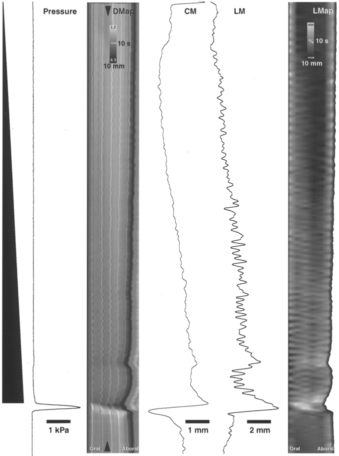
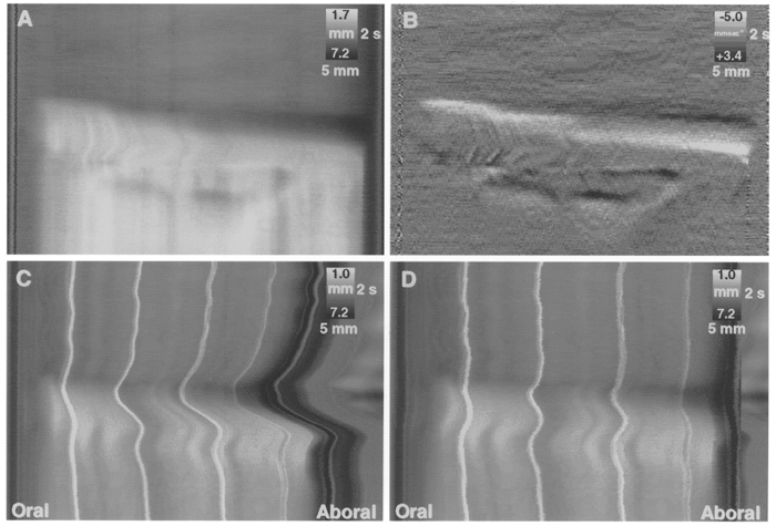
1. Peristalsis was evoked in guinea-pig small intestine by slow fluid infusion and recorded onto video and digitized. Spatio-temporal maps of diameter and longitudinal movement were constructed and parameters of motion were calculated. 2. During the filling of the isolated segments of intestine, rhythmic local longitudinal movements were observed at several points along the preparation. These phasic longitudinal muscle contractions were associated with small but significant local increases in diameter and probably reflect a passive mechanical coupling by connective tissue in the gut wall. In addition, occasional synchronized longitudinal muscle contractions caused net shortening of the preparation and always preceded the onset of peristaltic emptying. 3. Peristaltic emptying was characterized by a contraction of the circular muscle which usually started at the oral end of the preparation, that propagated aborally, propelling the contents. However, in 19 % of trials, the first circular muscle contraction occurred in the aboral half of the preparation. 4. The propagation of peristalsis consisted of separate sequential circular muscle contractions several centimetres long, particularly in the oral half of the preparation, giving a 'step-like' appearance to the spatio-temporal map. The gut was transiently distended aboral to the propagating circular muscle contraction due to the propulsion of contents. 5. At each point in the preparation, the longitudinal muscle remained contracted during the propulsive part of the circular muscle contraction. Only when the circular muscle contraction became lumen occlusive did lengthening of the longitudinal muscle take place. 6. Spatio-temporal maps are a powerful tool to visualize and analyse the complexity of gastrointestinal motility patterns.


Figures

Figure 1
Figure 1. Experimental arrangements used to study peristalsis
A segment of ileum (black), cannulated at each end, was placed in oxygenated warmed Krebs solution. Intraluminal pressure was monitored with a pressure transducer (P) and Krebs solution was infused via the oral cannula by a syringe pump. Movements of the preparation were recorded onto S-VHS video tape. The preparations emptied their luminal contents via the aboral cannula, through a one-way valve or into a vertical tube. In A, both ends of the preparation were fixed to prevent the overall shortening of the segment and the height of the outflow was adjusted to control outflow resistance. In B, the preparation was able to shorten during filling as the outflow cannula was mounted on a flexible tube. In C, the preparation emptied via the aboral cannula into a large diameter tube, thus maintaining intraluminal pressure constant. Fluid flowed back into the intestine after emptying.
Figure 2
Figure 2. Construction of spatio-temporal map of intestinal diameter (DMaps)
A single frame of a video recording of the intestine (A) was subjected to the threshold procedure (see Methods) and converted into a binary image (B). The number of black pixels in each column (corresponding to diameter of the intestine at each point along its length) was converted into a grey scale pixel (bar above preparation, in B). Multiple frames were treated in this way to produce a diameter map (C), showing the diameter at each point from the oral end of the preparation to the aboral end: the preparation was 55 mm long. In this map, the minimum diameter was 2.1 mm (represented by white pixels) and the maximum was 7.5 mm (black pixels) and the map represents a time period of approximately 18 s. Time starts at the top of the diameter map. The white dotted line corresponds to the frame shown in A and B. Using the diameter map, the profile of the intestine could be reconstructed at any moment (D).
Figure 3
Figure 3. Construction of spatio-temporal maps of longitudinal motion (LMaps)
A single frame of a video recording shows five black surface markers along the length of the preparation (A). A DMap, constructed from 20 s of recording (B) shows the movements of the surface markers as vertical, pale, wavy bands - the black dotted line represents the frame shown in A. A tracking routine was used to define the positions of the surface markers (black wavy lines, C) and the midpoint between each band was determined (grey lines, C). The distance between adjacent surface marker lines was then determined and represented as a grey scale pixel, at the calculated midpoint. Linear interpolation of grey scales was used to fill in the areas between the midpoints (D). The resulting LMap shows local shortening of the preparation, compared with resting conditions (prior to Krebs infusion) as pale grey. Local lengthening of the preparation is shown as dark grey. The right hand end of the LMap (D) represents the net shortening of the preparation in each video frame.
Figure 4
Figure 4. Example of peristalsis evoked by slow liquid infusion at 4.25 μl s−1
Infused fluid volume is shown schematically on the left as a black ramp. Time starts at the top of the figure. Intraluminal pressure is represented in the adjacent trace, showing a large, abrupt increase in pressure during peristaltic emptying. The diameter map (DMap) of the same period shows a gradual darkening during the course of the infusion, reflecting the gradual increase in diameter (see circular muscle diameter trace, CM). The arrowheads on the DMap show the point of the intestine at which circular muscle (CM) and longitudinal muscle (LM) traces were generated. Longitudinal muscle activity also increased during the preparatory phase (shown by the overall shortening of the preparation in the DMap and by the trace of longitudinal muscle activity, LM). The longitudinal map of the same period (right hand part of figure, LMap) shows a gradual shortening of the segment of intestine during the infusion (revealed as a gradual lightening of the map). The pattern of oblique stripes in the LMap during the preparatory phase corresponds to phasic changes in longitudinal muscle length (at approximately 37 c.p.m.) that occur with some delay along the length of the preparation, suggesting that the longitudinal muscle contractions propagate sometimes orally (sloping to the left) and at other times aborally (sloping to the right). There is a sudden net shortening of the longitudinal muscle just prior to the onset of peristalsis (visible in both the DMap and LMap). The circular muscle initially lengthens at the start of peristaltic emptying, then rapidly shortens (leftward deflection on CM trace). In contrast the longitudinal muscle, at the same point in the intestine, shortens prior to emptying (rightward deflection on LM trace).
Figure 5
Figure 5. Rate of change of diameter varied along the intestine during slow filling
Measurements of circular muscle diameter at the oral, middle and aboral ends of 5 preparations were used to calculate the mean rate of increase of diameter during slow filling. The oral end distended significantly more slowly during infusion than the aboral end (P < 0.05), indicating that the oral end is less distensible. This probably reflects greater inhibitory neuronal input to the circular muscle at the aboral end during filling.
Figure 6
Figure 6. Relationship between longitudinal and circular muscle during the preparatory phase
During slow filling of the intestine, the longitudinal muscle showed characteristic phasic contractions that appeared to propagate orally (LMap in B). These correlated with small phasic increases in overall diameter of the preparation revealed in DMaps (see dark bands in A). This suggests that localized longitudinal muscle shortening may cause a localized small increase in diameter.
Figure 9
Figure 9. Relationship between circular and longitudinal shortening during the emptying phase
Local changes in diameter and length (calculated from surface markers) were plotted for the oral, middle and aboral regions of a preparation during the preparatory and emptying phases of peristalsis. Values were calculated at intervals of 40 ms (each represented by a single dot). During filling, there were phasic contractions of the longitudinal muscle which correlated with phasic increases in diameter (between points a and b). At the onset of peristaltic emptying (point b), there was a sudden decrease in diameter, initially without lengthening, indicating that the longitudinal muscle did not relax during the initiation of peristalsis. Lengthening only commenced during the later stages of the circular muscle contraction (point c). Similarly, in the middle and aboral regions, there was no significant localized lengthening during the circular muscle contraction. However, particularly at the aboral end of the preparation there was a significant increase in diameter just prior to the circular muscle contraction due to fluid displaced from the contraction further orally.
Figure 7
Figure 7. Comparison of peristaltic emptying in preparations that were either fixed at both ends or free to move at the aboral end
A DMap of a preparation in which both oral and aboral ends were attached to fixed cannulae (A) shows an abrupt start to the emptying phase. The same map differentiated, to show the dynamic changes in circular muscle length (B), reveals that the emptying occurred as a sequence of three contractions at various points along its length, giving rise to ‘step-like’ propagation. In C, the aboral end of the preparation was attached to a freely moving cannula (see Fig. 1B) and surface markers were used to map the local shortening of the preparation. A pronounced shortening of the preparation occurred prior to the initial contraction of the circular muscle and the preparation lengthened during the ensuing emptying phase. The DMap was subsequently processed to maintain a constant overall length (D), making it possible to compare directly the emptying phase of fixed preparations (A) and preparations that were free to shorten (D), revealing that the rate of propagation of circular muscle contraction was not affected by shortening of the longitudinal muscle.
Figure 8
Figure 8. Dynamics of peristaltic emptying in preparations maintained at a fixed length
Three differentiated diameter maps from each of four animals (A, B, C and D) show features of propagation of the circular muscle contraction during the emptying phase. Most examples show a ‘step-like’ activation of the circular muscle contraction as it propagated aborally (B top and bottom, C top and D bottom). The initial circular muscle contraction usually occupied a small region at the oral end of the segment; however, occasionally the contraction occurred in a large region simultaneously (C and D middle). Occasionally, a circular muscle contraction occurred aborally before the peristaltic contraction was initiated at the oral end (B middle). The dark bands after the peristaltic contraction (B) are artefacts due to excessive bending caused by longitudinal muscle movement in a fixed preparation.

References

    1. Alvarez WC, Zimmermann A. The absence of inhibition ahead of peristaltic rushes. American Journal of Physiology. 1927;83:52–59.
    1. Baur M. Die Peristaltik des isolierten Meerschweinchendunndarms im Filmversuch. Archives of Experimental Pathology and Pharmacology. 1928;133:69–83.
    1. Bayliss WM, Starling EH. The movements and innervation of the small intestine. The Journal of Physiology. 1899;24:99–143. - PMC - PubMed
    1. Benard T, Bouchoucha M, Dupres M, Cugnenc PH. In vitro analysis of rat intestinal wall movements at rest and during propagated contraction - a new method. American Journal of Physiology. 1997;36:776–784. - PubMed
    1. Bennett A, Eley KG, Stockley HL. Inhibition of peristalsis in guinea-pig isolated ileum and colon by drugs that block prostaglandin synthesis. British Journal of Pharmacology. 1976;57:335–340. - PMC - PubMed

Publication types

LinkOut - more resources