. 1999 Jun;67(6):2804-9.
doi: 10.1128/IAI.67.6.2804-2809.1999.

CD4(+) T cells and the proinflammatory cytokines gamma interferon and interleukin-6 contribute to alveolar bone loss in mice

Affiliations

CD4(+) T cells and the proinflammatory cytokines gamma interferon and interleukin-6 contribute to alveolar bone loss in mice

P J Baker et al. Infect Immun. 1999 Jun.

Abstract

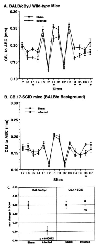
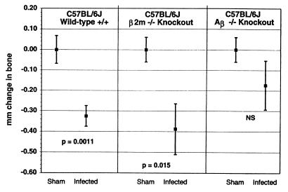
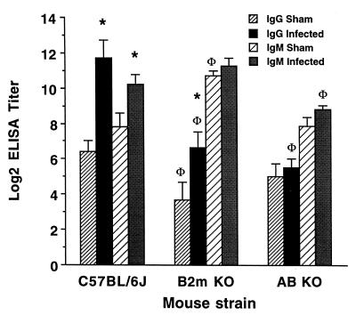
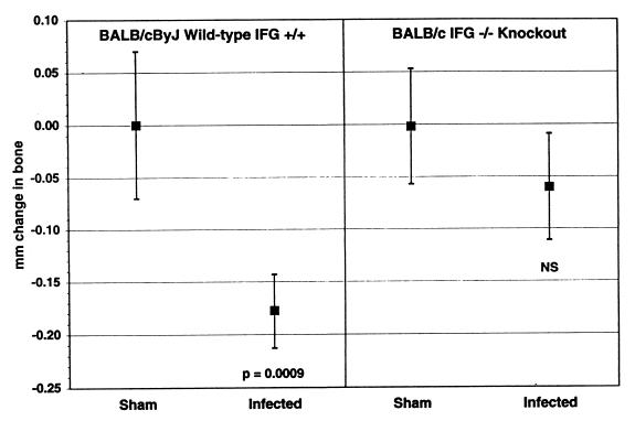
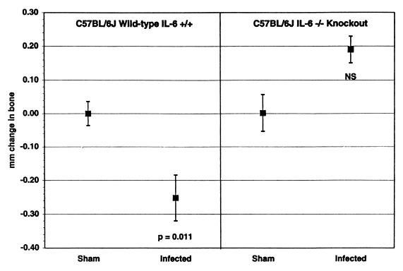
In this study, we used a mouse model to examine the role of the adaptive immune response in alveolar bone loss induced by oral infection with the human gram-negative anaerobic bacterium Porphyromonas gingivalis. Severe combined immunodeficient mice, which lack B and T lymphocytes, exhibited considerably less bone loss than did immunocompetent mice after oral infection, suggesting that lymphocytes contribute to this process. Bone loss after oral infection was decreased in mice deficient in major histocompatibility complex (MHC) class II-responsive CD4(+) T cells, but no change in bone loss was observed in mice deficient in MHC class I-responsive CD8(+) T cells or NK1(+) T cells. Mice lacking the cytokine gamma interferon or interleukin-6 also demonstrated decreased bone loss. These results suggest that the adaptive immune response, and in particular CD4(+) T cells and the proinflammatory cytokines that they secrete, are important effectors of bone loss consequent to P. gingivalis oral infection. The studies also reinforce the utility of the mouse oral infection model in dissecting the pathobiology of periodontal disease.


Figures

FIG. 1
FIG. 1
Bone loss after oral infection with P. gingivalis occurred in immunocompetent mice (A) but not in immunodeficient SCID mice (B). L, left; R, right. Sites 1 to 3 are on the first molar, sites 4 and 5 are on the second molar, and sites 5 and 6 are on the third molar. Data points represent the means from 13 mice ± 1 SEM. (A) BALB/cByJ wild-type, immunocompetent mice. The CEJ:ABC was greater in infected mice than in sham-infected mice at every site, indicating bone loss. ∗, values in infected mice significantly greater than in sham-infected controls (P < 0.05). When comparisons were made on the 14-site total CEJ:ABC, values in infected mice were also significantly greater than in the sham-infected mice (P = 0.0001). (B) CB.17-SCID mice on a BALB/cJ genetic background. At every site, the CEJ:ABC in infected SCID mice was equal to or less than the distances in the sham-infected mice, indicating no bone loss. At site R6, the values in sham-infected mice were greater than in infected mice. (▵; P < 0.05). (C) Transformation of data from panels A and B to give millimeter change in bone. The 14-site total CEJ:ABC for each mouse was subtracted from the mean CEJ:ABC from groups of sham-infected mice to produce the millimeter change in bone, where negative values indicate bone loss. Squares represent the means ± 1 SEM of 13 mice per group. There was a significantly greater change in bone in infected BALB/cByJ mice than in sham-infected BALB/cByJ mice, but in SCID mice infection did not induce a change in bone (NS, not significantly different from sham-infected mice at P ≥ 0.05).
FIG. 2
FIG. 2
Bone loss after oral infection with P. gingivalis occurred in wild-type, immunocompetent mice (C57BL/6J) and in mice lacking CD8+ and NK1+ T cells (B2m-knockout mice on a C57BL/6J background) but was diminished in mice deficient in CD4+ T lymphocytes (Aβ-knockout mice on a C57BL/6J background). The CEJ:ABC from 14 sites were summed for each mouse, and the total from each mouse was subtracted from the mean totals in the sham-infected mice to give the total millimeter of change in bone. Negative values of millimeter of change in bone indicate bone loss, which was significantly greater in infected wild-type and B2m-knockout mice than in sham-infected mice of the same strain at the P values shown. In Aβ-knockout mice, the bone change in infected mice was not significantly different (NS) than in sham-infected mice at P ≥ 0.05. Data points represent the means from nine mice ± 1 SEM. The expected changes in CD4+ and CD8+ lymphocyte populations were confirmed by flow cytometry (data not shown).
FIG. 3
FIG. 3
Influence of class I or class II proteins on the generation of P. gingivalis-specific antibodies. P. gingivalis-reactive IgG was increased in C57BL/6J and β2m-knockout (β2m KO) mice orally infected with P. gingivalis (∗, different from sham-infected controls at P < 0.05) but not in Aβ-knockout (Aβ KO) mice. P. gingivalis-reactive IgM was increased in orally infected C57BL/6J mice (∗) but not in infected β2m- or Aβ-knockout mice. Both sham-infected and infected β2m-knockout mice had less P. gingivalis-reactive IgG than sham-infected or infected C57BL/6J mice, as did infected Aβ-knockout mice (Φ, different from C57BL/6J at P < 0.05). Anti-P. gingivalis IgA was not found in any of the mouse strains tested (data not shown).
FIG. 4
FIG. 4
Alveolar bone response to oral infection in IFG-knockout BALB/cByJ mice compared to wild-type, immunocompetent BALB/cByJ mice. Infected BALB/cByJ mice lost bone (P = 0.0009 compared to sham-infected BALB/cByJ mice), but infected IFG-knockout mice did not (NS, not significantly different from IFG-knockout sham-infected mice at P ≥ 0.05). The CEJ:ABC from 14 sites were summed for each mouse, and the value from each mouse was subtracted from the mean totals from the sham-infected mice to give the total millimeters of bone lost. n = 20 ± 1 SEM.
FIG. 5
FIG. 5
Alveolar bone response to oral infection in IL-6-knockout C57BL/6J mice compared to wild-type, immunocompetent C57BL/6J mice. Infected C57BL/6J mice lost bone (P = 0.011 compared to sham-infected C57BL/6J), but infected IL-6-knockout mice did not (NS, not significantly different from IL-6-knockout sham-infected mice at P ≥ 0.05). The 14-site total CEJ:ABC for each mouse was subtracted from the mean CEJ:ABC from groups of sham-infected mice to produce the millimeter mm change in bone, where negative values indicate bone loss. n = 20 ± 1 SEM.

References

    1. Baker P J, Evans R T, Roopenian D C. Oral infection with Porphyromonas gingivalis and induced alveolar bone loss in immunocompetent and severe combined immunodeficient mice. Arch Oral Biol. 1994;39:1035–1040. - PubMed
    1. Baker, P. J., S. Carter, M. Dixon, R. T. Evans, and D. C. Roopenian. Serum antibody response to oral infection precedes but does not prevent Porphyromonas gingivalis-induced alveolar bone loss in mice. Oral Microbiol. Immunol., in press. - PubMed
    1. Baker, P. J., M. Dixon, and D. C. Roopenian. Genetic control of susceptibility to alveolar bone loss in mice. Submitted for publication. - PMC - PubMed
    1. Baron R, Vignery A, Horowitz M. Lymphocytes, macrophages, and the regulation of bone remodeling. In: Peck W A, editor. Bone and mineral research annual 2: a yearly survey of developments in the field of bone and mineral metabolism. New York, N.Y: Elsevier Science Publishers; 1983. pp. 175–243.
    1. Been V, Engel D. The effects of immunosuppressive drugs on periodontal inflammation in human renal allograft patients. J Periodontol. 1982;53:245–248. - PubMed

Publication types

LinkOut - more resources