. 1999 Jun;67(6):2874-83.
doi: 10.1128/IAI.67.6.2874-2883.1999.

Access of antibody or trypsin to an integral outer membrane protein (P66) of Borrelia burgdorferi is hindered by Osp lipoproteins

Affiliations

Access of antibody or trypsin to an integral outer membrane protein (P66) of Borrelia burgdorferi is hindered by Osp lipoproteins

J Bunikis et al. Infect Immun. 1999 Jun.

Abstract

The outer membrane of Borrelia burgdorferi, the Lyme disease agent, contains lipoproteins anchored by their lipid moieties and integral proteins with membrane-spanning regions. We used the techniques of in situ proteolysis, immunofluorescence, in vitro growth inhibition, and cross-linking with formaldehyde to characterize topological relationships between P66, an integral membrane protein, and selected Osp lipoproteins of B. burgdorferi. Protease treatment of intact spirochetes cleaved P66 and Osp proteins but not the periplasmic flagellin or the BmpA protein of the cytoplasmic membrane. P66 of cells lacking OspA, OspB, and OspC was more susceptible to trypsin cleavage than was P66 of cells with these Osp proteins. A monoclonal antibody against the surface loop of P66 bound, agglutinated, and inhibited the growth of viable spirochetes lacking OspA, OspB, OspC, and OspD but not of the cells that expressed OspA, OspC, and/or OspD. When cells were fixed, the antibody bound to cells that express OspD and OspC but still not to cells with OspA. The close association of OspA and P66 was confirmed by the crosslinking of the two proteins by formaldehyde. These results show that Osp proteins, particularly OspA, limit the access of antibody or trypsin to the surface loop region of P66. The proximity and possible contact between P66 and OspA (or other Osp proteins) may hinder the effectiveness of antibodies to what otherwise would be an appropriate vaccine target.


Figures
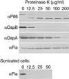
FIG. 1
FIG. 1
Western blot analysis of the effect of proteinase K on selected B. burgdorferi B31 proteins. Intact (top) or sonicated (bottom) cells were incubated in different concentrations of proteinase K. After being washed, the cells were subjected to PAGE (12% acrylamide), and blots of the cell lysates were probed with the monoclonal antibodies (α) against OspA (H5332), OspB (H6831), P66 (H914), or flagellin (H9724).
FIG. 2
FIG. 2
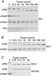
Western blot analysis of the effect of trypsin on selected proteins of B. burgdorferi isolates with different Osp phenotypes. Cells of B31 (A), B314 (B), and B313 and HB19R1 (C) were incubated with different concentrations of trypsin and subjected to PAGE (12% acrylamide). Blots of the cell lysates were probed with the monoclonal antibodies (α) against OspA (H5332), OspB (H6831), OspC, OspD (1C8), P66 (H1337), or flagellin (H9724).
FIG. 3
FIG. 3
Western blot analysis of the binding of monoclonal antibodies (α) for P66 (H1337) or OspB (H6831) to B. burgdorferi B31 cells. Different number of cells (range, 0.4 × 10⁹ to 16 × 109) were incubated in buffer with or without trypsin at 50 μg/ml. The cells were then subjected to PAGE (12% acrylamide), and blots of the cell lysates were probed with antibodies.
FIG. 4
FIG. 4
Photomicrographs under UV illumination of binding of monoclonal antibodies to methanol-fixed B. burgdorferi cells with different Osp phenotypes (Table 1). The isolates examined were B31, B314, HB19R1, and B313. Smears of spirochetes with sheep erythrocytes were incubated first with monoclonal antibodies (α) against P66 (H1337 or H914), OspA (H5332), OspC, OspD (1C8), or flagellin (H9724) and then with fluorescein-conjugated sheep anti-mouse immunoglobulin. Magnification, ×80.
FIG. 5
FIG. 5
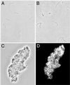
Photomicrographs under phase-contrast (A to C) or UV (D) microscopy of unfixed, suspended cells of B. burgdorferi isolates HB19R1 (A) or B313 (B to D) incubated with monoclonal antibodies H1337 (A, C, and D) or H914 (B). After incubation with the antibody, the cells were washed and incubated with fluorescein-conjugated sheep anti-mouse immunoglobulin. Magnification, ×400.
FIG. 6
FIG. 6
Western blot analysis of cross-linked proteins of B. burgdorferi B31 (A) and B313 and B314 (B). Intact or trypsin-treated cells were incubated in 1% (vol/vol) formaldehyde or buffer alone, washed, and lysed. The lysates were either directly subjected to Western blot analysis of a 10% acrylamide gel or first immunoprecipitated with monoclonal antibody H1337. The blots were probed with monoclonal antibodies H5532 (αOspA) or H1337 (αP66). Positions of monomers of OspA and full-length and trypsin-truncated (tr) P66 are indicated. Positions of putative heteromers of OspA and the full-length or truncated P66 are indicated by an asterisk and arrowhead, respectively. Molecular weight standards (MWS) in thousands (shown to the right) are myosin H-chain (200.0), phosphorylase B (97.4), bovine serum albumin (68.0), ovalbumin (43.0), carbonic anhydrase (29.0), and β-lactoglobulin (18.4).
FIG. 7
FIG. 7
Western blot analysis of the polyclonal antisera from mice immunized with whole-cell B. burgdorferi B31 and B313. B31 and B313 cell lysates were separated by PAGE (12% acrylamide), blotted, and probed with pooled antisera. Asterisks mark the positions of P66 and an approximately 20-kDa protein detected exclusively by antisera against Osp-less B313 cells. Molecular weight standards (MWS) in thousands (shown to the left) are the same as in Fig. 6.

References

    1. Anonymous. Recomendations for test performance and interpretation from the Second International Conference on Serologic Diagnosis of Lyme Disease. Morbid Mortal Weekly Rep. 1995;44:590–591. - PubMed
    1. Åsbrink E, Hovmark A, Hederstedt B. The spirochetal etiology of acrodermatitis chronica atrophicans Herxheimer. Acta Dermatol Venereol. 1984;64:506–512. - PubMed
    1. Barbour A G. Isolation and cultivation of Lyme disease spirochetes. Yale J Biol Med. 1984;57:521–525. - PMC - PubMed
    1. Barbour A G, Burgdorfer W, Grunwaldt E, Steere A C. Antibodies of patients with Lyme disease to components of the Ixodes dammini spirochete. J Clin Investig. 1983;72:504–515. - PMC - PubMed
    1. Barbour A G, Garon C F. Linear plasmids of the bacterium Borrelia burgdorferi have covalently closed ends. Science. 1987;237:409–411. - PubMed

Publication types

MeSH terms

LinkOut - more resources