

doi: 10.1128/IAI.67.6.3151-3154.1999.
Macrophages and epithelial cells respond differently to the Pseudomonas aeruginosa type III secretion system
Affiliations
- PMID: 10338535
- PMCID: PMC96636
- DOI: 10.1128/IAI.67.6.3151-3154.1999
Item in Clipboard
Macrophages and epithelial cells respond differently to the Pseudomonas aeruginosa type III secretion system
Infect Immun.
1999 Jun.
Abstract
The multiple effects of Pseudomonas aeruginosa type III secretion have largely been attributed to variations in cytotoxin expression between strains. Here we show that the target cell type is also important. While lung epithelial cells showed significant changes in morphology but not viability when infected with P. aeruginosa, macrophages were efficiently killed by P. aeruginosa. Both responses were dependent on the type III secretion system.
Figures
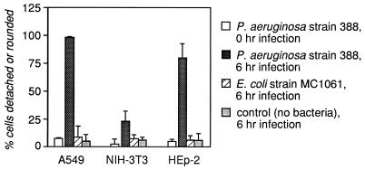
Human lung epithelial cells are susceptible to P. aeruginosa intoxication. The human epithelial cell lines A549 (lung carcinoma derived) and HEp-2 (laryngeal carcinoma derived) and the murine fibroblast line NIH 3T3 were infected for various times with E. coli MC1061 or with P. aeruginosa 388 at an MOI of 10 (bacterium/cell ratio = 10) or with the medium without bacteria. Detachment was scored with a hemocytometer, and rounding was scored visually at a magnification of ×100. Shown are the means plus standard deviations of four fields in two wells for each condition; data are representative of two independent experiments. Data are shown for only the 0- and 6-h points.
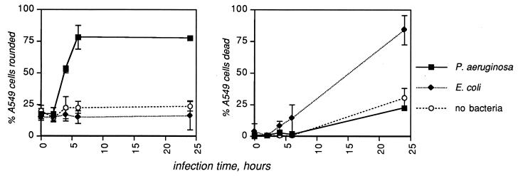
P. aeruginosa causes rounding but not death of human lung epithelial cells. Layers of the human lung epithelial cell line A549 were infected with the medium control, WT P. aeruginosa 388, or E. coli MC1061, at an MOI of 10 bacteria/cell. Rounding was assessed visually at a magnification of ×100; death was assessed by trypan blue exclusion. Shown are the means plus standard deviations of four fields in each of three replicate wells. Data are representative of several independent experiments.
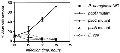
The type III secretion system is required for intoxication of A549 lung epithelial cells by P. aeruginosa. A panel of mutants in various components of the type III secretion apparatus (15, 17) was tested as described for Fig. 1 and 2. The results shown were obtained with an MOI of 10 for each bacterial strain; similar results were obtained at an MOI of 1. Shown are the means plus standard deviations of four fields in each of three replicate wells.
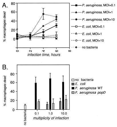
Macrophages are killed by P. aeruginosa. Primary cultures of bone marrow-derived macrophages from strain A/J mice were infected as described for Fig. 1 and 2 with either P. aeruginosa or E. coli. Death was quantitated by trypan blue exclusion. Changes in gross morphology were assessed visually, but no significant differences between infection conditions were observed. (A) Time course of toxicity in response to different MOI of P. aeruginosa and E. coli; shown are the means plus standard deviations of four fields in three wells. (B) Toxicity is dependent on the type III secretion system. The data shown are the means plus standard deviations of six fields in two wells for the 6-h point only.
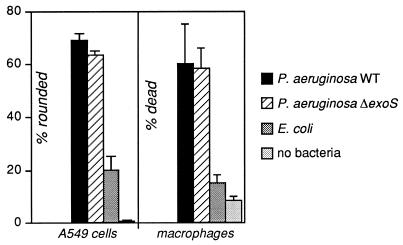
The A549 epithelial cell line and macrophages are intoxicated to the same extent by WT and exoS mutant strains of P. aeruginosa. Layers of each cell type were infected for 6 h at an MOI of 10 with the WT P. aeruginosa 388, with a derivative of that strain in which the gene encoding ExoS had been disrupted (11), with E. coli MC1061, or with no bacteria. Cell rounding was assessed visually at a magnification of ×100. Cell death was assessed by trypan blue exclusion. The A549 data represent the means plus standard deviations of four fields in three wells; the macrophage data are from six fields in two wells.
References
-
- Coburn J, Wyatt R T, Iglewski B H, Gill D M. Several GTP-binding proteins, including p21c-H-ras, are preferred substrates of Pseudomonas aeruginosa exoenzyme S. J Biol Chem. 1989;264:9004–9008. - PubMed
-
- Finck-Barbançon V, Goranson J, Zhu L, Sawa T, Wiener-Kronish J P, Fleiszig S M, Wu C, Mende-Mueller L, Frank D W. ExoU expression by Pseudomonas aeruginosa correlates with acute cytotoxicity and epithelial injury. Mol Microbiol. 1997;25:547–557. - PubMed
-
- Finck-Barbançon, V., and D. W. Frank. Unpublished hybridization data.
Publication types
MeSH terms
Substances
Grants and funding
LinkOut - more resources
Full Text Sources
