

Review
doi: 10.1073/pnas.96.11.5891.
Bacterial cell division: a moveable feast
Affiliations
- PMID: 10339512
- PMCID: PMC34200
- DOI: 10.1073/pnas.96.11.5891
Item in Clipboard
Review
Bacterial cell division: a moveable feast
Proc Natl Acad Sci U S A.
.
No abstract available
Figures
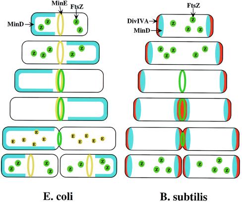
Models for division-site selection in E.
coli (Left) and B. subtilis
(Right). MinD is in blue, MinE in yellow, FtsZ in green
and DivIV in red. Shown are different stages of the cell cycle,
beginning with a newborn cell and finishing with cell division that
produces two daughter cells. (Left) In E.
coli, MinE localizes to a ring-like structure at or near the
middle of the cell early in the division cycle. MinD accumulates
alternately at the membrane periphery on either side of the MinE ring
(3). The alternation of MinD localization from one pole to the other
occurs at a frequency of the order of tens of seconds. The rapid
relocation of MinD ensures that no FtsZ ring is assembled at either the
¼ or ¾ sites in the cell halves. The presence of MinE
at midcell prevents the MinD inhibitory activity at this site, allowing
assembly of the FtsZ ring at this site. The MinE ring disassembles
before completion of constriction. (Right) In B.
subtilis, DivIVA and MinD are localized to the cell poles in a
newborn cell, and therefore the presence of the MinD inhibitor prevents
the formation of the FtsZ ring at these sites. Later, presumably after
completion of DNA replication, a new potential division site is created
at midcell. The sequestration of the MinD inhibitor to the poles allows
assembly of the FtsZ ring at midcell and recruitment of other cell
division proteins. At this point, the division machinery presumably
becomes resistant to the MinD inhibition. DivIVA and MinD proteins then
are recruited to the midcell. Constriction then is initiated. When
constriction is completed, the FtsZ ring disassembles, but DivIVA and
MinD remain at the newly formed poles, preventing further divisions
from taking place in these polar sites.
Comment on
-
Rapid pole-to-pole oscillation of a protein required for directing division to the middle of Escherichia coli.Proc Natl Acad Sci U S A. 1999 Apr 27;96(9):4971-6. doi: 10.1073/pnas.96.9.4971. Proc Natl Acad Sci U S A. 1999. PMID: 10220403 Free PMC article.
References
Publication types
MeSH terms
Substances
LinkOut - more resources
Full Text Sources
Other Literature Sources
