. 1999 May 25;96(11):6072-7.
doi: 10.1073/pnas.96.11.6072.

A role for the light-dependent phosphorylation of visual arrestin

Affiliations

A role for the light-dependent phosphorylation of visual arrestin

P G Alloway et al. Proc Natl Acad Sci U S A. .

Abstract

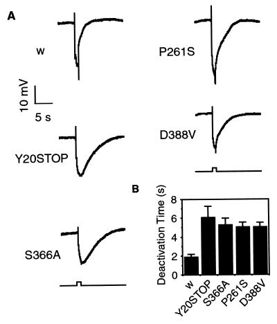
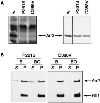
Arrestins are regulatory proteins that participate in the termination of G protein-mediated signal transduction. The major arrestin in the Drosophila visual system, Arrestin 2 (Arr2), is phosphorylated in a light-dependent manner by a Ca2+/calmodulin-dependent protein kinase and has been shown to be essential for the termination of the visual signaling cascade in vivo. Here, we report the isolation of nine alleles of the Drosophila photoreceptor cell-specific arr2 gene. Flies carrying each of these alleles underwent light-dependent retinal degeneration and displayed electrophysiological defects typical of previously identified arrestin mutants, including an allele encoding a protein that lacks the major Ca2+/calmodulin-dependent protein kinase site. The phosphorylation mutant had very low levels of phosphorylation and lacked the light-dependent phosphorylation observed with wild-type Arr2. Interestingly, we found that the Arr2 phosphorylation mutant was still capable of binding to rhodopsin; however, it was unable to release from membranes once rhodopsin had converted back to its inactive form. This finding suggests that phosphorylation of arrestin is necessary for the release of arrestin from rhodopsin. We propose that the sequestering of arrestin to membranes is a possible mechanism for retinal disease associated with previously identified rhodopsin alleles in humans.


Figures

Figure 1
Figure 1
Flies carrying arr2 missense mutations have prolonged deactivation kinetics that are indistinguishable from those of an arr2 null allele. (A) Representative electroretinogram recordings of white-eyed control (w) and arr2 mutant flies exposed to a 1-s pulse of blue light (480 nm). Electroretinograms were performed as described (18). (B) Histogram of the time to 85% deactivation for white-eyed control (w) and arr2 mutant flies. For w control flies, t85 = 1.87 ± 0.3 s; for Y20STOP flies, t85 = 6.07 ± 1.2 s; for S366A flies, t85 = 5.27 ± 0.7 s; for P261S flies, t85 = 5.05 ± 0.5 s; for D388V flies, t85 = 5.03 ± 0.5 s (n = 20–25). Data are means ± SD.
Figure 2
Figure 2
Phosphorylation assays of retinal proteins indicate that the S366A mutant Arr2 protein is not phosphorylated in vitro and fails to undergo light-induced phosphorylation in vivo. (A, Left) Autoradiograph of γ-32P-labeled proteins isolated from Drosophila head lysates in the presence of 0.5 mM Ca2+ (lanes 1, 3, and 4) or 2 mM EGTA, a calcium-specific chelator (lane 2). (A, Right) The same gel transferred to nitrocellulose and probed with antibodies specific for Arr1 and Arr2 (18), as well as Rh1 (Developmental Studies Hybridoma Bank, University of Iowa). Y20STOP represents a null mutation in Arr2. w, white-eyed control flies. (B, Left) Autoradiographs of in vivo γ-32P-labeled retinal proteins from dark-reared white-eyed control (w) and S366A flies either kept in the dark (D) or exposed to light for 30 min (L). (B, Right) The same gels transferred to nitrocellulose and probed with antibodies specific for Arr2 and Arr1. Note the lack of light-induced phosphorylation of Arr2 in the flies expressing the S366A transgene.
Figure 3
Figure 3
S366A flies retain the ability to bind to rhodopsin in a light-dependent manner. (A) Isolated wild-type fly heads were exposed to 5 min of either orange (580 nm) or blue (480 nm) light, homogenized in the dark, and centrifuged (13,000 × g for 5 min) Pellet (P) and supernatant (S) fractions were subjected to SDS/PAGE and Western analysis with antibodies directed against Arr2 and Rh1. (B) Western blot of white-eyed control (w), ninaEI17, S366A, and S366A;ninaEI17 heads that were exposed to 5 min of blue light and then fractionated and centrifuged as described in A. ninaEI17 represents a null mutation in the structural gene for the major rhodopsin, Rh1
Figure 4
Figure 4
Phosphorylated Arr2 protein is found as a non-membrane-associated soluble factor. (Left) Autoradiograph of γ-32P-labeled proteins isolated from fractionated wild-type Drosophila head lysates in the presence of 0.5 mM Ca2+ (lanes 1 and 2) or 2 mM EGTA (lanes 3 and 4). S, supernatant fraction; P, pellet fraction. (Right) The same gel transferred to nitrocellulose and probed with antibodies specific for Arr1, Arr2, and Rh1.
Figure 5
Figure 5
Mutant flies that have reduced phosphorylation of Arr2 have defects in the release of Arr2 from rhodopsin. Western blot of fractionated white-eyed control (w), S366A, norpAP41 (norpA), and cam352/camn339 (cam) mutant heads that were exposed to 10 s of blue light (B) or 10 s of blue light followed by 60 s of orange light (BO). Pellet (P) and supernatant (S) fractions were subjected to SDS/PAGE and Western analysis with antibodies directed against Arr2 and Rh1. cam352 and camn339 represent hypomorphic and null mutations, respectively, in the gene encoding calmodulin (37), whereas norpAP41 (43) is a strong loss-of-function mutation in the eye-specific phospholipase C gene.
Figure 6
Figure 6
The P261S and D388V missense Arr2 proteins are not phosphorylated in vitro and have defects in releasing from rhodopsin. (A, Left) Autoradiograph of γ-32P-labeled proteins isolated from Drosophila head lysates in the presence of 0.5 mM Ca2+; (A, Right) The same gel transferred to nitrocellulose and probed with antibodies specific for Arr2. P261S and D388V represent missense mutations in Arr2. w, white-eyed control flies. (B) Western blot of fractionated P261S and D388V mutant heads that were exposed to 10 s of blue light (B) or 10 s of blue light followed by 60 s of orange light (BO). Pellet (P) and supernatant (S) fractions were subjected to SDS/PAGE and Western analysis with antibodies directed against Arr2 and Rh1.

Similar articles

Cited by

References

    1. Baldwin J M. Curr Opin Cell Biol. 1994;6:180–190. - PubMed
    1. Ji T H, Grossmann M, Ji I. J Biol Chem. 1998;273:17299–17302. - PubMed
    1. Ferguson S S, Barak L S, Zhang J, Caron M G. Can J Physiol Pharmacol. 1996;74:1095–1110. - PubMed
    1. Palczewski K, Saari J C. Curr Opin Neurobiol. 1997;7:500–504. - PubMed
    1. Carman C, Benovic J. Curr Opin Neurobiol. 1998;8:335–344. - PubMed

Publication types

MeSH terms

LinkOut - more resources