. 1999 May 25;96(11):6090-5.
doi: 10.1073/pnas.96.11.6090.

Recognition of nonhybridizing base pairs during nucleotide excision repair of DNA

Affiliations

Recognition of nonhybridizing base pairs during nucleotide excision repair of DNA

N Buschta-Hedayat et al. Proc Natl Acad Sci U S A. .

Abstract

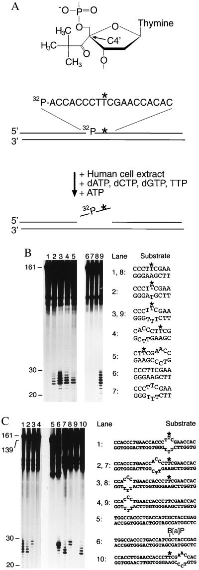
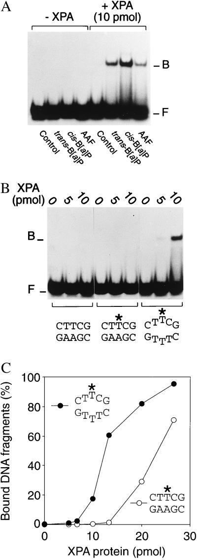
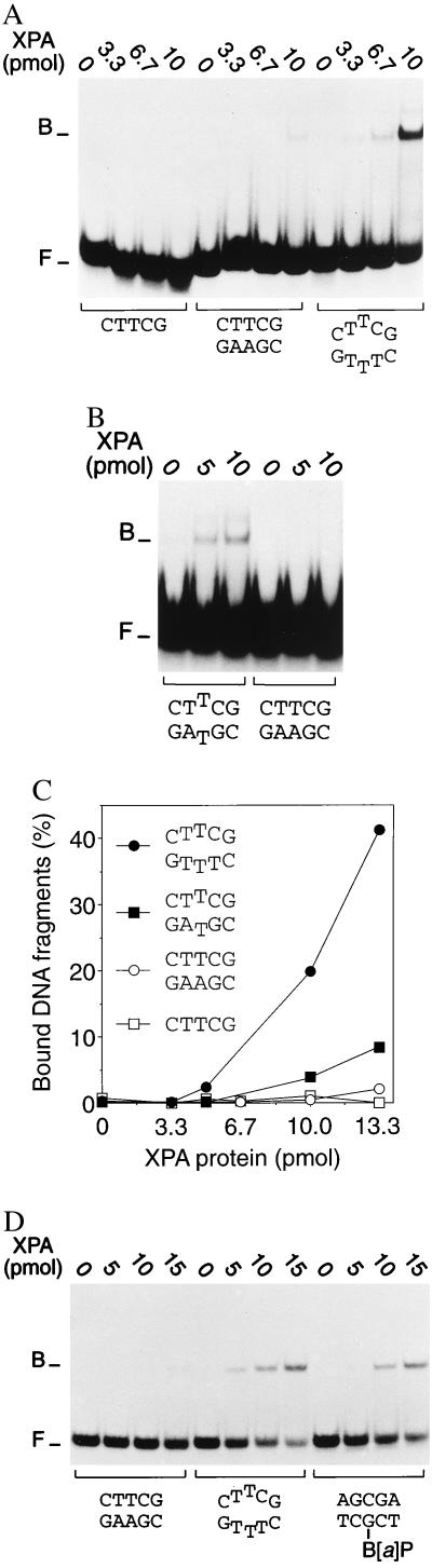
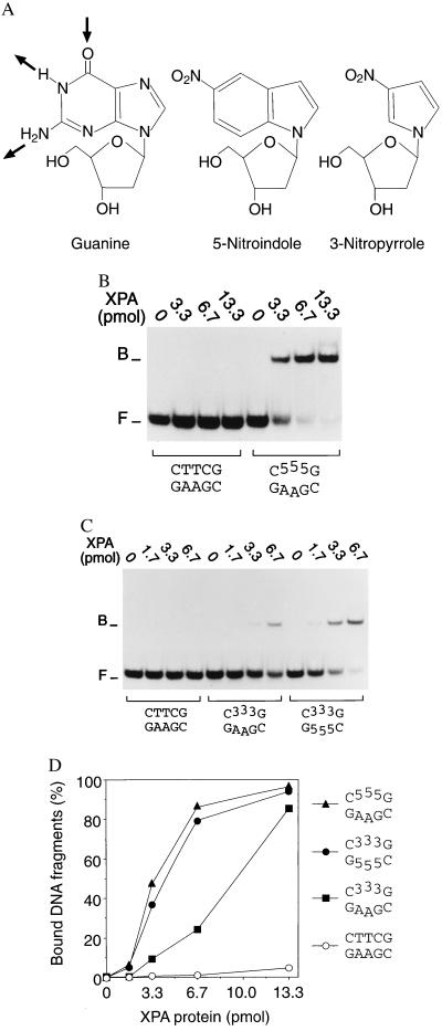
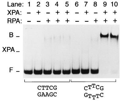
Nondistorting C4' backbone adducts serve as molecular tools to analyze the strategy by which a limited number of human nucleotide excision repair (NER) factors recognize an infinite variety of DNA lesions. We have constructed composite DNA substrates containing a noncomplementary site adjacent to a nondistorting C4' adduct to show that the loss of hydrogen bonding contacts between partner strands is an essential signal for the recruitment of NER enzymes. This specific conformational requirement for excision is mediated by the affinity of xeroderma pigmentosum group A (XPA) protein for nonhybridizing sites in duplex DNA. XPA recognizes defective Watson-Crick base pair conformations even in the absence of DNA adducts or other covalent modifications, apparently through detection of hydrophobic base components that are abnormally exposed to the double helical surface. This recognition function of XPA is enhanced by replication protein A (RPA) such that, in combination, XPA and RPA constitute a potent molecular sensor of denatured base pairs. Our results indicate that the XPA-RPA complex may promote damage recognition by monitoring Watson-Crick base pair integrity, thereby recruiting the human NER system preferentially to sites where hybridization between complementary strands is weakened or entirely disrupted.


Figures

Figure 1
Figure 1
Recruitment of human NER activity depends on defective hybridization. (A) Structure of the nondistorting C4′ pivaloyl adduct (Top), basic design of the 161-mer substrates (Middle) and oligonucleotide excision assay (Bottom). The C4′ pivaloyl modification is indicated by ∗, and a 32P-labeled residue is situated on the 5′ side of this lesion. (B) Determination of human NER activity in HeLa cell extract. After incubations of 40 min at 30°C, excision products were visualized by denaturing gel electrophoresis and autoradiography. All reactions contained the same amount of radioactive substrate (5 fmol), and oligonucleotide lengths were estimated by using appropriate markers. These incubations in cell extract also generate nonspecific degradation products, visible in the upper part of each lane, which do not interfere with detection of excised oligomers. The sequence environment in the central portion of each substrate is indicated, with the asterisks denoting the C4′ pivaloyl adduct. (C) Probing of NER activity using composite substrates. The central portion of each double-stranded fragment is outlined, again with ∗ denoting the C4′ modification. Lane 6 shows a control reaction with a (−)-cis-B[a]P-modified substrate of 139 bp (12).
Figure 2
Figure 2
Binding of human XPA protein to covalently modified DNA. The indicated amounts of XPA protein were coincubated with DNA duplexes of 19 residues (40 fmol per reaction), followed by analysis of protein–DNA interactions by electrophoretic mobility-shift assays. One strand of each substrate was 32P-labeled at its 5′ end. The positions of XPA–DNA complexes (B) and of free DNA fragments (F) in the gel autoradiographs are indicated. (A) DNA fragments were modified by a site-directed carcinogen–DNA adduct. (B) DNA fragments carried a C4′ pivaloyl adduct (denoted by ∗) in the center. (C) Mean percentages of bound DNA fragments obtained from quantitative densitometric scanning of 3–5 experiments.
Figure 3
Figure 3
Recognition of nonhybridizing base pairs. Protein–DNA interactions were analyzed by mobility-shift assays, and the positions of XPA–DNA complexes (B) and free DNA fragments (F) are indicated. (A) Preferential interaction with 32P-labeled 19-mer DNA duplexes (40 fmol) containing artificially denatured sites that were generated by insertion of three consecutive mismatches. (B) Preferential binding to 19-mer duplexes containing a single mismatch. (C) Diagram representing the mean quantitative values (percentage of bound fragments) of at least 3 experiments. (D) Side-by-side comparison of XPA binding to homoduplex DNA, duplex DNA with three mispaired bases, or homoduplex DNA fragments containing a trans-B[a]P-modified guanine in a central position.
Figure 4
Figure 4
XPA protein is guided to nonhybridizing sites by hydrophobic attractions. (A) Nucleoside analogs containing 5-nitroindole or 3-nitropyrrole preserve normal backbone structure, retain aromatic interactions but, in contrast to natural bases such as guanine, lack donor and acceptor groups for Watson–Crick hydrogen bonding. The arrows indicate the hydrogen acceptor and donors employed in Watson–Crick pairing between guanine and cytosine. (B) Preferential binding of XPA to 32P-labeled DNA duplexes (40 fmol) containing 5-nitroindole (“5”). (C) Preferential binding of XPA to DNA duplexes containing 3-nitropyrrole analogs (“3”), or both 5-nitroindoles in one strand and 3-nitropyrroles at the corresponding positions of the complementary strand. (D) Mean percentages of bound fragments obtained by densitometric scanning of three independent experiments. The composition in the center of each double-stranded 19-mer substrate is indicated.
Figure 5
Figure 5
Cooperative binding of XPA and RPA to nonhybridizing base pairs. XPA and RPA (2 pmol) were incubated with 43-mer DNA fragments (20 fmol), either homoduplexes (lanes 1–5) or partial duplexes containing three mismatches in the center (lanes 6–10), and protein–DNA interactions were analyzed by mobility shift gels. The positions of XPA–RPA–DNA complexes (B) and of free DNA fragments (F) is indicated. Additionally, the figure shows the position of complexes formed by XPA alone (XPA). Because of the low amount of XPA protein, these binary XPA–DNA complexes are visible in lane 7 only on overexposure of the gel.

References

    1. Friedberg E C, Walker G C, Siede W. DNA Repair and Mutagenesis. Washington, DC: Am. Soc. Microbiol.; 1995. pp. 634–649.
    1. Hoeijmakers J H J. Eur J Cancer. 1994;30:1912–1921. - PubMed
    1. Hanawalt P C. Science. 1994;266:1957–1958. - PubMed
    1. Sancar A. Annu Rev Biochem. 1996;65:43–81. - PubMed
    1. Wood R D. Annu Rev Biochem. 1996;65:135–167. - PubMed

Publication types

LinkOut - more resources