. 1999 May 25;96(11):6096-101.
doi: 10.1073/pnas.96.11.6096.

Cell cycle-dependent regulation of RNA polymerase I transcription: the nucleolar transcription factor UBF is inactive in mitosis and early G1

Affiliations

Cell cycle-dependent regulation of RNA polymerase I transcription: the nucleolar transcription factor UBF is inactive in mitosis and early G1

J Klein et al. Proc Natl Acad Sci U S A. .

Abstract

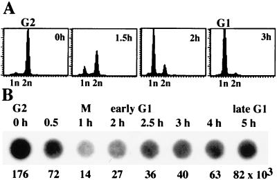
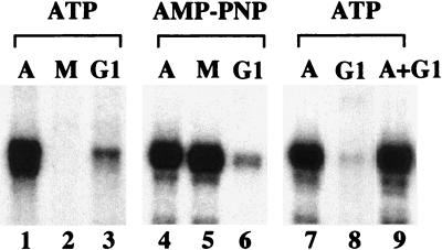
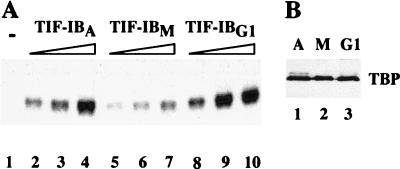
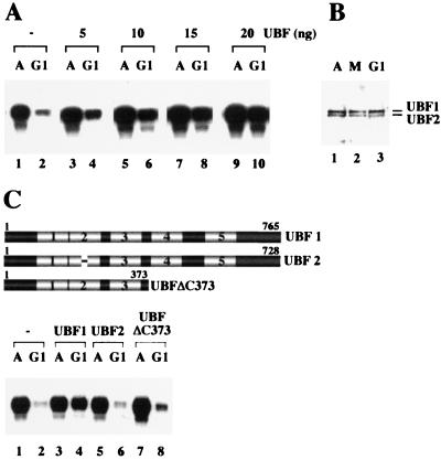
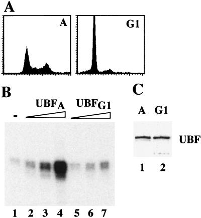
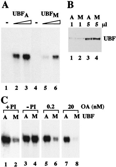
Transcription of ribosomal RNA genes by RNA polymerase (pol) I oscillates during the cell cycle, being maximal in S and G2 phase, repressed during mitosis, and gradually recovering during G1 progression. We have shown that transcription initiation factor (TIF)-IB/SL1 is inactivated during mitosis by cdc2/cyclin B-directed phosphorylation of TAFI110. In this study, we have monitored reactivation of transcription after exit from mitosis. We demonstrate that the pol I factor UBF is also inactivated by phosphorylation but recovers with different kinetics than TIF-IB/SL1. Whereas TIF-IB/SL1 activity is rapidly regained on entry into G1, UBF is reactivated later in G1, concomitant with the onset of pol I transcription. Repression of pol I transcription in mitosis and early G1 can be reproduced with either extracts from cells synchronized in M or G1 phase or with purified TIF-IB/SL1 and UBF isolated in the presence of phosphatase inhibitors. The results suggest that two basal transcription factors, e.g., TIF-IB/SL1 and UBF, are inactivated at mitosis and reactivated by dephosphorylation at the exit from mitosis and during G1 progression, respectively.


Figures

Figure 1
Figure 1
Measurement of pre-rRNA synthesis during the cell cycle. (A) FACS analysis. FT210 cells were synchronized in G2 by shifting to 39°C for 18 h and released from the G2 block by shifting to the permissive temperature (33°C). Aliquots of cells were subjected to FACS analysis at the times indicated. (B) Quantitation of nascent pre-rRNA chains. Nascent RNA was extracted from synchronized cells at the times indicated, dotted onto a membrane, and hybridized to a labeled murine rDNA probe covering nucleotides from −168 to +3,101. Quantitation of the hybridization signals by a PhosphorImager is shown below the blots.
Figure 2
Figure 2
In vitro reproduction of transcriptional repression in extracts from M and G1 phase cells. Standard transcription assays contained 25 μg of extract from asynchronous (A), mitotic (M), or G1 cells and either ATP and GTP (lanes 1–3, 7–9) or AMP-PNP/GMP-PNP (lanes 4–6). To exclude that early G1 extracts contain a dominant repressor of pol I transcription, equal amounts of asynchronous and G1 extract were assayed alone (lanes 7, 8) or simultaneously (lane 9).
Figure 3
Figure 3
TIF-IB/SL1 is reactivated after exit from mitosis. (A) In vitro transcription. Extracts from asynchronous (A), mitotic (M), and early G1 FT210 cells were preincubated with 2.5 μM adenosine 5′-[γ-thio]triphosphate for 30 min at 30°C, and then TIF-IB was immunoprecipitated with α-mTAFI95 antibodies immobilized on sheep anti-rabbit IgG Dynabeads. After stringent washing, bead-bound TIF-IB activity was monitored in a reconstituted transcription system. Lane 1 shows transcription in the absence of TIF-IB, lanes 2–4 in the presence of increasing amounts (50–200 pg) of TIF-IB from asynchronous cells (TIF-IBA), lanes 5–7 from mitotic cells (TIF-IBM), and lanes 8–10 from G1 cells (TIF-IBG1). (B) Western blot. Bead-bound TIF-IB precipitated from asynchronous (lane 1), mitotic (lane 2) and G1 cell extracts (lane 3) were subjected to Western blot analysis by using anti-TBP antibodies.
Figure 4
Figure 4
Transcription in early G1 cell extracts is rescued by UBF. (A) Exogenous UBF stimulates transcriptional activity of early G1 extracts. Transcription assays contained 25 μg of protein from asynchronous (A) and G1 cells (G1) and increasing amounts of recombinant UBF1 added at the onset of transcription (lanes 3–10). (B) The amount of cellular UBF does not change throughout the cell cycle. Protein (100 μg) extracted from asynchronous (A, lane 1), mitotic (M, lane 2), and early G1 cells (lane 3) were analyzed on immunoblots by using anti-UBF antiserum. (C) Rescue of transcription in G1 extracts requires transcriptionally active UBF. Asynchronous and G1 extracts were assayed in the absence of UBF (lanes 1, 2) and in the presence of 20 ng of UBF1 (lanes 3, 4), UBF2 (lanes 5, 6), or UBFΔC373 (lanes 7, 8). A scheme representing UBF1, UBF2, and the deletion mutant UBFΔC373 is shown above. The HMG-box motifs are numbered.
Figure 5
Figure 5
Impaired transcriptional activity of UBF isolated from G1 cells. (A) FACS analysis. Serum-starved NIH 3T3 cells were synchronized in early G1 by serum stimulation for 3 h. (B) In vitro transcription. UBF was immunopurified from asynchronous (UBFA, lanes 2–4) or early G1 phase (UBFG1, lanes 5–7) NIH 3T3 cells expressing Flag-epitope tagged UBF1. Purified UBF (1, 2.5, and 4 ng) was assayed in an UBF-responsive reconstituted transcription system. (C) Western blot. Flag-UBF1 (10 μl; 15 ng) isolated from asynchronous (lane 1) and G1 cells (lane 2) were subjected to Western blot analysis by using anti-Flag antibodies (mAb M2).
Figure 6
Figure 6
UBF from mitotic cells is inactive. (A) Comparison of the transcriptional activity of UBF from asynchronous and mitotic FT210 cells. UBF was isolated by chromatography on DEAE- and Q-Sepharose and assayed for transcriptional activity. Shown is transcription in the absence of UBF (lanes 1 and 4) and in the presence of 4 (lanes 2 and 3) or 8 (lanes 5 and 6) ng of UBF from asynchronous (UBFA) and mitotic cells (UBFM). Reactions contained AMP-PNP, GMP-PNP, and nonspecific phosphatase inhibitors to prevent modification of UBF during transcription. (B) Western blot. Q-Sepharose fractions (1 and 5 μl) purified from asynchronous (lanes 1, 3) and mitotic (lanes 2, 4) extracts were subjected to Western blot analysis by using anti-UBF serum. (C) An okadaic acid-sensitive phosphatase counteracts mitotic inactivation of UBF. Equal amounts (10 ng) of UBF purified from asynchronous (A) and mitotic (M) FT210 cells were assayed in a partially purified transcription system either in the presence of 2 mM 2-glycerophosphate/0.2 mM sodium orthovanadate (+PI; lanes 1 and 2), in the absence of phosphatase inhibitors (−PI; lanes 3 and 4) or in the presence of 0.2 nM (lanes 5 and 6) and 20 nM okadaic acid (lanes 7 and 8). The assays were performed in the presence of AMP-PNP and GMP-PNP to prevent de novo phosphorylation of UBF during transcription.

References

    1. Gottesfeld J M, Forbes D J. Trends Biochem Sci. 1997;22:197–202. - PubMed
    1. Segil N, Guermah M, Hoffmann A, Roeder R G, Heintz N. Genes Dev. 1996;10:2389–2400. - PubMed
    1. Long J L, Leresche A, Kriwacki R W, Gottesfeld J M. Mol Cell Biol. 1998;18:1467–1476. - PMC - PubMed
    1. Akoulitechev S, Reinberg D. Genes Dev. 1998;12:3541–3550. - PMC - PubMed
    1. Cisek L J, Corden J L. Nature (London) 1989;339:679–684. - PubMed

Publication types

MeSH terms

LinkOut - more resources