. 1999 May 25;96(11):6161-5.
doi: 10.1073/pnas.96.11.6161.

Regulation of the OxyR transcription factor by hydrogen peroxide and the cellular thiol-disulfide status

Affiliations

Regulation of the OxyR transcription factor by hydrogen peroxide and the cellular thiol-disulfide status

F Aslund et al. Proc Natl Acad Sci U S A. .

Abstract

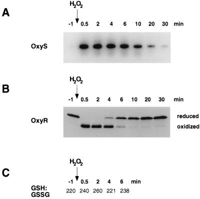
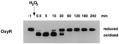
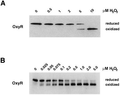
The Escherichia coli transcription factor OxyR is activated by the formation of an intramolecular disulfide bond and subsequently is deactivated by enzymatic reduction of the disulfide bond. Here we show that OxyR can be activated by two possible pathways. In mutants defective in the cellular disulfide-reducing systems, OxyR is constitutively activated by a change in the thiol-disulfide redox status in the absence of added oxidants. In wild-type cells, OxyR is activated by hydrogen peroxide. By monitoring the presence of the OxyR disulfide bond after exposure to hydrogen peroxide in vivo and in vitro, we also show that the kinetics of OxyR oxidation by low concentrations of hydrogen peroxide is significantly faster than the kinetics of OxyR reduction, allowing for transient activation in an overall reducing environment. We propose that the activity of OxyR in vivo is determined by the balance between hydrogen peroxide levels and the cellular redox environment.


Figures

Figure 1
Figure 1
OxyR activity in thioredoxin and glutaredoxin mutants. (A) Total RNA was isolated from untreated and hydrogen peroxide (200 μM)-treated wild-type (DHB4), trxA (WP570), trxA gorA (FÅ378), trxA gshA (WP612), trxA grxA (WP813), and katG ahpCF (FÅ369) cells, and the levels of the OxyS RNA were assayed by primer extension. (B) The levels of alkaline phosphatase activity in midlogarithmically growing cells were determined as described (7).
Figure 2
Figure 2
OxyS RNA expression, redox status of OxyR, and GSH/GSSG ratio after treatment with hydrogen peroxide. (A) Midlogarithmically growing wild-type cells (FÅ371) were treated with 200 μM hydrogen peroxide, and then aliquots were taken at 0.5, 2, 4, 6, 10, 20, and 30 min. Total RNA was isolated and the levels of OxyS expression were analyzed by primer extension. (B) Aliquots of the above cells also were precipitated with TCA, treated with AMS, and subjected to SDS/PAGE and immunoblot analysis. (C) The GSH and GSSG levels in aliquots of the indicated cultures were determined by the DTNB-glutathione reductase recycling assay (8).
Figure 3
Figure 3
Transient OxyR oxidation by hydrogen peroxide in vitro. OxyR4CA (1 μM final concentration) was reduced fully by incubation with a buffer containing 25 mM GSH, 0.1 mM GSSG, and 10 μM Grx1. Hydrogen peroxide (2 μM) was added, and the redox status of OxyR was assayed at 0.5, 10, 30, 60, 120, 180, and 240 min. Samples were mixed with a 1/10 vol of 100% TCA and then treated with AMS. Separation and detection of OxyR were achieved by SDS/PAGE and immunoblot analysis.
Figure 4
Figure 4
Minimum concentrations of hydrogen peroxide required to oxidized OxyR. (A) Midlogarithmically growing wild-type cells (FÅ371) were treated with 0, 0.5, 1, 2, 5, and 10 μM hydrogen peroxide. (B) OxyR4CA (0.01 μM final concentration) was reduced fully by incubation with a buffer containing 10 μM glutaredoxin 1, 25 mM GSH, and 0.1 mM GSSG. Aliquots were removed and treated with hydrogen peroxide to achieve 0, 0.025, 0.05, 0.075, 0.1, 0.2, 0.5, 1, 2, and 5 μM final concentrations. After 30 sec, the samples in both A and B were acidified with TCA and then treated with AMS. Again, separation and detection of OxyR was achieved by SDS/PAGE and immunoblot analysis.
Figure 5
Figure 5
Pathways of OxyR oxidation and reduction. Two different reactions can determine the redox status of OxyR. During normal growth, when hydrogen peroxide concentrations are low, the reaction rate for the second pathway is low, so the first pathway dominates. Under these conditions in wild-type strains, the cellular GSH/GSSG ratio favors reduced OxyR. However, in mutant strains with a low GSH/GSSG ratio, oxidized OxyR is formed. After exposure to ≥5 μM external hydrogen peroxide, the second pathway dominates and oxidized OxyR becomes the major species.

Similar articles

Cited by

References

    1. Zheng M, Åslund F, Storz G. Science. 1998;279:1718–1721. - PubMed
    1. Gilbert H F. Adv Enzymol Relat Areas Mol Biol. 1990;63:69–172. - PubMed
    1. Holmgren A, Fagerstedt M. J Biol Chem. 1982;257:6926–6930. - PubMed
    1. Hwang C, Sinskey A J, Lodish H F. Science. 1992;257:1496–1502. - PubMed
    1. Chomczynski P, Sacchi N. Anal Biochem. 1987;162:156–159. - PubMed

Publication types

LinkOut - more resources