. 1999 May 25;96(11):6223-8.
doi: 10.1073/pnas.96.11.6223.

Poly-ADP ribose polymerase activates nuclear proteasome to degrade oxidatively damaged histones

Affiliations

Poly-ADP ribose polymerase activates nuclear proteasome to degrade oxidatively damaged histones

O Ullrich et al. Proc Natl Acad Sci U S A. .

Abstract

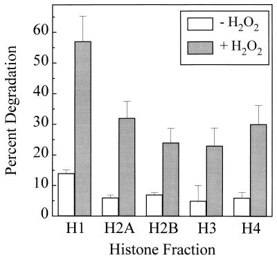
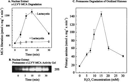
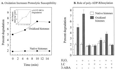
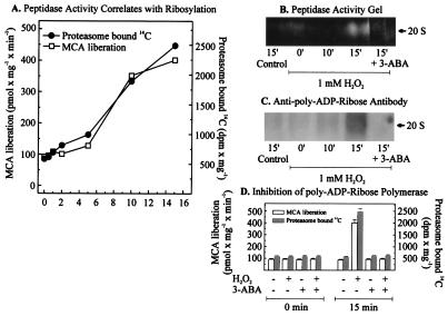
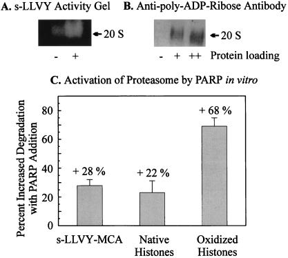
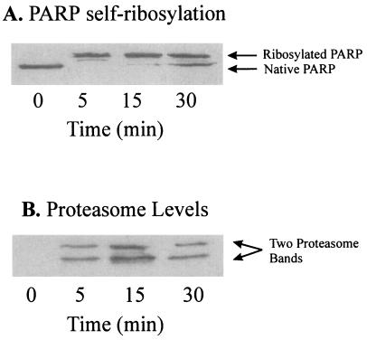
The 20S proteasome has been shown to be largely responsible for the degradation of oxidatively modified proteins in the cytoplasm. Nuclear proteins are also subject to oxidation, and the nucleus of mammalian cells contains proteasome. In human beings, tumor cells frequently are subjected to oxidation as a consequence of antitumor chemotherapy, and K562 human myelogenous leukemia cells have a higher nuclear proteasome activity than do nonmalignant cells. Adaptation to oxidative stress appears to be one element in the development of long-term resistance to many chemotherapeutic drugs and the mechanisms of inducible tumor resistance to oxidation are of obvious importance. After hydrogen peroxide treatment of K562 cells, degradation of the model proteasome peptide substrate suc-LLVY-MCA and degradation of oxidized histones in nuclei increases significantly within minutes. Both increased proteolytic susceptibility of the histone substrates (caused by modification by oxidation) and activation of the proteasome enzyme complex occur independently during oxidative stress. This rapid up-regulation of 20S proteasome activity is accompanied by, and depends on, poly-ADP ribosylation of the proteasome, as shown by inhibitor experiments, 14C-ADP ribose incorporation assays, immunoblotting, in vitro reconstitution experiments, and immunoprecipitation of (activated) proteasome with anti-poly-ADP ribose polymerase antibodies. The poly-ADP ribosylation-mediated activated nuclear 20S proteasome is able to remove oxidatively damaged histones more efficiently and therefore is proposed as an oxidant-stimulatable defense or repair system of the nucleus in K562 leukemia cells.


Figures

Figure 1
Figure 1
Degradation of histones in intact human K562 cells stressed by exposure to hydrogen peroxide. K562 cells were grown in RPMI medium 1640 supplemented with 10% FCS, with the addition of [3H]leucine for 5 days. Next, the cells were washed and either exposed or sham-exposed to 1.0 mM H2O2 in PBS. After 30 min of exposure, new culture medium was added, and the cells were harvested, lysed, and subjected to acetic acid-urea-Triton-PAGE as described (25). The gels were Coomassie-stained, and the band patterns were compared with those of isolated histones (Boehringer Mannheim). The band for each histone was excised from the gels and counted by liquid scintillation. Two electrophoresis gels were run for each sample and averaged. Four such independent experiments were conducted, and the values shown for each histone are the grand means ± SE of the four experiments.
Figure 2
Figure 2
Activation of nuclear proteolysis and increased proteolytic susceptibility of histones after hydrogen peroxide treatment. K562 cells were grown and exposed to H2O2 as described in the legend to Fig. 1. Cell nuclei then were isolated and lysed, and degradation of the fluorogenic peptide suc-LLVY-MCA was measured as described in Materials and Methods. (A) The lactacystin-sensitive suc-LLVY-MCA degradation as a function of time of K562 cell exposure to 1 mM hydrogen peroxide. The values represent the means of six independent measurements with SD less than 10%. (B) A representative suc-LLVY-MCA degradation activity stain of a one-dimensional polyacrylamide nondenaturing proteasome electrophoresis gel, according to ref. , also as a function of time of K562 cell exposure to 1 mM hydrogen peroxide. (C) The proteolytic susceptibility of H2O2-modified histones, incubated with the purified 20S proteasome. [3H]-labeled histones (at 2.0 mg protein/ml, Boehringer Mannheim) were radiolabeled and exposed (or sham-exposed) to hydrogen peroxide in vitro as described in Materials and Methods. The x axis shows the concentrations of hydrogen peroxide used to oxidize histones. After extensive dialysis, proteasome was added for a 2-h incubation, and proteolysis was measured by the fluorescamine assay for release of free amino acids, after incubation of 40 μg histone substrate protein with 0.6 μg proteasome (see Materials and Methods). All values in C represent the means ± SD of three independent experiments.
Figure 3
Figure 3
Proteasome-dependent selective degradation of oxidized histones in nuclear extracts and the role of poly-ADP ribosylation. (A) The degradation of undamaged and H2O2-modified histones and suc-LLVY-MCA by lysates of H2O2-treated isolated nuclei from K562 hematopoietic cells. Nuclei were isolated, exposed to 1 mM H2O2 for times from 0 to 15 min (or sham exposed), lysed, and used to measure the proteolytic susceptibility of both untreated and H2O2-treated [3H]-labeled histones (added to the nuclear lysates) exactly as described in Materials and Methods. Isolated [3H]-labeled histones were oxidatively modified (or used as unmodified “native” histones) as described in Materials and Methods. The time scale represents the length of nuclei exposure (or sham-exposure) to H2O2 and the data points represent the means of three independent experiments (for which SD were always less than 10%). (Inset) The increase in the suc-LLVY-MCA degrading activity of nuclear lysates after treatment of nuclei with 1 mM H2O2 (for details see Fig. 2). (B) The degradation of H2O2-modified histones by K562 cell nuclear lysates is stimulated by pretreatment of nuclei with H2O2 but inhibited by both lactacystin and 3-ABA. Nuclei were isolated, exposed to 1 mM H2O2 for 15 min (or sham exposed), lysed, and used to measure the proteolytic susceptibility of both untreated and H2O2-treated [3H]-labeled histones, as described in Materials and Methods, in the presence or absence of the inhibitors 5 μM lactacystin (LC) or 1 mM 3-ABA. Where used, oxidized histones were treated with 15 mM hydrogen peroxide. All values represent the means ± SD of three independent experiments.
Figure 4
Figure 4
Poly-ADP ribosylation of the proteasome in nuclei of K562 human hematopoietic cells after hydrogen peroxide treatment increases its activity. (A) Proteasome peptidase activity correlated with the degree of poly-ADP ribosylation, measured as incorporated 14C from 14C-NAD+ after bolus addition of 1 mM hydrogen peroxide to isolated nuclei from K562 cells. Nuclei of K562 cells were isolated by a modified method of Emig et al. (20) and incubated with 1 mM hydrogen peroxide in 100 mM phosphate buffer (pH 7.8), 200 μM NAD+, 10 mM MgCl2, and 5 mM DTT, containing 10,000 dpm 14C-labeled NAD+. (B and C) The analysis of lysates from K562 cell nuclei after H2O2 treatment. The lysates were analyzed by nondenaturing one-dimensional polyacrylamide electrophoresis according to ref. . (B) An activity staining and (C) an immunoblot of the proteasome, using an anti-poly-ADP ribose antibody (Biomol, Plymouth Meeting, PA). (D) The effect of 3-ABA on the proteasome activity and the [14C] incorporation, after hydrogen peroxide treatment. Experimental conditions were as in Fig. 3A except for the use of 3-ABA (1 mM).
Figure 5
Figure 5
Activation of the isolated 20S proteasome by in vitro poly-ADP ribosylation. (A) Activation of the isolated 20S proteasome after in vitro poly-ADP ribosylation. In vitro poly-ADP ribosylation of the 20S proteasome was performed according to Banasik et al. (27). Purified proteasome (0.15 mg/ml) was incubated with 1 μg/ml of PARP and 10 μg/ml of DNA (sonicated 10 times for 20 s) in a medium consisting of 100 mM Tris, 200 μM NAD+, 10 mM MgCl2, and 5 mM DTT. Controls were incubated without NAD+. After 10 min the reaction was stopped by addition of 1 mM 3-ABA, and the mixture was rapidly cooled on ice. Peptidase activity was determined after nondenaturing one-dimensional polyacrylamide electrophoresis as described in Fig. 4. (A) The activation of the peptidase activity of the proteasome after overlaying the gel with 200 μM suc-LLVY-MCA. (B) The immunoblot analysis of the gel after blotting on a nitrocellulose membrane by using the anti-ADP ribose antibody (Biomol) used in Fig. 4. The lanes indicated by + and ++ are the same probes loaded with different amounts of protein. Proteolytic activities in C were analyzed before and after in vitro poly-ADP ribosylation, using undamaged and H2O2-modified histones and the peptide suc-LLVY-MCA as substrates (see Figs. 1–4).
Figure 6
Figure 6
Immunoprecipitation of the proteasome with a monoclonal anti-DBD-PARP antibody in nuclear extracts from H2O2-treated K562 cells. Intact K562 human chronic myelogenous leukemia cells were grown, harvested, treated with 1 mM H2O2, and then subjected to isolation and lysis of nuclei, as described in Materials and Methods. Immunoprecipitation was conducted with 100 μg of lysate protein, 100 μl of Tris/NaCl buffer (containing 50 mM Tris, plus 100 μM NaCl, at pH 8.0), and 1 μl of a mAb directed against the DBD (anti-DBD) of PARP (at 5 mg/ml), by a modification of the procedure of Zwilling et al. (30). The primary antibody reaction was performed in 50 mM Tris (pH 7.8) containing 150 mM NaCl (Tris buffered saline), 0.1% BSA, and 1.0 μl of rabbit-anti-DBD-IgG (5 μg/μl stock) for 1 h at 4°C in a total volume of 200 μl. For secondary antibody reactions 1.0 μl of donkey-anti-rabbit-IgG (5 μg/μl stock) was added and incubated for 1 h at 4°C. Binding of the secondary antibody was detected by using an ECL kit. Immunoprecipitation was initiated by addition of 20 μl of protein A-Sepharose (equilibrated 1:1 with a pH 8.0 solution of 50 mM Tris, 150 mM NaCl, and 0.1% BSA) for a 1-h incubation at 4°C. Controls were incubated with the secondary antibody and the protein A-Sepharose only. The Sepharose was centrifuged (12,000 × g for 30 min) and washed five times with Tris buffered saline. Sepharose-bound proteins were separated by 15% SDS/PAGE, transblotted onto nitrocellulose membranes, and incubated either with a polyclonal goat-anti-PARP-IgG (A) or a polyclonal rabbit-anti-core-proteasome-IgG (B). Control samples containing secondary antibody plus Sepharose A, or Sepharose A alone, produced no visible bands in the gels (data not shown). The results shown are a representative immunoblot from a series of several independent experimental repeats.

References

    1. Sinha B K, Mimnaugh E G. Free Radical Biol Med. 1990;8:567–581. - PubMed
    1. Davies K J A, Doroshow J H. J Biol Chem. 1986;261:3060–3067. - PubMed
    1. Doroshow J H, Davies K J A. J Biol Chem. 1986;261:3068–3074. - PubMed
    1. Marcillat O, Zhang Y, Davies K J A. Biochem J. 1989;259:181–189. - PMC - PubMed
    1. Feinstein E, Canaani E, Weiner L M. Biochemistry. 1993;32:13156–13161. - PubMed

Publication types

MeSH terms

LinkOut - more resources