Metalloprotease-mediated ligand release regulates autocrine signaling through the epidermal growth factor receptor
- PMID: 10339571
- PMCID: PMC26865
- DOI: 10.1073/pnas.96.11.6235
Metalloprotease-mediated ligand release regulates autocrine signaling through the epidermal growth factor receptor
Abstract
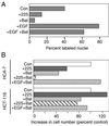
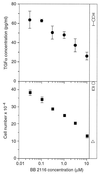
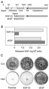
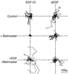
Ligands that activate the epidermal growth factor receptor (EGFR) are synthesized as membrane-anchored precursors that appear to be proteolytically released by members of the ADAM family of metalloproteases. Because membrane-anchored EGFR ligands are thought to be biologically active, the role of ligand release in the regulation of EGFR signaling is unclear. To investigate this question, we used metalloprotease inhibitors to block EGFR ligand release from human mammary epithelial cells. These cells express both transforming growth factor alpha and amphiregulin and require autocrine signaling through the EGFR for proliferation and migration. We found that metalloprotease inhibitors reduced cell proliferation in direct proportion to their effect on transforming growth factor alpha release. Metalloprotease inhibitors also reduced growth of EGF-responsive tumorigenic cell lines and were synergistic with the inhibitory effects of antagonistic EGFR antibodies. Blocking release of EGFR ligands also strongly inhibited autocrine activation of the EGFR and reduced both the rate and persistence of cell migration. The effects of metalloprotease inhibitors could be reversed by either adding exogenous EGF or by expressing an artificial gene for EGF that lacked a membrane-anchoring domain. Our results indicate that soluble rather than membrane-anchored forms of the ligands mediate most of the biological effects of EGFR ligands. Metalloprotease inhibitors have shown promise in preventing spread of metastatic disease. Many of their antimetastatic effects could be the result of their ability to inhibit autocrine signaling through the EGFR.
Figures


Similar articles
-
Removal of the membrane-anchoring domain of epidermal growth factor leads to intracrine signaling and disruption of mammary epithelial cell organization.J Cell Biol. 1998 Nov 30;143(5):1317-28. doi: 10.1083/jcb.143.5.1317. J Cell Biol. 1998. PMID: 9832559 Free PMC article.
-
The autocrine loop of epidermal growth factor receptor-epidermal growth factor / transforming growth factor-alpha in malignant rhabdoid tumor cell lines: heterogeneity of autocrine mechanism in TTC549.Jpn J Cancer Res. 2001 Mar;92(3):269-78. doi: 10.1111/j.1349-7006.2001.tb01091.x. Jpn J Cancer Res. 2001. PMID: 11267936 Free PMC article.
-
Metalloprotease disintegrin-mediated ectodomain shedding of EGFR ligands promotes intestinal epithelial restitution.Am J Physiol Gastrointest Liver Physiol. 2004 Dec;287(6):G1213-9. doi: 10.1152/ajpgi.00149.2004. Epub 2004 Jul 29. Am J Physiol Gastrointest Liver Physiol. 2004. PMID: 15284022
-
Epidermal growth factor-related peptides and the epidermal growth factor receptor in normal and malignant prostate.World J Urol. 1995;13(5):290-6. doi: 10.1007/BF00185972. World J Urol. 1995. PMID: 8581000 Review.
-
TACE/ADAM17 processing of EGFR ligands indicates a role as a physiological convertase.Ann N Y Acad Sci. 2003 May;995:22-38. doi: 10.1111/j.1749-6632.2003.tb03207.x. Ann N Y Acad Sci. 2003. PMID: 12814936 Review.
Cited by
-
The membrane-anchoring domain of epidermal growth factor receptor ligands dictates their ability to operate in juxtacrine mode.Mol Biol Cell. 2005 Jun;16(6):2984-98. doi: 10.1091/mbc.e04-11-0994. Epub 2005 Apr 13. Mol Biol Cell. 2005. PMID: 15829568 Free PMC article.
-
Mitochondrial reactive oxygen species mediate GPCR-induced TACE/ADAM17-dependent transforming growth factor-alpha shedding.Mol Biol Cell. 2009 Dec;20(24):5236-49. doi: 10.1091/mbc.e08-12-1256. Mol Biol Cell. 2009. PMID: 19846666 Free PMC article.
-
Expression in mammalian cell cultures reveals interdependent, but distinct, functions for Star and Rhomboid proteins in the processing of the Drosophila transforming-growth-factor-alpha homologue Spitz.Biochem J. 2002 Apr 15;363(Pt 2):347-52. doi: 10.1042/0264-6021:3630347. Biochem J. 2002. PMID: 11931664 Free PMC article.
-
HER/ErbB receptor interactions and signaling patterns in human mammary epithelial cells.BMC Cell Biol. 2009 Oct 31;10:78. doi: 10.1186/1471-2121-10-78. BMC Cell Biol. 2009. PMID: 19878579 Free PMC article.
-
Paracrinicity: the story of 30 years of cellular pituitary crosstalk.J Neuroendocrinol. 2008 Jan;20(1):1-70. doi: 10.1111/j.1365-2826.2007.01616.x. J Neuroendocrinol. 2008. PMID: 18081553 Free PMC article. Review.
References
-
- Miettinen P J, Berger J E, Meneses J, Phung Y, Pedersen R A, Werb Z, Derynck R. Nature (London) 1995;376:337–341. - PubMed
-
- Threadgill D W, Dlugosz A A, Hansen L A, Tennenbaum T, Lichti U, Yee D, LaMantia C, Mourton T, Herrup K, Harris R C, et al. Science. 1995;269:230–234. - PubMed
-
- Derynck R. Adv Cancer Res. 1992;58:27–52. - PubMed
-
- Shoyab M, Plowman G D, McDonald V L, Bradley J G, Todaro G J. Science. 1989;243:1074–1076. - PubMed
-
- Higashiyama S, Abraham J A, Miller J, Fiddes J C, Klagsbrun M. Science. 1991;251:936–939. - PubMed
Publication types
MeSH terms
Substances
LinkOut - more resources
Full Text Sources
Other Literature Sources
Research Materials
Miscellaneous

