. 1999 May 25;96(11):6273-8.
doi: 10.1073/pnas.96.11.6273.

beta-Trcp couples beta-catenin phosphorylation-degradation and regulates Xenopus axis formation

Affiliations

beta-Trcp couples beta-catenin phosphorylation-degradation and regulates Xenopus axis formation

C Liu et al. Proc Natl Acad Sci U S A. .

Abstract

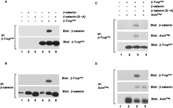
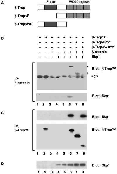
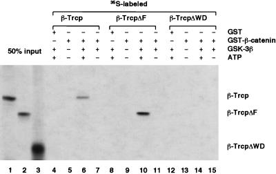
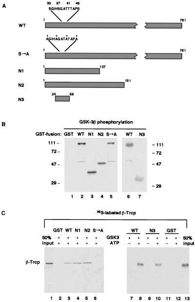
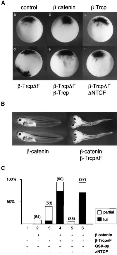
Regulation of beta-catenin stability is essential for Wnt signal transduction during development and tumorigenesis. It is well known that serine-phosphorylation of beta-catenin by the Axin-glycogen synthase kinase (GSK)-3beta complex targets beta-catenin for ubiquitination-degradation, and mutations at critical phosphoserine residues stabilize beta-catenin and cause human cancers. How beta-catenin phosphorylation results in its degradation is undefined. Here we show that phosphorylated beta-catenin is specifically recognized by beta-Trcp, an F-box/WD40-repeat protein that also associates with Skp1, an essential component of the ubiquitination apparatus. beta-catenin harboring mutations at the critical phosphoserine residues escapes recognition by beta-Trcp, thus providing a molecular explanation for why these mutations cause beta-catenin accumulation that leads to cancer. Inhibition of endogenous beta-Trcp function by a dominant negative mutant stabilizes beta-catenin, activates Wnt/beta-catenin signaling, and induces axis formation in Xenopus embryos. Therefore, beta-Trcp plays a central role in recruiting phosphorylated beta-catenin for degradation and in dorsoventral patterning of the Xenopus embryo.


Figures

Figure 1
Figure 1
β-Trcp forms a complex with β-catenin and Axin in vivo. (A and B) Association between β-Trcp and β-catenin. (A) Immunoprecipitation of β-Trcpmyc with an anti-myc antibody. Embryo extracts were from stage 9 embryos expressing β-catenin, β-catenin (S→A), or β-Trcpmyc alone or in combination. The precipitates were examined for β-catenin (Upper) and for β-Trcpmyc (Lower). A longer exposure revealed that β-Trcpmyc also coprecipitated endogenous β-catenin (data not shown). (B) Immunoprecipitation of β-catenin using the same embryo extracts as in A. The precipitates were examined for β-Trcpmyc (Upper) and for β-catenin (Lower). Endogenous β-catenin is seen in lanes 1 and 4. RNA injected per embryo: 1 ng each for β-catenin, β-catenin (S→A), and β-Trcpmyc. (C and D) A complex formation between β-Trcp and Axin mediated by β-catenin. (C) Immunoprecipitation of β-Trcpmyc. The extracts were from stage 9 embryos expressing β-Trcpmyc plus AxinFlag in the presence or absence of β-catenin or β-catenin (S→A). The precipitates were examined for β-catenin (Top), AxinFlag (Middle), and β-Trcpmyc (Bottom). (D) Immunoprecipitation of AxinFlag using the same extracts as in C. The precipitates were examined for β-Trcpmyc (Top), β-catenin (Middle), and AxinFlag (Bottom). RNA injected per embryo: same as in A and B except 2 ng for AxinFlag.
Figure 2
Figure 2
β-Trcp forms a complex with both β-catenin and Skp1. (A) Schematic diagram of wild-type β-Trcp, β-TrcpΔF, and β-TrcpΔWD. (B) Immunoprecipitation of β-catenin. The precipitates were examined for β-Trcp (Upper) and Skp1 (Lower). A longer exposure revealed that endogenous β-catenin also precipitated β-Trcpmyc (data not shown). (C) Immunoprecipitation of β-Trcpmyc using the same embryo extracts as in B. The precipitates were examined for Skp1 (Upper) and β-Trcp (Lower). (D) Immunoblot of Skp1 in the same embryo extracts as in B. RNA injected per embryo: 1 ng each for β-Trcpmyc and for deletion mutants β-catenin and Skp1.
Figure 3
Figure 3
β-Trcp WD40-repeat domain recognizes GSK-3β-phosphorylated β-catenin in vitro. In this GST-pull-down assay, purified GST or GST-β-catenin was examined for its ability to bind 35S-labeled β-Trcp. GST-β-catenin on beads was either untreated or were treated with purified GSK-3β in the presence or absence of ATP before incubation with β-Trcp. Note that, in this experiment, GSK-3β and ATP were removed from phosphorylated GST-β-catenin by extensively washing the GST-β-catenin on beads after phosphorylation and before incubation with β-Trcp. Lanes 1–3 represent 50% of the input 35S-labeled β-Trcp or β-Trcp mutants used in each GST-pull-down assay.
Figure 4
Figure 4
Phosphorylation of the amino-terminal region of β-catenin is necessary and sufficient for recognition by β-Trcp in vitro. (A) Schematic diagram of the wild-type and mutant derivatives of β-catenin. The four critical serine/threonine residues (S33, S37, T41, and S45), alanine substitutions of these residues in the S→A mutant, and surrounding residues are highlighted. Note that the Arm-repeat region that is required for Axin or TCF binding starts from residue 131. (B) Phosphorylation of β-catenin and its mutant derivatives (as GST-fusion proteins) by purified recombinant GSK-3β. Note that GST was not a substrate for GSK-3β. (C) On phosphorylation, N1, N2, and N3, but not β-catenin (S→A) were effectively recognized by β-Trcp. Lanes 1 and 13 represent 50% input of 35S-labeled β-Trcp in each GST-pull-down assay.
Figure 5
Figure 5
Inhibition of endogenous β-Trcp function by β-TrcpΔF induces ectopic Xnr3 expression and dorsal axis duplication. (A) Ventral injection of β-TrcpΔF RNA induced Xnr3 expression as examined by whole-mount in situ hybridization. All panels shown were ventral injection except in the control panel (a) (no injection). RNA amount injected per embryo: 1 ng for β-Trcp or β-TrcpΔF each, 100 pg for β-catenin, and 500 pg for ΔNTCF. The numbers of embryos with ectopic Xnr3 expression were 0 of 10 (number of embryos examined) in a; 0 of 8 in b; 10 of 10 for β-catenin (at 500 pg RNA per embryo; data not shown); 0 of 10 in c; 7 of 10 in d; 8 of 8 for β-TrcpΔF (at 4 ng RNA per embryo; data not shown); 0 of 10 in e; and 0 of 10 in f. (B and C) Axis duplication by β-TrcpΔF or β-catenin plus β-TrcpΔF. All shown were ventral injection except the control with no injection. Partial, partial axis duplication, defined as lacking eyes and other anterior structures in the duplicated axis. Full, complete axis duplication, defined as having eyes and other anterior structures in the duplicated axis. Number of injected embryos are indicated above each bar. RNA injected per embryo: β-catenin, 100 pg; β-TrcpΔF, 1 ng; GSK-3β, 1 ng; ΔNTCF, 500 pg.
Figure 6
Figure 6
Inhibition of endogenous β-Trcp function by β-TrcpΔF stabilizes β-catenin in vivo. Accumulation of β-cateninFlag or β-catenin (S→A)Flag protein was examined in stage 9 embryo extracts by immunoblotting with an anti-Flag antibody. Immunoblotting of tubulin was used as a protein loading control (Lower). This experiment were performed twice with similar results. RNA injected per embryo: 50 pg for β-cateninFlag or β-catenin (S→A)Flag; 4 ng for β-Trcp or β-TrcpΔF.

References

    1. Cadigan K M, Nusse R. Genes Dev. 1997;11:3286–3305. - PubMed
    1. Moon R T, Brown J D, Torres M. Trends Genet. 1997;13:157–162. - PubMed
    1. Kinzler K W, Vogelstein B. Cell. 1996;87:159–170. - PubMed
    1. Peifer M. Science. 1997;275:1752–1754. - PubMed
    1. Behrens J, Von Kries J P, Kuhl M, Bruhn L, Wedlich D, Grosschedl R, Birchmeier W. Nature (London) 1996;382:638–642. - PubMed

Publication types

MeSH terms

Substances

LinkOut - more resources