. 1999 May 25;96(11):6353-8.
doi: 10.1073/pnas.96.11.6353.

Monoclonal antibodies against oxidized low-density lipoprotein bind to apoptotic cells and inhibit their phagocytosis by elicited macrophages: evidence that oxidation-specific epitopes mediate macrophage recognition

Affiliations

Monoclonal antibodies against oxidized low-density lipoprotein bind to apoptotic cells and inhibit their phagocytosis by elicited macrophages: evidence that oxidation-specific epitopes mediate macrophage recognition

M K Chang et al. Proc Natl Acad Sci U S A. .

Abstract

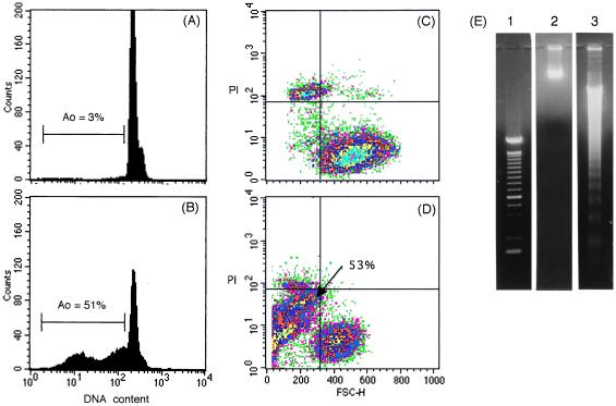
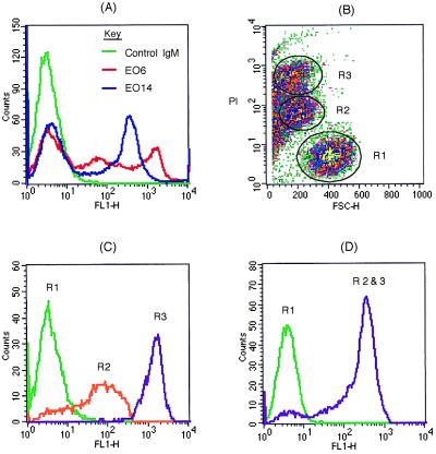
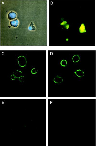
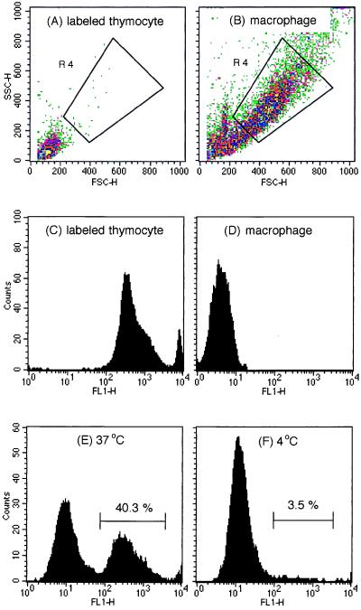
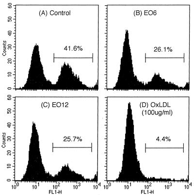
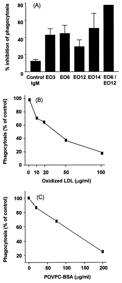
Apoptosis is recognized as important for normal cellular homeostasis in multicellular organisms. Although there have been great advances in our knowledge of the molecular events regulating apoptosis, much less is known about the receptors on phagocytes responsible for apoptotic cell recognition and phagocytosis or the ligands on apoptotic cells mediating such recognition. The observations that apoptotic cells are under increased oxidative stress and that oxidized low-density lipoprotein (OxLDL) competes with apoptotic cells for macrophage binding suggested the hypothesis that both OxLDL and apoptotic cells share oxidatively modified moieties on their surfaces that serve as ligands for macrophage recognition. To test this hypothesis, we used murine monoclonal autoantibodies that bind to oxidation-specific epitopes on OxLDL. In particular, antibodies EO6 and EO3 recognize oxidized phospholipids, including 1-palmitoyl 2-(5-oxovaleroyl) phosphatidylcholine (POVPC), and antibodies EO12 and EO14 recognize malondialdehyde-lysine, as in malondialdehyde-LDL. Using FACS analysis, we demonstrated that each of these EO antibodies bound to apoptotic cells but not to normal cells, whereas control IgM antibodies did not. Confocal microscopy demonstrated cell-surface expression of the oxidation-specific epitopes on apoptotic cells. Furthermore, each of these antibodies inhibited the phagocytosis of apoptotic cells by elicited peritoneal macrophages, as did OxLDL. In addition, an adduct of POVPC with BSA also effectively prevented phagocytosis. These data demonstrate that apoptotic cells express oxidation-specific epitopes-including oxidized phospholipids-on their cell surface, and that these serve as ligands for recognition and phagocytosis by elicited macrophages.


Figures

Figure 1
Figure 1
FACS analysis of PAECs: DNA content was measured by DNA staining. (A) Normal PAECs show mostly diploid DNA. (B) Apoptosis-induced PAECs show that DNA content of 51% of PAECs are below Go/G1 (Ao). Morphologic change of (C) normal PAECs and (D) apoptosis-induced PAEC were analyzed with X parameter of FSC and Y parameter of PI fluorescence. (E) Agarose gel electrophoresis of DNA obtained from normal PAECs (lane 2) and apoptosis-induced PAECs (lane 3). The migration positions of 100-bp marker DNA are indicated (Left, lane 1). Apoptosis-induced PAECs show the characteristic ladder pattern of fragmented DNA.
Figure 2
Figure 2
FACS analysis of EO antibody and control IgM binding to apoptosis-induced PAECs. (A) Binding of EO6 and EO14 to the PAECs shows two and one peak shifts, respectively, above the fluorescence of control IgM. (B) Apoptosis-induced PAECs were gated into three populations according to PI intensity and FSC; normal (R1), dim (R2), and bright PI staining (R3). (C) EO6 and (D) EO14 staining according to three gated regions shown in B. Note that EO6 and EO14 do not bind to cells in region1 (R1) but bind to cells in dim PI staining region (R2) and bright PI staining region (R3).
Figure 3
Figure 3
Immunofluorescence microscopy of apoptosis-induced PAECs. (A) DNA staining with Hoechst dye showed normal cell with single nucleus (open arrow) and apoptotic cell with characteristic fragmented nuclei (closed arrow). Note apoptotic body below. (B) Binding of EO6 to the cells in the same field observed under a fluorescence microscope. The upper normal cell showed diffuse nonspecific fluorescence, whereas the apoptotic cells and body showed specific punctuate fluorescence. Confocal microscopy of apoptosis-induced PAECs: (C) EO6 and (D) EO14 bound to surface of apoptotic cells but not to (E) normal PAEC. (F) Apoptotic PAEC stained with control mouse IgM showed nonspecific fluorescence.
Figure 4
Figure 4
Phagocytosis assay by using flow cytometry. (A) Fluorescence of calcein-AM labeled apoptotic thymocytes and (B) autofluorescence of macrophages by parameter of FSC and SSC (side scatter, which represent granularity). Fluorescence of (C) calcein-AM-labeled apoptotic thymocytes and (D) macrophages. Region 4 (R4) was gated to analyze calcein-AM fluorescence of engulfed thymocytes by macrophages. Gated macrophages were analyzed after the macrophages were incubated with calcein-AM labeled apoptotic thymocytes at (E) 37°C and (F) 4°C and then trypsinized to remove bound thymocytes.
Figure 5
Figure 5
Inhibition of phagocytosis of labeled apoptotic thymocytes. (A) Demonstrates the percentage of macrophages that have phagocytosed labeled apoptotic thymocytes in the absence of competitor, (B) apoptotic thymocytes preincubated with 1 mg/ml EO6, (C) 1 mg/ml of EO12, or (D) 100 μg/ml of OxLDL.
Figure 6
Figure 6
Competition for macrophage phagocytosis of apoptotic cells. (A) Shown is a summary bar graph of the percentage inhibition of phagocytosis of apoptotic cells. Each value is mean ± SD of one to four separate experiments. (B) OxLDL dose-dependently inhibited the phagocytosis of apoptotic thymocytes. (C) POVPC-BSA dose-dependently inhibited the phagocytosis of apoptotic thymocytes.

References

    1. Cohen J J. Immunol Today. 1993;14:126–130. - PubMed
    1. Dixit V M, Green D R, Reed J C, Thornberry N A, Lazebnik Y, Evan G, Littlewood T, Adams J M, Cory S. Science. 1998;281:1305–1326. - PubMed
    1. Fadok V A, Bratton D L, Konowal A, Freed P W, Westcott J Y, Henson P M. J Clin Invest. 1998;101:890–898. - PMC - PubMed
    1. Savill J. Brit Med Bull. 1997;53:491–508. - PubMed
    1. Fadok V A, Voelker D R, Campbell P A, Cohen J J, Bratton D L, Henson P M. J Immunol. 1992;148:2207–2216. - PubMed

Publication types

MeSH terms