. 1999 May 25;96(11):6439-44.
doi: 10.1073/pnas.96.11.6439.

Regulation of RpoS proteolysis in Escherichia coli: the response regulator RssB is a recognition factor that interacts with the turnover element in RpoS

Affiliations

Regulation of RpoS proteolysis in Escherichia coli: the response regulator RssB is a recognition factor that interacts with the turnover element in RpoS

G Becker et al. Proc Natl Acad Sci U S A. .

Abstract

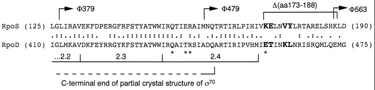
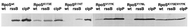
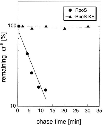
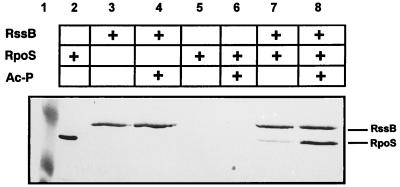
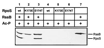
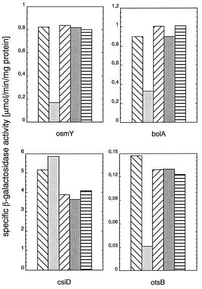
The degradation of the RpoS (sigmaS) subunit of RNA polymerase in Escherichia coli is a prime example of regulated proteolysis in prokaryotes. RpoS turnover depends on ClpXP protease, the response regulator RssB, and a hitherto uncharacterized "turnover element" within RpoS itself. Here we localize the turnover element to a small element (around the crucial amino acid lysine-173) directly downstream of the promoter-recognizing region 2.4 in RpoS. Its sequence as well as its location identify the turnover element as a unique proteolysis-promoting motif. This element is shown to be a site of interaction with RssB. Thus, RssB is functionally unique among response regulators as a direct recognition factor in ClpXP-dependent RpoS proteolysis. Binding of RssB to RpoS is stimulated by phosphorylation of the RssB receiver domain, suggesting that environmental stress affects RpoS proteolysis by modulating RssB affinity for RpoS. Initial evidence indicates that lysine-173 in RpoS, besides being essential of RpoS proteolysis, may play a role in promoter recognition. Thus the same region in RpoS is crucial for proteolysis as well as for activity as a transcription factor.


Figures

Figure 1
Figure 1
Alignment of the turnover element-containing region of RpoS with the corresponding region of RpoD. Partial amino acid sequences of RpoS and RpoD between the positions indicated are given. Amino acids at positions that were mutated in the present study are given in larger and bold capital letters. Regions important for transcription initiation as well as RpoD residues that are included in a partial crystal structure (34) are indicated. Amino acids in RpoD with a direct role in promoter recognition are indicated by ∗. Arrows indicate fusion joints of translational rpoSlacZ fusions (5, 8). In addition, the position of an in-frame deletion that interferes with RpoS proteolysis is shown (8).
Figure 2
Figure 2
Cellular levels of various mutant forms of RpoS in exponential phase. Cells expressing wild-type or mutant versions of RpoS from pBAD18 in wild-type, rssBcat, or clpPcat backgrounds were grown in minimal medium M9 with 0.4% glycerol. Immunoblots are shown demonstrating cellular RpoS levels during exponential phase (at an OD578 of 0.6).
Figure 3
Figure 3
RpoSK173E is not subject to proteolysis. Cells expressing RpoSwt (●) or RpoSK173E (▴) from pBAD18 were grown in minimal medium M9 with 0.4% glycerol. RpoS degradation was demonstrated by pulse–chase labeling and immunoprecipitation of exponential phase samples (harvested at an OD578 of 0.6) as detailed in Materials and Methods.
Figure 4
Figure 4
Coelution of RssB and RpoS from S-protein agarose in the presence and absence of acetyl phosphate. Equimolar amounts (0.5 nmol) of purified S-TRX-His6-RssB and/or His6-RpoS were incubated for 1 hr at room temperature in the presence or absence of 50 mM acetyl-phosphate. Proteins adsorbed to S-protein agarose (added for another 30 min) were washed and eluted with sodium citrate (pH 2). Proteins were separated by SDS/PAGE and visualized by immunoblotting using Penta-His antibodies. Lane 2 shows purified His6-RpoS. Size standard proteins (49.5 and 32 kDa) are shown in lane 1.
Figure 5
Figure 5
RpoSK173E does not interact with RssB adsorbed to S-protein agarose. Equimolar amounts (0.5 nmol) of S-TRX-His6-RssB and His6-RpoSwt, His6-RpoSK173E, or His6-RpoSE174T (lanes 1–3) were incubated with 50 mM acetyl phosphate, adsorbed to S-protein agarose, washed, eluted, and visualized as described in the legend to Fig. 4. As controls, either S-TRX-His6-RssB (lane 7) or the different His6-RpoS proteins alone (lanes 4–6) were subject to the same treatment.
Figure 6
Figure 6
The K173E mutation in RpoS differentially affects the expression of various RpoS-dependent genes. Strains carrying rssBcat, rpoS∷Tn10, and lacZ fusions in the RpoS-controlled genes osmY, bolA, csiD, and otsB, which express different RpoS variants from pBAD18-derived plasmids, were grown in LB. Two hours after entry into stationary phase, specific β-galactosidase activities were determined. In each panel representing one of the reporter gene fusions, the bars represent strains carrying the following RpoS variants (from left to right): RpoSwt, RpoSK173E, RpoSE174T, RpoSV177K, and RpoSY178L. Values given are averages of two independent cultures, each of which was sampled in triplicate.

References

    1. Hengge-Aronis R. In: Escherichia coli and Salmonella typhimurium: Cellular and Molecular Biology. Neidhardt F C, editor. Washington, DC: Am. Soc. Microbiol.; 1996. pp. 1497–1512.
    1. Loewen P C, Hengge-Aronis R. Annu Rev Microbiol. 1994;48:53–80. - PubMed
    1. Bearson S M D, Benjamin W H, Jr, Swords W E, Foster J W. J Bacteriol. 1996;178:2572–2579. - PMC - PubMed
    1. Lange R, Hengge-Aronis R. Genes Dev. 1994;8:1600–1612. - PubMed
    1. Muffler A, Traulsen D D, Lange R, Hengge-Aronis R. J Bacteriol. 1996;178:1607–1613. - PMC - PubMed

Publication types

MeSH terms

LinkOut - more resources