. 1999 May 25;96(11):6445-50.
doi: 10.1073/pnas.96.11.6445.

Plasmodium falciparum subtilisin-like protease 2, a merozoite candidate for the merozoite surface protein 1-42 maturase

Affiliations

Plasmodium falciparum subtilisin-like protease 2, a merozoite candidate for the merozoite surface protein 1-42 maturase

J C Barale et al. Proc Natl Acad Sci U S A. .

Abstract

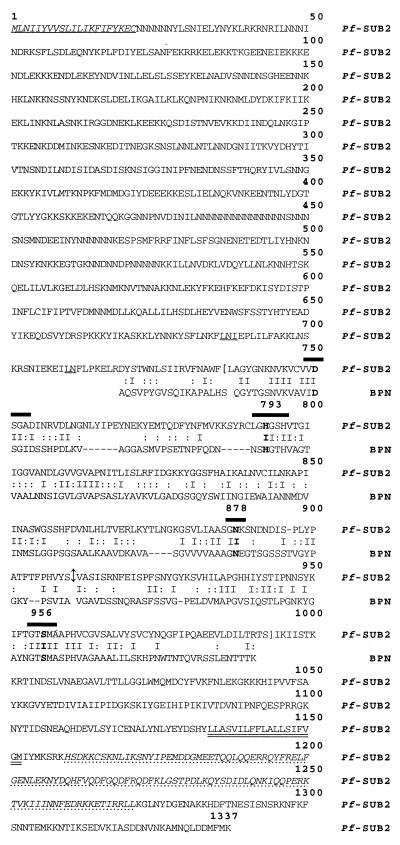
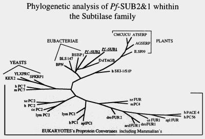
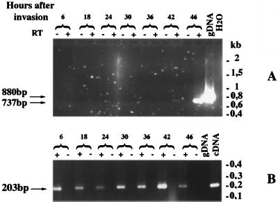
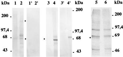
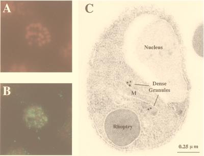
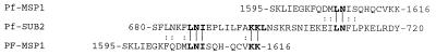
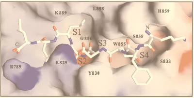
The process of human erythrocyte invasion by Plasmodium falciparum parasites involves a calcium-dependent serine protease with properties consistent with a subtilisin-like activity. This enzyme achieves the last crucial maturation step of merozoite surface protein 1 (MSP1) necessary for parasite entry into the host erythrocyte. In eukaryotic cells, such processing steps are performed by subtilisin-like maturases, known as proprotein convertases. In an attempt to characterize the MSP1 maturase, we have identified a gene that encodes a P. falciparum subtilisin-like protease (PfSUB2) whose deduced active site sequence resembles more bacterial subtilisins. Therefore, we propose that PfSUB2 belongs to a subclass of eukaryotic subtilisins different from proprotein convertases. Pfsub2 is expressed during merozoite differentiation and encodes an integral membrane protein localized in the merozoite dense granules, a secretory organelle whose contents are believed to participate in a late step of the erythrocyte invasion. PfSUB2's subcellular localization, together with its predicted enzymatic properties, leads us to propose that PfSUB2 could be responsible for the late MSP1 maturation step and thus is an attractive target for the development of new antimalarial drugs.


Figures

Figure 1
Figure 1
Predicted protein sequence of PfSUB2 (GenBank accession no. AJ132006) and the alignment of its predicted catalytic domain with B. amyloliquefaciens BPN subtilisin (GenBank accession no. X00165). The translation initiation and stop codons are in bold and underlined. PfSUB2 protein sequence begins with a signal peptide shown in bold. The four residues that compose the subtilase active site are heavily overlined, and the C-terminal transmembrane segment is double underlined. The interruption of the ORF by an intron is shown by ↕. Brackets define the 259 residues of the catalytic domain used to perform the phylogenetic analysis, and the sequence corresponding to the GST-fusion protein is presented in italics and dashed underlined. The two proposed activation sites are underlined.
Figure 2
Figure 2
Pfsub2 gene specifically hybridizes to chromosome 11. Chromosome blocks from P. falciparum Palo Alto strain were probed with 1,027 bp of cDNA corresponding to the catalytic domain of PfSUB2.
Figure 3
Figure 3
Phylogenetic tree analysis based on sequence alignment of catalytic domains of subtilases from various organisms. PfSUB2 and PfSUB1 appear to diverge from known eukaryotic subtilases and to be more similar to the bacterial enzymes. DdTAGB and human S1P/SKI-1 are the most related eukaryotic subtilases and with PfSUB2 and PfSUB1 define a subclass of eukaryotic subtilases.
Figure 4
Figure 4
Analysis of Pfsub2 mRNA expression through the P. falciparum asexual cycle. RT-PCR was performed on total RNA isolated between 6 and 46 hr after invasion of erythrocytes by merozoites of P. falciparum Palo Alto strain cultivated in vitro. (A) Pfsub2 oligonucleotides have been chosen to flank the Pfsub2 intron. The expected sizes of the amplified fragments are 880 bp and 737 bp on genomic DNA and cDNA, respectively. (B) RT-PCR was performed by using two Pfrab6 oligonucleotides, where one oligonucleotide crossed the junction of the two first exons of Pfrab6 and therefore only cDNA was amplified. As negative controls RT-PCR on water were performed.
Figure 5
Figure 5
Western blot analysis on protein extracts prepared from P. falciparum segmented schizonts and merozoites. Lanes 1 and 2 correspond to Na2CO3-soluble and Na2CO3-insoluble protein extracts prepared from segmented schizonts. Lanes 1′ and 2′ contain the same proportion of Na2CO3-soluble and –insoluble protein extracts prepared from noninfected RBCs. Lanes 3 and 3′ and lanes 4 and 4′ show Western blot analysis performed on Na2CO3-soluble and -insoluble merozoite protein extracts. Lanes 1 and 2, lanes 1′ and 2′, and lanes 3 and 4 have been incubated with anti-GST-PfSUB2 antibodies, and lanes 3′ and 4′ have been incubated with antibodies to GST alone. Lanes 5 and 6 correspond to the immunoprecipitation of segmented schizont S35-labeled proteins performed with mouse anti-GST-PfSUB2 and anti-GST.
Figure 6
Figure 6
Localization of PfSUB2 protein by immunofluorescence on segmented schizonts and by IEM on fixed merozoites. (A and B) An air-dried fixed segmented schizont incubated with 25 μg/ml of propidium iodide to label the parasite nuclei (A) and mouse anti-GST-PfSUB2 antibodies (B). Bound antibodies then were detected with FITC-coupled anti-mouse antibodies (magnification: ×1,500). (C) The IEM analysis of thin sections of resin-embedded merozoites probed with mouse anti-GST-PfSUB2 antibodies and revealed with gold-labeled anti-mouse IgG antibody. M, mitochondrion.
Figure 7
Figure 7
Alignment of the 22 residues surrounding the Leu↕Asn MSP1–42 maturation site with the 38 residues corresponding to the predicted PfSUB2 auto-activation site region. Identities are shown by vertical bars, and residues of the same group are shown by double dots.
Figure 8
Figure 8
Homology model of PfSUB2 predicted catalytic domain in complex with MSP1–42 cleavage site (Nterm-K-F-Q-D-M-L-N-I-Cterm). Residues involved in the PfSUB2 predicted S1, S2, S3, and S4 subsites are numbered. Modeling was carried out with computer programs quanta and charmm by using the atomic coordinates of B. amyloliquefaciens BPN subtilisin in complex with the eglin inhibitor (PDB code 2SNI) and B. licheniformis Carlsberg’s subtilisin in complex with eglin C (PDB code 2SEC) as templates. Framework residues differing between proteins were mutated, and all required insertions (Fig. 1) were modeled in hypothetical conformations taken from the loop library implemented in quanta. These insertions occur in surface-exposed loops located far from the substrate-binding region, with the possible exception of the segment preceding the catalytic His793 residue (including Arg789), which may influence the local structure of the S1′-S2′ subsites. The model was manually adjusted to remove unreasonable contacts and subjected to energy minimization by using the program charmm.

References

    1. Aikawa M. In: Malaria: Principles and Practice of Malariology. Wernsdorfer W H, McGregor I, editors. Vol. 1. Edinburgh: Churchill Livingstone; 1988. pp. 97–129.
    1. Bannister L H, Mitchell G H. J Protozool. 1989;36:362–367. - PubMed
    1. Braun-Breton C, Pereira da Silva L. Parasitol Today. 1993;9:92–96. - PubMed
    1. Perrin L H, Ramirez E, Lambert P H, Miescher P. Nature (London) 1981;289:301–303. - PubMed
    1. Torii M, Adams J H, Miller L H, Aikawa M. Infect Immun. 1989;57:3230–3233. - PMC - PubMed

Publication types

MeSH terms

Substances

Associated data

LinkOut - more resources