. 1999 May 25;96(11):6468-72.
doi: 10.1073/pnas.96.11.6468.

Agrin in Alzheimer's disease: altered solubility and abnormal distribution within microvasculature and brain parenchyma

Affiliations

Agrin in Alzheimer's disease: altered solubility and abnormal distribution within microvasculature and brain parenchyma

J E Donahue et al. Proc Natl Acad Sci U S A. .

Abstract

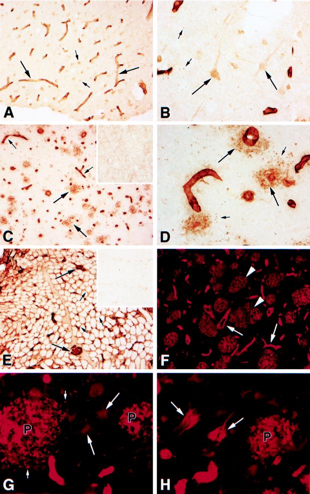
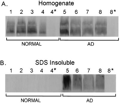
Agrin is a heparan sulfate proteoglycan that is widely expressed in neurons and microvascular basal lamina in the rodent and avian central nervous system. Agrin induces the differentiation of nerve-muscle synapses, but its function in either normal or diseased brains is not known. Alzheimer's disease (AD) is characterized by loss of synapses, changes in microvascular architecture, and formation of neurofibrillary tangles and senile plaques. Here we have asked whether AD causes changes in the distribution and biochemical properties of agrin. Immunostaining of normal, aged human central nervous system revealed that agrin is expressed in neurons in multiple brain areas. Robust agrin immunoreactivity was observed uniformly in the microvascular basal lamina. In AD brains, agrin is highly concentrated in both diffuse and neuritic plaques as well as neurofibrillary tangles; neuronal expression of agrin also was observed. Furthermore, patients with AD had microvascular alterations characterized by thinning and fragmentation of the basal lamina. Detergent extraction and Western blotting showed that virtually all the agrin in normal brain is soluble in 1% SDS. In contrast, a large fraction of the agrin in AD brains is insoluble under these conditions, suggesting that it is tightly associated with beta-amyloid. Together, these data indicate that the agrin abnormalities observed in AD are closely linked to beta-amyloid deposition. These observations suggest that altered agrin expression in the microvasculature and the brain parenchyma contribute to the pathogenesis of AD.


Figures

Figure 1
Figure 1
Localization of agrin in aged normal and AD brain. Sections were immunohistochemically stained with either diaminobenzidine-labeled tertiary antibody (AE) or immunofluorescence-stained with CY3-labeled secondary antibody (FH) as described in Materials and Methods. (A) Aged control brain section labeled with antiagrin antibody. Agrin immunoreactivity is prominent within the cerebral microvasculature (large arrows) and also is evident in selected neurons (small arrows). Prefrontal cortex, A10, ×200. (B) A higher magnification of the control brain section in A. Agrin immunoreactivity is evident within the cytoplasm of the neuronal soma and processes (large arrows). Occasional neurons also demonstrate staining of the nucleus. Note the presence of rare, agrin-immunoreactive puncta (small arrows) in the neuropil, which often are adjacent to blood vessels. (A10, ×600.) (C) Prefrontal cortex (A10) of a patient with AD immunostained with anti-agrin antibody. Note the robust staining of neuritic and diffuse plaques (large arrows) and blood vessels. In contrast to aged control cases (e.g., A), blood vessels in AD had attenuated diameters and a more ragged profile (small arrows). No immunoreactivity was observed if the antisera was preabsorbed with 10−6 M agrin protein (Inset). (×200.) (D) A higher magnification of AD brain illustrating two neuritic plaques with surrounding puncta of agrin immunoreactivity (large arrows). Circumferential puncta of immunoreactivity also can be seen in plaques surrounding and adjacent to cerebral capillaries (small arrows). (A10, ×600.) (E) Normal infant skeletal muscle labeled with anti-agrin antibody. Note the uniform agrin immunoreactivity of the basement membranes surrounding individual muscle fibers (small arrows) and capillaries (large arrows). (Inset) The same skeletal muscle after the primary antibody was preabsorbed with 10−6 M agrin protein. There is essentially complete abolishment of agrin immunoreactivity. (Quadriceps muscle, ×200.) (F) Amygdala of a patient with AD labeled with anti-agrin antibody. Note the robust staining of neuritic and diffuse plaques (arrowheads) and blood vessels. Blood vessels in this AD case have attenuated diameters and ragged profiles (arrows). (×200.) (G) A higher magnification of the AD amygdala seen in F illustrating two neuritic plaques (P) with surrounding puncta of agrin immunoreactivity (small arrows). Agrin immunoreactivity also may be seen in reactive gemistocytic astrocytes and their stellate processes (large arrows). (×600.) (H) Another high-magnification photomicrograph of the AD amygdala seen in F showing agrin immunoreactivity within two neurofibrillary tangles (arrows). Note the fine wisps of paired helical filaments conforming to the shape of the neurons they are within. An agrin-stained neuritic plaque (P) also is present. (×600.)
Figure 2
Figure 2
Western blot analysis of agrin expression in normal aged and AD) brain. Equal volumes of total homogenates or of the SDS-insoluble fractions from normal (–4) or AD brain (–8) prefrontal cortex (area A10) were probed with anti-agrin antisera. All samples were solubilized in 0.2 M NaOH before electrophoresis (see Materials and Methods). (A) The anti-agrin antibody recognized a polypeptide with an apparent mobility of ≈500 kDa in both normal and AD brains. No polypeptide was detected when normal rabbit IgG was substituted for the first layer (lanes 4* and 8*). (B) Virtually all of the agrin from normal brains was efficiently solubilized in 1% SDS at neutral pH (lanes 1–4). However, a portion (see Results) of the agrin from AD brains was insoluble under these conditions (lanes 5–8). No polypeptide was detected when normal rabbit IgG was substituted for the first layer (lanes 4* and 8*).

References

    1. Tsen G, Halfter W, Kroger S, Cole G J. J Biol Chem. 1995;270:3392–3399. - PubMed
    1. Nitkin R M, Smith M A, Magill C, Fallon J R, Yao Y M, Wallace B G, McMahan U J. J Cell Biol. 1987;195:2471–2478. - PMC - PubMed
    1. Reist N E, Magill C, McMahan U J. J Cell Biol. 1987;105:2457–2469. - PMC - PubMed
    1. Wallace B G. J Neurosci. 1989;9:1294–1302. - PMC - PubMed
    1. Gautam M, Noakes P G, Moscoso L, Rupp F, Scheller R H, Merlie J P, Sanes J R. Cell. 1996;85:525–535. - PubMed

Publication types