PAX6 expression in the developing human eye
- PMID: 10340984
- PMCID: PMC1723067
- DOI: 10.1136/bjo.83.6.723
PAX6 expression in the developing human eye
Abstract
Aims: To investigate the changes in PAX6 expression in the developing human eye.
Methods: Six developing human eyes from 6 to 22 weeks' gestation were evaluated. Frozen sections were immunohistochemically stained with monoclonal antibody to chick Pax6 (amino acids 1-223). To verify antibody specificity, western blot analysis was carried out using cell lysates from P19 cells transfected with the human PAX6 gene.
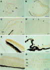
Results: Western blot analysis demonstrated that the antibody reacted to human PAX6 protein. Positive immunostainings for PAX6 were seen in the surface ectoderm, lens vesicle, inner and outer layers of the optic cup, and optic stalk at 6 weeks, and in the corneal epithelia and conjunctiva, lens, and non-pigmented ciliary epithelia from 8 to 22 weeks. In the retina, positive cells were seen in the entire retina from 8 to 10 weeks, and were restricted to the ganglion cell layer and the inner and outer portions of the inner nuclear layer after 21 weeks.
Conclusions: PAX6 is expressed on the surface and neuroectoderms at an early stage, then in the differentiating cells in the cornea, lens, ciliary body, and retina through development. PAX6 may play a role in determining cell fate in the morphogenesis of various human ocular tissue.
Figures
Comment in
-
Science! Why should the clinician care?Br J Ophthalmol. 1999 Jun;83(6):638-9. doi: 10.1136/bjo.83.6.638. Br J Ophthalmol. 1999. PMID: 10340966 Free PMC article. No abstract available.
References
MeSH terms
Substances
LinkOut - more resources
Full Text Sources
Other Literature Sources


