

Review
doi: 10.1136/bjo.83.6.748.
Amniotic membrane transplantation
Affiliations
- PMID: 10340988
- PMCID: PMC1723092
- DOI: 10.1136/bjo.83.6.748
Item in Clipboard
Review
Amniotic membrane transplantation
Br J Ophthalmol.
1999 Jun.
No abstract available
Figures

Transmission electron microscopy of the amnion. The apical border of the amniotic epithelial cells contains a great number of microvilli. The cytoplasm contains numerous vesicles. Basal cell processes (pedicels) extend into the basement membrane. The underlying connective tissue has a homogeneous structure.
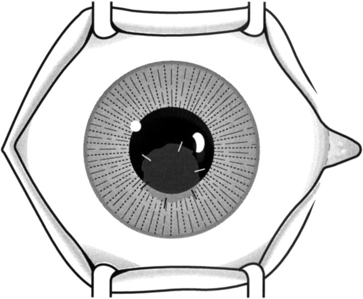
Surgical technique. The diagram illustrates the amniotic membrane sutured to the cornea and covering a paracentral corneal epithelial defect.
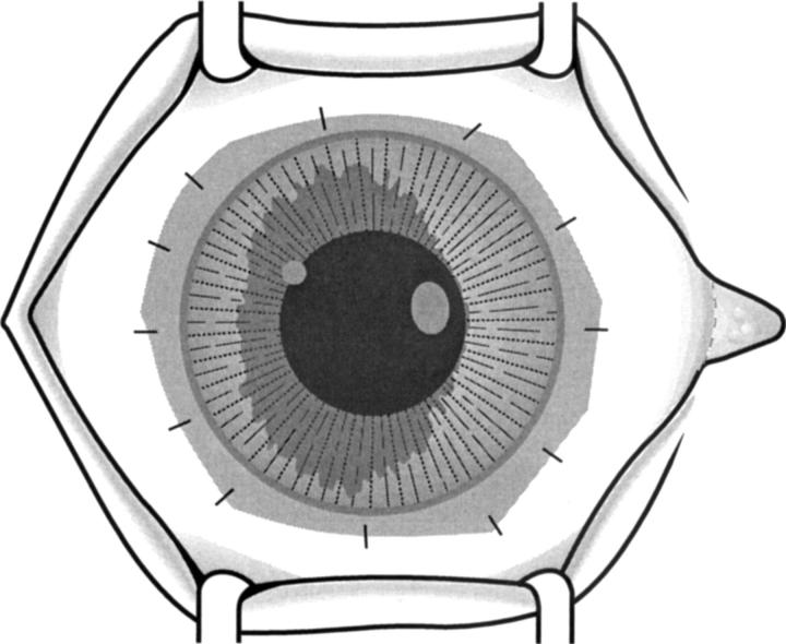
Surgical technique. The amniotic membrane is sutured to perilimbal episclera and to the edge of the conjunctiva (after peritomy) covering the whole corneal surface.

Surgical technique. The amniotic membrane can be used to cover a conjunctival defect after releasing adhesions during symblepharon surgery, and in a similar manner (nasally or temporally) after excision of pterygium.
References
Publication types
MeSH terms
LinkOut - more resources
Full Text Sources
Other Literature Sources
Medical