. 1999 Jun 1;19(11):4200-10.
doi: 10.1523/JNEUROSCI.19-11-04200.1999.

Electron microscopic evidence against apoptosis as the mechanism of neuronal death in global ischemia

Affiliations

Electron microscopic evidence against apoptosis as the mechanism of neuronal death in global ischemia

F Colbourne et al. J Neurosci. .

Abstract

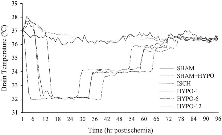
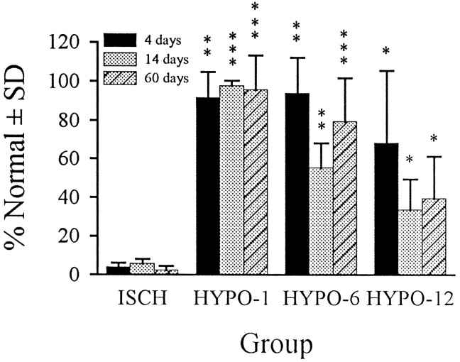
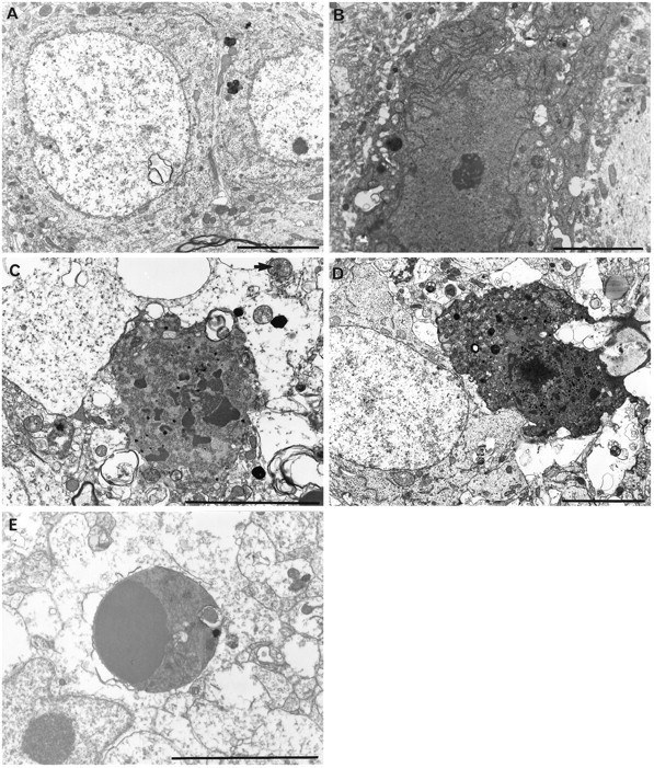
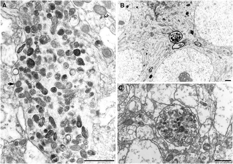
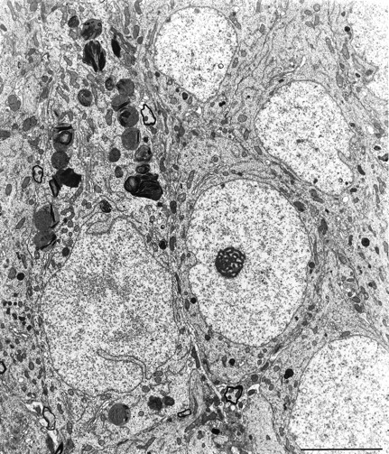
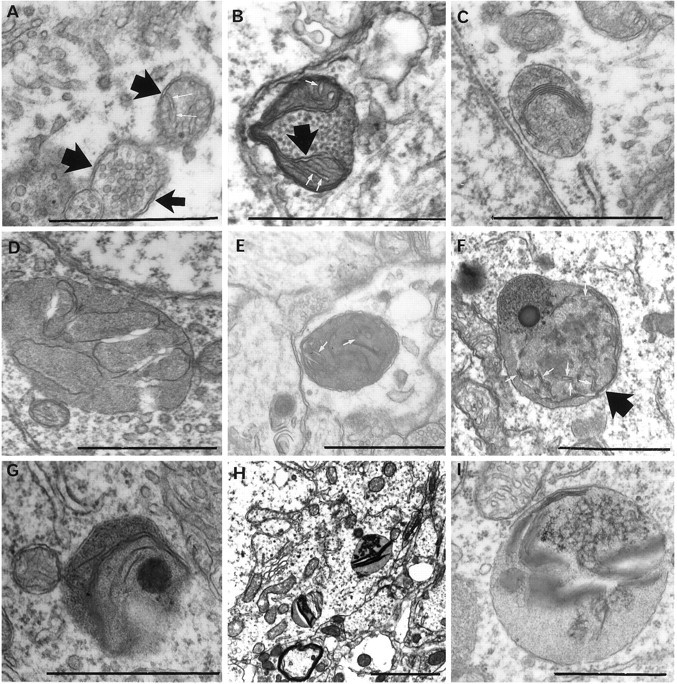
It has been repeatedly claimed that neuronal death in the hippocampal CA1 sector after untreated global ischemia occurs via apoptosis. This is based largely on DNA laddering, nick end labeling, and light microscopy. Delineation of apoptosis requires fine structural examination to detect morphological events of cell death. We studied the light and ultrastructural characteristics of CA1 injury after 5 min of untreated global ischemia in gerbils. To increase the likelihood of apoptosis, some ischemic gerbils were subjected to delayed postischemic hypothermia, a treatment that mitigates injury and delays the death of some neurons. In these gerbils, 2 d of mild hypothermia was initiated 1, 6, or 12 hr after ischemia, and gerbils were killed 4, 14, or 60 d later. Ischemia without subsequent cooling killed 96% of CA1 neurons by day 4, whereas all hypothermia-treated groups had significantly reduced injury at all survival times (2-67% loss). Electron microscopy of ischemic neurons with or without postischemic hypothermia revealed features of necrotic, not apoptotic, neuronal death even in cells that died 2 months after ischemia. Dilated organelles and intranuclear vacuoles preceded necrosis. Unique to the hypothermia-treated ischemic groups, some salvaged neurons were persistently abnormal and showed accumulation of unusual, morphologically complex secondary lysosomes. These indicate selective mitochondrial injury, because they were closely associated with normal and degenerate mitochondria, and transitional forms between mitochondria and lysosomes occurred. The results show that untreated global ischemic injury has necrotic, not apoptotic, morphology but do not rule out programmed biochemical events of the apoptotic pathway occurring before neuronal necrosis.


Figures

Fig. 1.
Fig. 1.
Brain temperature averaged every hour starting after ischemia/sham occlusion to 4 d later. Data were collected every 20 or 30 sec but plotted as averaged 1 hr intervals. All ischemic groups and the SHAM+HYPO group were regulated for 2 d after ischemia (see Materials and Methods), whereas SHAM animals were not manipulated. Servo-control was achieved in awake, freely moving gerbils by independent use of fan, lamp, and a water-misting system. See Table 1 for baseline, occlusion, and first hour mean temperatures.
Fig. 2.
Fig. 2.
CA1 cell counts expressed as a mean percentage of normal (SHAM and SHAM+HYPO groups) ±SD. Cell counts in SHAM and SHAM+HYPO groups were the same and thus were combined. Likewise, cell counts in the three ISCH sub groups were very similar (differed by only a few cells) and were combined into one group for statistical comparisons. Hypothermia significantly blunted injury in all groups (*p < 0.05, **p < 0.01, ***p < 0.001), but the greatest benefit occurred with the 1 hr intervention delay where CA1 injury was almost totally abolished. There was no overlap between CA1 counts in any HYPO group and the ISCH groups.
Fig. 3.
Fig. 3.
Light microscopic CA1 neuronal findings. Dark neurons were rare but were found in all groups, as is characteristic of aldehyde-fixed tissue. The one illustrated (A) is from a SHAM animal. Hypothermia-treated ischemic animals had robust CA1 neuroprotection (B), but occasional neurons (arrow) had granular inclusions (also see Figs. 7, 8). Untreated ischemia produced near-total CA1 loss by 4 d after ischemia (C), and neuronal cytolysis and karyorrhectic debris (arrow) predominate in the field. Necrotic neurons from HYPO groups had similar light microscopic morphology as from ISCH groups. Large clumps of karyorrhectic debris might be confused with apoptotic bodies under lower magnification. However, no apoptotic body was found in any ISCH or HYPO group. For control, an apoptotic body (compacted chromatin enclosed within a membrane) is shown (D) from the tectum of a neonatal gerbil. The same apoptotic body in D is shown with briefer exposure to illustrate the chromatin mass within the dense body (inset). Tissue was embedded in Epon, and sections were stained with toluidine blue. Scale bar (shown in Dapplies to A–D): 20 μm.
Fig. 4.
Fig. 4.
CA1 neurons from control animals (SHAM) showing small lysosomes and some intranuclear vacuoles (A) and a dark but undamaged neuron (B). CA1 cell necrosis in untreated ischemia (ISCH) showed nuclear and plasma membrane breaks, clumped tigroid chromatin, and amorphous organelles with mitochondrial flocculent densities after 4 d survival (C). Although less prevalent after hypothermic-treated ischemia (D, HYPO-12, 4 d survival), neuronal necrosis has an identical ultrastructural appearance. Necrotic neurons had variable electron density. Apoptotic bodies, like those found in neonatal gerbil striatum (E), were never found in any adult gerbil examined by light and electron microscopy. Scale bars, 5 μm.
Fig. 5.
Fig. 5.
In contrast to the terminal necrotic changes shown in Fig. 4, some of the remaining neurons showed sublethal alterations that were either unique or present far in excess of their incidence in controls. Dilated organelles, including RER, Golgi apparatus, and mitochondria as well as extensive intranuclear vacuoles (arrows) (A, ISCH 4 d survival; B, HYPO-6, 14 d survival) and large clusters of primary and secondary lysosomes (C, HYPO-12, 2 month survival) were seen in untreated and hypothermia-treated ischemic groups. Rarely, neurons contained stacks of proliferated RER (D, HYPO-12, 4 d survival). Scale bars, 5 μm.
Fig. 6.
Fig. 6.
Dendritic (A, HYPO-12, 14 d survival) and axonal (B, HYPO-1, 2 month survival) pyknotic inclusions as found in HYPO and ISCH groups. Similar clusters were found in PD6 gerbils (C, colliculus). Because normal (A, arrow) and degenerate mitochondria can be seen with these clusters, these dense inclusions are likely degenerating mitochondria. Scale bars, 1 μm.
Fig. 7.
Fig. 7.
Some CA1 neurons of all HYPO groups at all survival times showed large numbers of accumulating bodies, probably derived from mitochondria (see Fig. 9). The neuron containing this large number of autolysosomes was otherwise healthy looking (HYPO-1, 14 d survival). Adjacent neurons look normal. Scale bar, 5 μm.
Fig. 8.
Fig. 8.
View of early (A) and late (B) mitochondrial evolution into lysosomes. On the left are seen a group of mitochondria with variably disorganized cristae and a tubulovesicular internal structure. The appearance of homogenous electron dense streaks suggests lipid concentration within them. Other groups of cytoplasmic bodies (B) appear more electron dense throughout most of their internal structure, apart from the small, round body left of center, which has a double membrane (arrows) indicating a mitochondrial origin. Scale bars, 1 μm.
Fig. 9.
Fig. 9.
Progression of mitochondrial injury to autolysosomes. All of these forms were found only in hypothermia-treated ischemic animals at all survival periods. Double membranes (broad arrows, A, B, F) as well as remnant cristae (white arrows, A, B, E, F) attest to the mitochondrial origin of these structures. Collapsing internal membranes are seen (C, D, G). Increased internal electron density (F–I) suggests eventual accumulation of lipid or lipofuscin-like material. Scale bars, 1 μm.

Similar articles

Cited by

References

    1. Abe K, Aoki M, Kawagoe J, Yoshida T, Hattori A, Kogure K, Itoyama Y. Ischemic delayed neuronal death. A mitochondrial hypothesis. Stroke. 1995;26:1478–1489. - PubMed
    1. Auer RN. Structural neurotoxicologic investigation of the glycine antagonist 5-nitro-6,7-dichloroquinoxalinedione (ACEA1021). Neurotoxicology. 1997;18:53–62. - PubMed
    1. Bicknell GR, Cohen GM. Cleavage of DNA to large kilobase pair fragments occurs in some forms of necrosis as well as apoptosis. Biochem Biophys Res Commun. 1995;207:40–47. - PubMed
    1. Bonfoco E, Krainc D, Ankarcrona M, Nicotera P, Lipton SA. Apoptosis and necrosis: two distinct events induced, respectively, by mild and intense insults with N-methyl-d-aspartate or nitric oxide/superoxide in cortical cell cultures. Proc Natl Acad Sci USA. 1995;92:7162–7166. - PMC - PubMed
    1. Bonnekoh P, Barbier A, Oschlies U, Hossmann KA. Selective vulnerability in the gerbil hippocampus: morphological changes after 5-min ischemia and long survival times. Acta Neuropathol (Berl) 1990;80:18–25. - PubMed

Publication types

LinkOut - more resources