

Localization of type I inositol 1,4,5-triphosphate receptor in the outer segments of mammalian cones
- PMID: 10341226
- PMCID: PMC6782613
- DOI: 10.1523/JNEUROSCI.19-11-04221.1999
Localization of type I inositol 1,4,5-triphosphate receptor in the outer segments of mammalian cones
Abstract
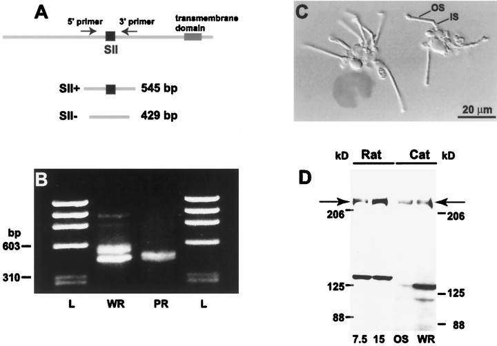
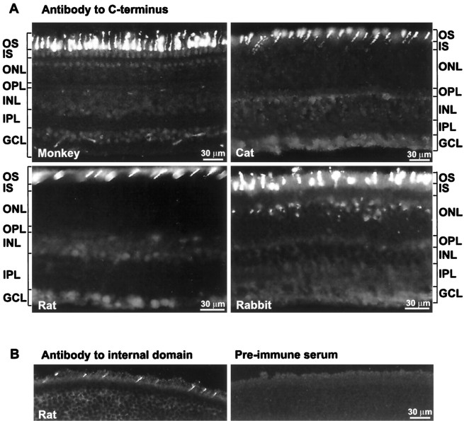
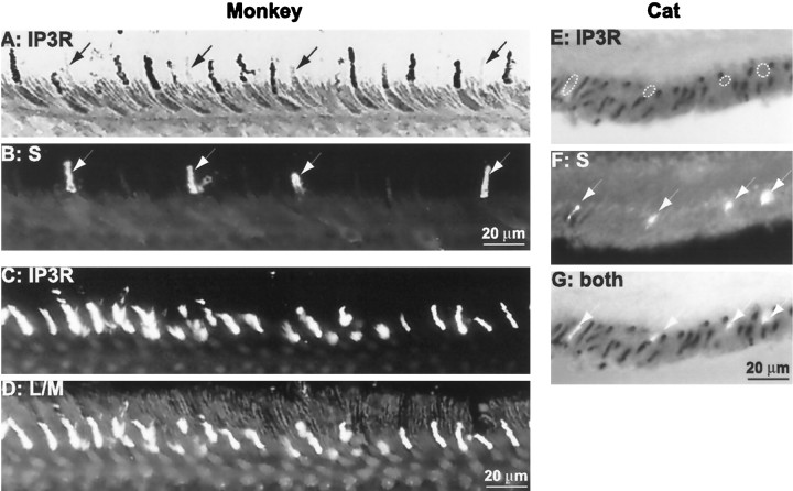
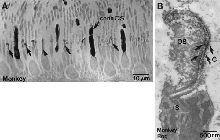
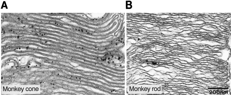
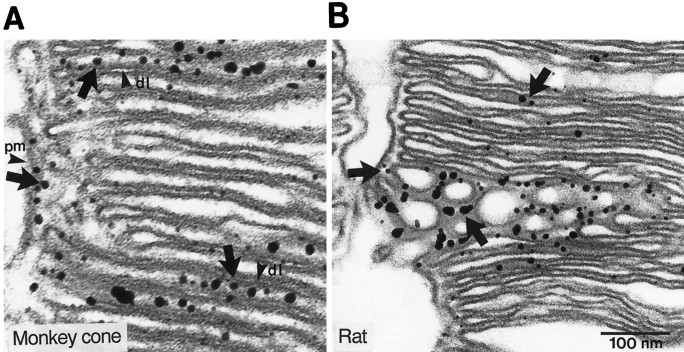
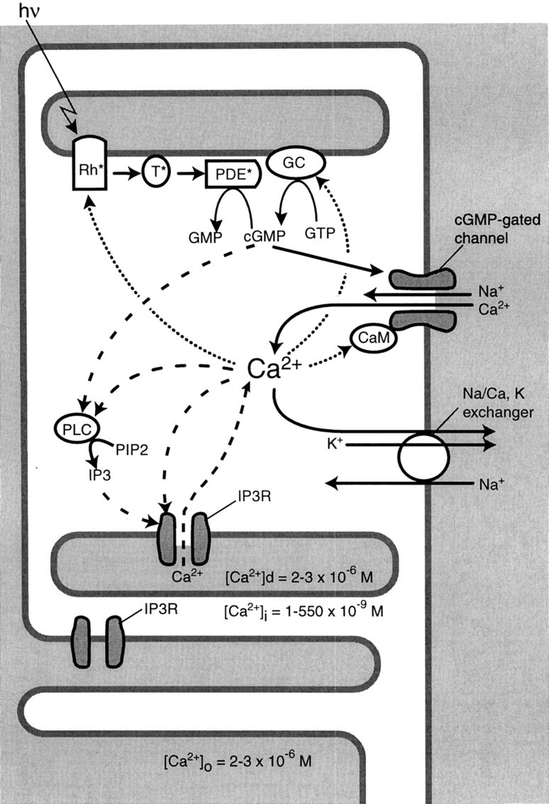
Calcium enters the outer segment of a vertebrate photoreceptor through a cGMP-gated channel and is extruded via a Na/Ca, K exchanger. We have identified another element in mammalian cones that might help to control cytoplasmic calcium. Reverse transcription-PCR performed on isolated photoreceptors identified mRNA for the SII- splice variant of the type I receptor for inositol 1,4,5-triphosphate (IP3), and Western blots showed that the protein also is expressed in outer segments. Immunocytochemistry showed type I IP3 receptor to be abundant in red-sensitive and green-sensitive cones of the trichromatic monkey retina, but it was negative or weakly expressed in blue-sensitive cones and rods. Similarly, the green-sensitive cones expressed the receptor in dichromatic retina (cat, rabbit, and rat), but the blue-sensitive cones did not. Immunostain was localized to disk and plasma membranes on the cytoplasmic face. To restore sensitivity after a light flash, cytoplasmic cGMP must rise to its basal level, and this requires cytoplasmic calcium to fall. Cessation of calcium release via the IP3 receptor might accelerate this fall and thus explain why the cone recovers much faster than the rod. Furthermore, because its own activity of the IP3 receptor depends partly on cytoplasmic calcium, the receptor might control the set point of cytoplasmic calcium and thus affect cone sensitivity.
Figures
Similar articles
-
Tuning outer segment Ca2+ homeostasis to phototransduction in rods and cones.Adv Exp Med Biol. 2002;514:179-203. doi: 10.1007/978-1-4615-0121-3_11. Adv Exp Med Biol. 2002. PMID: 12596922 Review.
-
Identification and localization of an immunoreactive AMPA-type glutamate receptor subunit (GluR4) with respect to identified photoreceptor synapses in the outer plexiform layer of goldfish retina.J Neurocytol. 1997 Oct;26(10):651-66. doi: 10.1023/a:1018597811439. J Neurocytol. 1997. PMID: 9368879
-
Preservation of endoplasmic reticulum (ER) Ca2+ stores by deletion of inositol-1,4,5-trisphosphate receptor type 1 promotes ER retrotranslocation, proteostasis, and protein outer segment localization in cyclic nucleotide-gated channel-deficient cone photoreceptors.FASEB J. 2021 Jun;35(6):e21579. doi: 10.1096/fj.202002711R. FASEB J. 2021. PMID: 33960001 Free PMC article.
-
Inositol 1,4,5-trisphosphate receptors: immunocytochemical localization in the dorsal cochlear nucleus.J Comp Neurol. 1995 Jul 17;358(1):102-18. doi: 10.1002/cne.903580107. J Comp Neurol. 1995. PMID: 7560273
-
Ca2+ flux in retinal rod and cone outer segments: differences in Ca2+ selectivity of the cGMP-gated ion channels and Ca2+ clearance rates.Cell Calcium. 1995 Oct;18(4):285-300. doi: 10.1016/0143-4160(95)90025-x. Cell Calcium. 1995. PMID: 8556768 Review.
Cited by
-
Synaptic transmission at retinal ribbon synapses.Prog Retin Eye Res. 2005 Nov;24(6):682-720. doi: 10.1016/j.preteyeres.2005.04.002. Prog Retin Eye Res. 2005. PMID: 16027025 Free PMC article. Review.
-
Repeated spike exposure to the insecticide chlorpyrifos interferes with the recovery of visual sensitivity in rats.Doc Ophthalmol. 2005 Jan;110(1):79-90. doi: 10.1007/s10633-005-7347-8. Doc Ophthalmol. 2005. PMID: 16249959
-
Inhibitory effect of somatostatin-14 on L-type voltage-gated calcium channels in cultured cone photoreceptors requires intracellular calcium.J Neurophysiol. 2009 Sep;102(3):1801-10. doi: 10.1152/jn.00354.2009. Epub 2009 Jul 15. J Neurophysiol. 2009. PMID: 19605612 Free PMC article.
-
Calcium stores in vertebrate photoreceptors.Adv Exp Med Biol. 2012;740:873-89. doi: 10.1007/978-94-007-2888-2_39. Adv Exp Med Biol. 2012. PMID: 22453974 Free PMC article. Review.
-
Calcium regulation in photoreceptors.Front Biosci. 2002 Sep 1;7:d2023-44. doi: 10.2741/A896. Front Biosci. 2002. PMID: 12161344 Free PMC article. Review.
References
-
- Anderson DH, Fisher SK. The photoreceptors of diurnal squirrels: outer segment structure disc shedding and protein renewal. J Ultrastruct Res. 1976;55:119–141. - PubMed
-
- Berridge MJ. Inositol triphosphate and calcium signaling. Nature. 1993;361:315–325. - PubMed
-
- Bezprozvanny I, Watras J, Ehrlich BE. Bell-shaped calcium–response curves in Ins 1,4,5-P3- and calcium-gated channels from endoplasmic reticulum of cerebellum. Nature. 1991;351:751–754. - PubMed
-
- Brown JE, Blazynski C, Cohen AI. Light induces a rapid and transient increase in inositol triphosphate in toad rod outer segments. Biochem Biophys Res Commun. 1987;146:1392–1396. - PubMed
Publication types
MeSH terms
Substances
Grants and funding
LinkOut - more resources
Full Text Sources
Molecular Biology Databases
Miscellaneous