. 1999 Jun 1;19(11):4263-9.
doi: 10.1523/JNEUROSCI.19-11-04263.1999.

High tolerance and delayed elastic response of cultured axons to dynamic stretch injury

Affiliations

High tolerance and delayed elastic response of cultured axons to dynamic stretch injury

D H Smith et al. J Neurosci. .

Abstract

Although axonal injury is a common feature of brain trauma, little is known of the immediate morphological responses of individual axons to mechanical injury. Here, we developed an in vitro model system that selectively stretches axons bridging two populations of human neurons derived from the cell line N-Tera2. We found that these axons demonstrated a remarkably high tolerance to dynamic stretch injury, with no primary axotomy at strains <65%. In addition, the axolemma remained impermeable to small molecules after injury unless axotomy had occurred. We also found that injured axons exhibited the behavior of "delayed elasticity" after injury, going from a straight orientation before injury to developing an undulating course as an immediate response to injury, yet gradually recovering their original orientation. Surprisingly, some portions of the axons were found to be up to 60% longer immediately after injury. Subsequent to returning to their original length, injured axons developed swellings of appearance remarkably similar to that found in brain-injured humans. These findings may offer insight into mechanical-loading conditions leading to traumatic axonal injury and into potential mechanisms of axon reassembly after brain trauma.


Figures
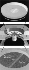
Fig. 1.
Fig. 1.
Schematic illustration of the technique and apparatus used to induce dynamic stretch injury to axons spanning two populations of human neurons. Top, Stainless steel well with a thin transparent deformable membrane in the center on which neurons (NT2N) are plated is shown. A 1.5 × 18 mm cell-free gap is formed in the middle of the membrane that is bridged only by axons. Middle, The well is placed in the stretch device (cut away in the figure to reveal components) that consists of an aluminum cover block with a quartz viewing window and a stainless steel plate on the bottom with a machined 1.5 × 18 mm slit that aligns with the gap region on the membrane. For continuous observation of the axons via an inverted microscope, the apparatus is bolted to the microscope stage that also creates a sealed chamber. Bottom, Compressed air is introduced into the chamber causing a downward deflection of only the gap region of the membrane, thereby inducing uniaxial stretch of the axons.
Fig. 2.
Fig. 2.
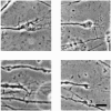
Phase-contrast photomicrographs of four examples of primary axotomy immediately after dynamic stretch injury. Primary axotomy, found only at strains >65%, is represented by severed axons shown in the middle of each panel. Note the torn or shredded appearance of the axonal terminal stumps with possible cytoskeletal remnants trailing out. Scale bar, 10 μm.
Fig. 3.
Fig. 3.
Phase-contrast photomicrographs of two examples (left, right) of the temporal evolution of the delayed elastic response of axons to dynamic stretch injury. As labeled, axons change from a straight orientation before injury to developing large undulations immediately at the time of injury yet gradually reassume their original orientation within 1 hr. Scale bar, 10 μm.
Fig. 4.
Fig. 4.
Quantitative temporal maps of the delayed elastic response of axons to dynamic stretch injury. Top, The temporal change in percent initial total length of two individual axons (circles, triangles) after stretch injury. Bottom, Average percent change in length of 11 axons over time after injury (*p < 0.001 compared with preinjury length).
Fig. 5.
Fig. 5.
Accumulation of neurofilament in axons by 2 hr after dynamic stretch injury. Confocal (top) and deconvolution (bottom) microscopy demonstrates multiple swellings along injured axons immunostained to reveal neurofilament protein. Both confocal and deconvolution microscopy reveals what appears to be a central core of neurofilament in the swellings represented by more intense immunostaining. In some of the swellings, this central core appears disturbed from its original orientation with a tortuous course, even though the axons as a whole had returned to their straight preinjury orientation.
Fig. 6.
Fig. 6.
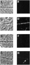
Representative images of axolemmal permeability shown under phase (left) and fluorescence (right) microscopy. These axons were exposed to a fluorescent dye, the uptake of which demonstrates the membrane permeability of small molecules. A, B, No dye is taken up in the absence of either stretch injury or chemical permeabilization of membranes. C, D, Dye is readily taken up after chemical permeabilization of the axolemma of axons. E, F, No dye is taken up by stretch-injured axons that are distorted but not severed.G, H, A detectable amount of dye is taken up in stretch-injured severed (primary axotomy) axons.

References

    1. Adams JH, Graham DI, Murray LS, Scott G. Diffuse axonal injury due to nonmissile head injury in humans: an analysis of 45 cases. Ann Neurol. 1982;12:557–563. - PubMed
    1. Adams JH, Doyle D, Graham DI. Diffuse axonal injury in head injuries caused by a fall. Lancet. 1984;2:1420–1422. - PubMed
    1. Adams JH, Doyle D, Ford I, Gennarelli TA, Graham DI, McClellan DR. Diffuse axonal injury in head injury: definition, diagnosis, and grading. Histopathology. 1989;15:49–59. - PubMed
    1. Burgoyne P. The neuronal cytoskeleton. Wiley; New York: 1991.
    1. De Gennes P-G. Scaling concepts in polymer physics. Cornell UP; Ithaca, NY: 1979.

Publication types

Substances

LinkOut - more resources