. 1999 Jun 1;19(11):4280-92.
doi: 10.1523/JNEUROSCI.19-11-04280.1999.

Cloning and characterization of Aplysia neutral endopeptidase, a metallo-endopeptidase involved in the extracellular metabolism of neuropeptides in Aplysia californica

Affiliations

Cloning and characterization of Aplysia neutral endopeptidase, a metallo-endopeptidase involved in the extracellular metabolism of neuropeptides in Aplysia californica

J P Zappulla et al. J Neurosci. .

Abstract

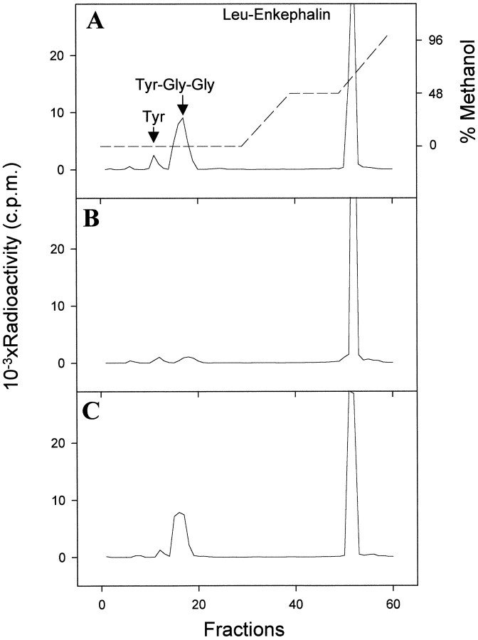
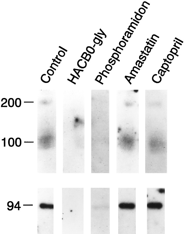
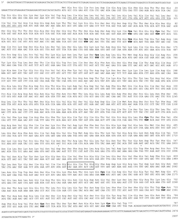
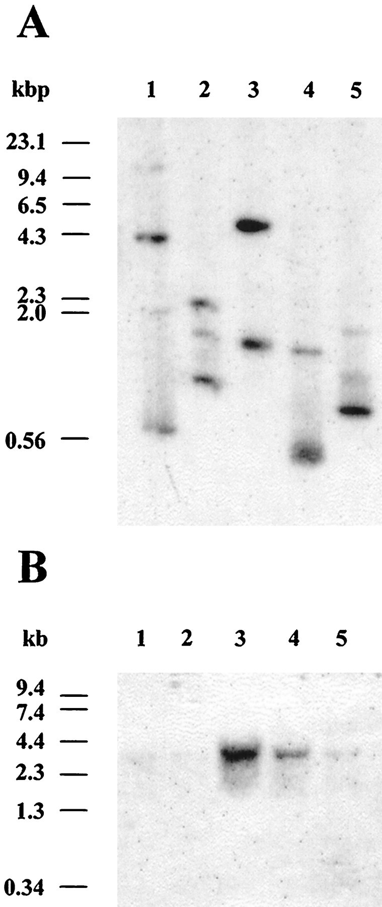
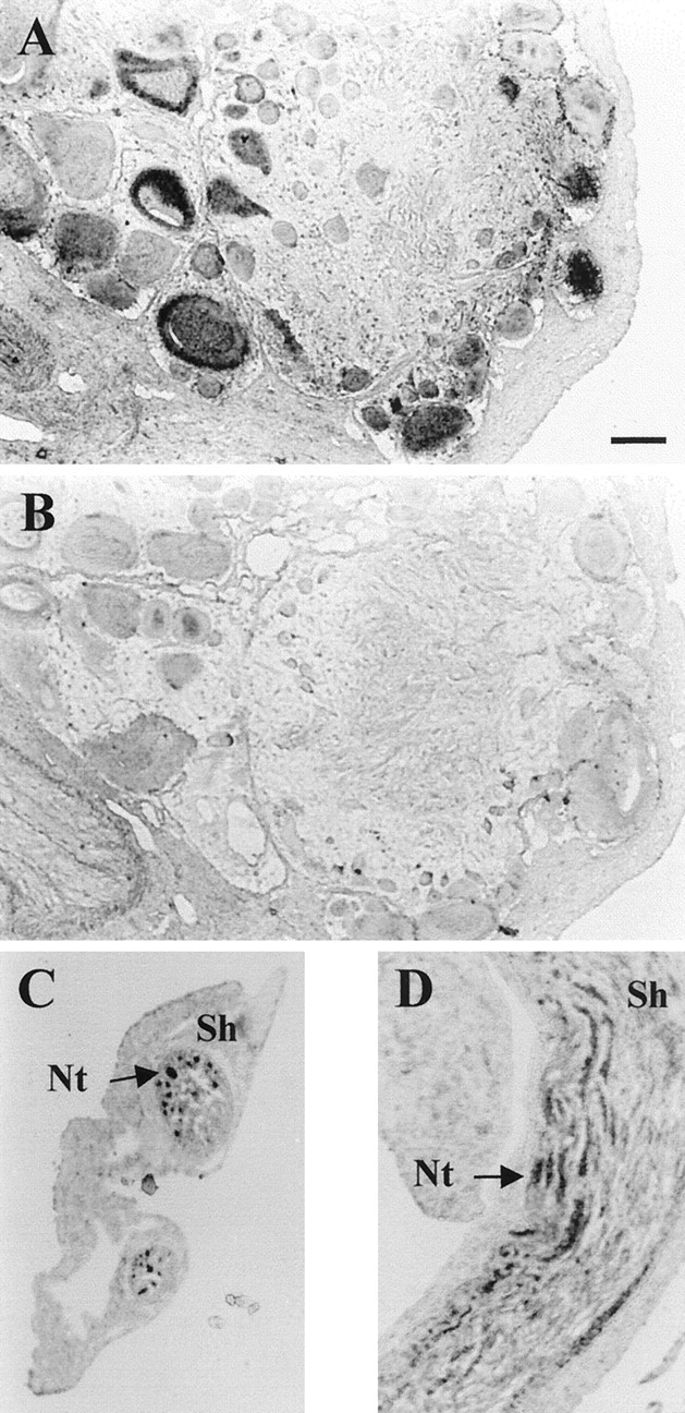
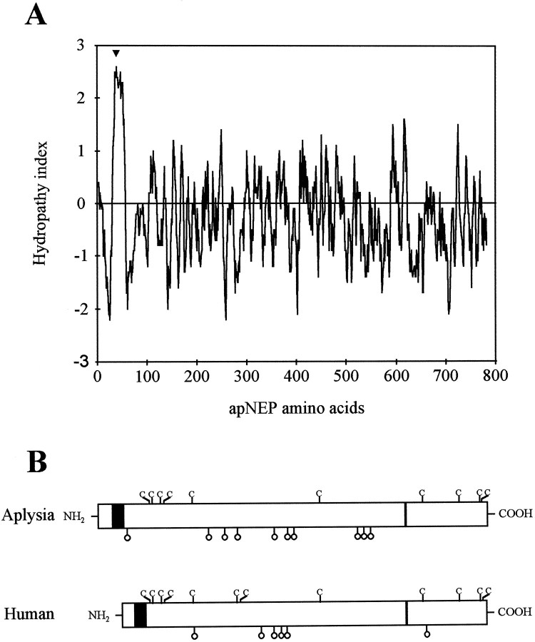
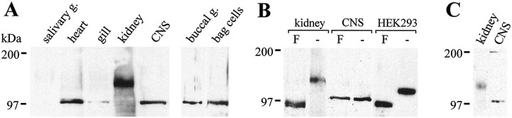
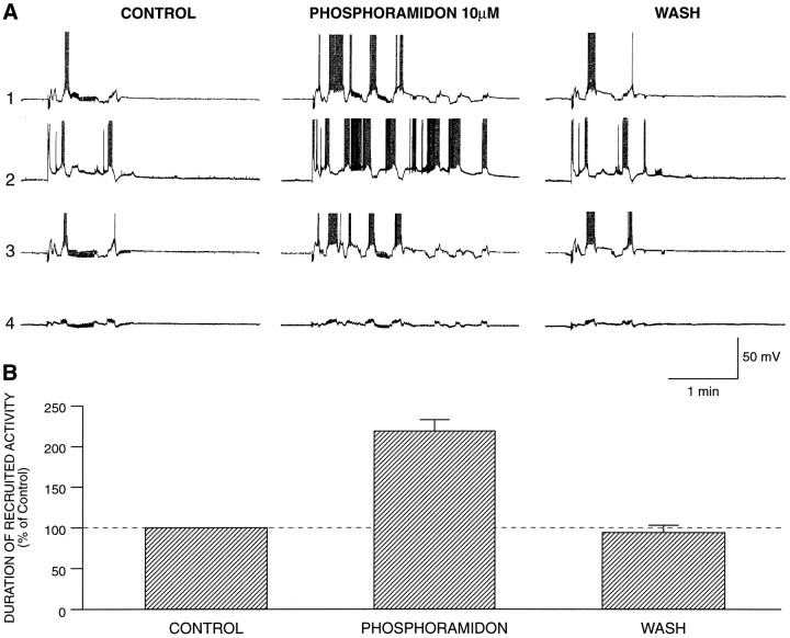
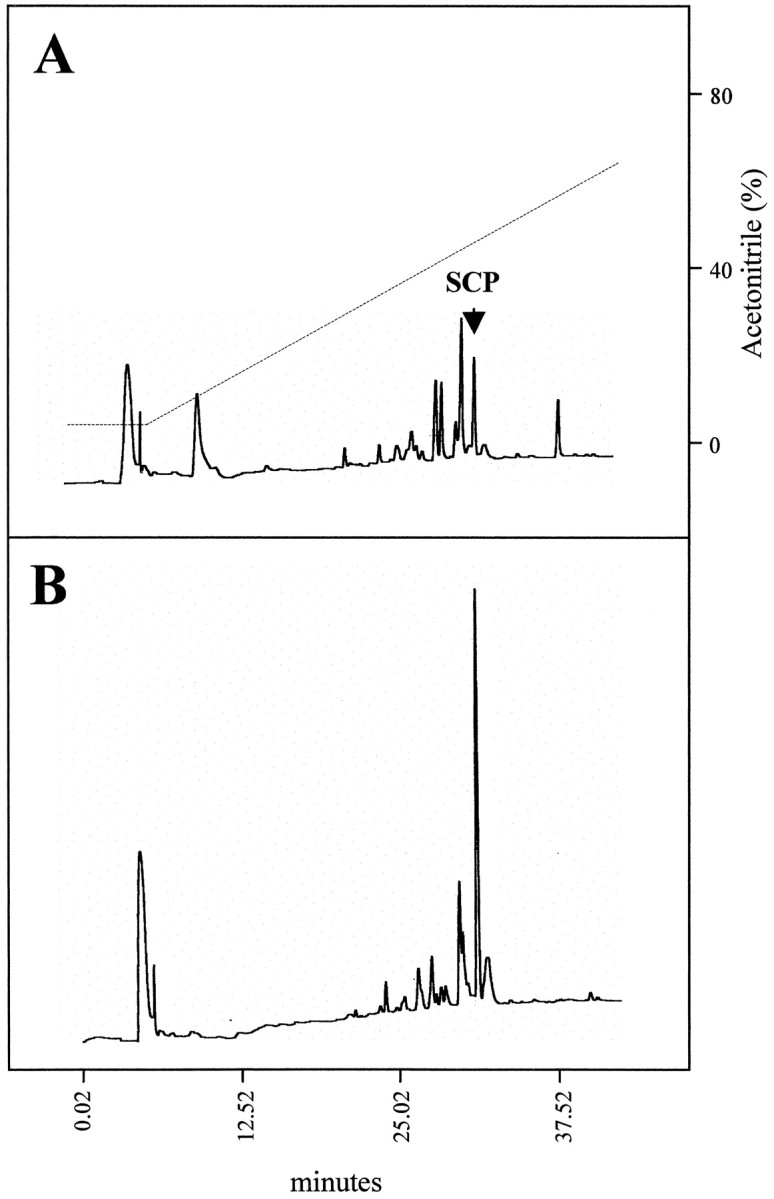
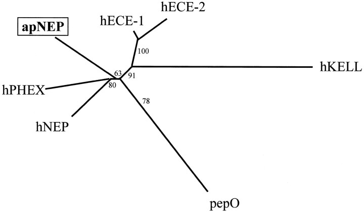
Cell surface metallo-endopeptidases play important roles in cell communication by controlling the levels of bioactive peptides around peptide receptors. To understand the relative relevance of these enzymes in the CNS, we characterized a metallo-endopeptidase in the CNS of Aplysia californica, whose peptidergic pathways are well described at the molecular, cellular, and physiological levels. The membrane-bound activity cleaved Leu-enkephalin at the Gly3-Phe4 bond with an inhibitor profile similar to that of the mammalian neutral endopeptidase (NEP). This functional homology was supported by the molecular cloning of cDNAs from the CNS, which demonstrated that the Aplysia and mammalian NEPs share all the same amino acids that are essential for the enzymatic activity. The protein is recognized both by specific anti-Aplysia NEP (apNEP) antibodies and by the [125I]-labeled NEP-specific inhibitor RB104, demonstrating that the apNEP gene codes for the RB104-binding protein. In situ hybridization experiments on sections of the ganglia of the CNS revealed that apNEP is expressed in neurons and that the mRNA is present both in the cell bodies and in neurites that travel along the neuropil and peripheral nerves. When incubated in the presence of a specific NEP inhibitor, many neurons of the buccal ganglion showed a greatly prolonged physiological response to stimulation, suggesting that NEP-like metallo-endopeptidases may play a critical role in the regulation of the feeding behavior in Aplysia. One of the putative targets of apNEP in this behavior is the small cardioactive peptide, as suggested by RP-HPLC experiments. More generally, the presence of apNEP in the CNS and periphery may indicate that it could play a major role in the modulation of synaptic transmission in Aplysia and in the metabolism of neuropeptides close to their point of release.


Figures

Fig. 1.
Fig. 1.
RP-HPLC analysis of degradation metabolites of [3H][Leu]enkephalin in theAplysia CNS. The substrate was incubated with CNS plasma membranes, in the absence (A) or presence of 1 μm RB104 (B) or 1 μm captopril (C).Arrows indicate the elution position of standard peptides. The dashed line represents the methanol gradient used in the HPLC.
Fig. 2.
Fig. 2.
Inhibitor gel electrophoresis with [125I]RB104 and different peptidase inhibitors. Solubilized Aplysia CNS membrane proteins (top panel) and purified rabbit NEP (bottom panel) were separated by SDS-PAGE and transferred onto nitrocellulose membranes. NEP-like proteins were labeled with 100 pm [125I]RB104 in the presence or absence of peptidase inhibitors: absence of inhibitor (Control); HACBO-gly at 10 μm; Phosphoramidon at 10 μm;Amastatin at 10 μm;Captopril at 10 μm.
Fig. 3.
Fig. 3.
Comparison of the apNEP (λNEPg1 clone), hNEP, hECE1, hKELL, and hPHEX exons that code for the zinc-binding domain. Nucleotide sequences of exons and flanking introns are shown incapital and small letters, respectively. Splicing consensus sequences are underlined. The deduced amino acid sequence of the apNEP exon is shown above the nucleotide sequence. The codons for identical amino acids are inbold type, and the pentapeptide consensus sequences (His-Glu-Xaa-Xaa-His) that are part of the metalloprotease zinc-binding domain are boxed.
Fig. 4.
Fig. 4.
Nucleotide and deduced amino acid sequence of theAplysia neutral endopeptidase. Amino acids are numbered starting at the first ATG of the open reading frame. The putative transmembrane region is underlined. Potential sites ofN-glycosylation are indicated by anasterisk, and the cysteine residues arebold. The zinc-binding signature HEXXH isboxed. The nucleotide sequence has been submitted to the GenBank Data Bank with accession number AF104361.
Fig. 5.
Fig. 5.
Southern and Northern blot analysis of theapNEP gene. A, Genomic DNA was isolated from ovotestis and digested with either BglII (lane 1), EcoRI (lane 2),HindIII (lane 3), SacI (lane 4), or XbaI (lane 5). Digested DNA (10 μg/lane) was run on a 0.8% agarose gel, transferred to a nitrocellulose membrane, and hybridized at high stringency with the [32P]-labeledHindIII–AccI apNEP fragment (nucleotides 1142–1458) as described previously (Wickham and DesGroseillers, 1991). DNA molecular weight markers are indicated in kilobase pairs (kbp) on the left. B, Northern blot analysis of the apNEP transcript. Total RNA was extracted from different tissues, and poly(A+) RNA (5 μg) isolated from gill (lane 1), heart (lane 2), ovotestis (lane 3), kidney (lane 4), and CNS (lane 5) was fractionated on a 1% formaldehyde/agarose gel, blotted to a nitrocellulose membrane, and hybridized at high stringency with the [32P]-(HindIII–AccI) apNEP fragment, as performed previously (Auclair et al., 1994). RNA molecular weight markers are indicated in kilobases (kb) on the left. To control the amounts of RNA in each lane, filters were stripped and rehybridized with an Aplysiaactin probe (data not shown).
Fig. 6.
Fig. 6.
In situ hybridization of apNEP on paraffined sections of Aplysia ganglia. Sections of the abdominal ganglion (A, B) and of a buccal ganglion nerve (C, D) were hybridized with either an apNEP cRNA antisense (A, C, D) or sense (B) probe. Positive signal is seen in neurons (A) and neurites (Nt) extending into the nerve. No signal is detected in the sheath (Sh). The same results were obtained with sections from all the major ganglia. Scale bar, 100 μm.
Fig. 7.
Fig. 7.
Molecular structure of apNEP. A, Hydropathy analysis of apNEP. The 787 amino acid-long apNEP sequence was scanned using the computer program of Kyte and Doolittle (1982).Numbers on the horizontal axis refer to the amino acid sequence. Negative values correspond to hydrophilic regions and positive values to hydrophobic regions. The arrowheadindicates the only potential membrane-spanning segment of apNEP.B, Schematic representation of the primary sequences of the human and Aplysia NEP proteins. The cysteine residues in the two proteins are indicated by the one-letter codeC. The black rectangle represents the transmembrane region, and the thin rectangle represents the HEXXH gluzincin domain. The position of the possibleN-glycosylation sites is indicated by open lollipops.
Fig. 8.
Fig. 8.
Immunoblot analysis of the expression and glycosylation of apNEP in different A. californicatissues. A, Twenty micrograms of solubilized membrane proteins (salivary gland, heart, gill, kidney, and CNS) and 30 μg of total protein extracts (buccal ganglion and bag cells) were separated on a 6% SDS-polyacrylamide gel under reducing conditions, blotted, and detected with an anti-apNEP antisera. B, Plasma membrane protein extracts isolated from Aplysia tissues (kidney, CNS) or from transiently transfected mammalian HEK293 cells (HEK293) were incubated in the absence (−) or presence (F) of PNGase F, before loading on the gel.C, SDS-PAGE under nonreducing conditions. Thearrow indicates the position of the 200 kDa band.
Fig. 9.
Fig. 9.
Phosphoramidon prolongs the responses of the buccal neurons to radula nerve stimulation. A, Simultaneous recordings from four neurons before, during, and after exposure to phosphoramidon (10 μm). In all cases the activity evoked is prolonged: trace 1, B neuron; trace2, A neuron; trace 3, B neuron; trace4, unidentified cell (see Results for details).B, Summary of five experiments (18 neurons) with phosphoramidon (10 or 100 μm). Prolongation of the responses evoked by radula nerve stimulation was observed in all the monitored neurons. The duration of the recruited activity was normalized to each respective control. The percentage average of every neuron in one experiment contributed to the average score of that experiment.
Fig. 10.
Fig. 10.
SCPB is degraded by anAplysia CNS NEP-like enzyme. SCPB was incubated with CNS plasma membranes, in the absence (A) or presence of 10 μmphosphoramidon (B). The arrowindicates the elution position of the uncleaved SCPB. Thedashed line represents the acetonitrile gradient used in the HPLC.
Fig. 11.
Fig. 11.
Phylogenetic analysis of the members of the NEP-like family. Sequences were aligned using the Clustal V program (Thompson et al., 1994). The phylogenetic tree was constructed using the Neighbor Joining method (Saitou and Nei, 1987) with a bootstrap analysis that calculates the probability of occurrence of the presented branching for 100 possible trees (Felsenstein, 1993).hNEP, Human neutral endopeptidase (accession numberM26605); hECE-1, human endothelin-converting enzyme 1 (accession number Z35307); hECE-2, human endothelin-converting enzyme 2 (accession number AB011179);apNEP, A. californica neutral endopeptidase (accession number AF104361); hPHEX, human phosphate-regulating gene with homologies to endopeptidases on the X-chromosome (accession number Y10196); hKELL, human kell blood group protein (accession number M64934);pepO, lactococcus lactis PepO gene (accession number L04938). Sequences were aligned, and only the peptide regions that could be aligned with the PepO sequence were retained for the analysis; this roughly corresponds to the extracellular parts of the human and mollusk enzymes.

Similar articles

Cited by

References

    1. Almenoff J, Teirstein AS, Thornton JC, Orlowski M. Identification of a thermolysin-like metallo-endopeptidase in serum: activity in normal subjects and in patients with sarcoidosis. J Lab Clin Med. 1984;103:420–431. - PubMed
    1. Ambron RT, Schachen S, Rayport SG. Proteins rapidly transported to the synapses of a single identified neuron of Aplysia californica. J Neurosci. 1985;5:2866–2873. - PMC - PubMed
    1. Angers A, Storozhuk MV, Duchaine T, Catellucci VF, DesGroseillers L. Cloning and functional expression of an Aplysia 5-HT receptor negatively coupled to adenylate cyclase. J Neurosci. 1998;18:5586–5593. - PMC - PubMed
    1. Auclair D, Lang BF, Forest P, DesGroseillers L. Analysis of genes encoding highly conserved lysine-rich proteins in Aplysia californica and Saccharomyces cerevisiae. Eur J Biochem. 1994;220:997–1003. - PubMed
    1. Barnes K, Turner AJ, Kenny AJ. Electromicroscopic immunocytochemistry of pig brain shows that endopeptidase-24.11 is localized in neuronal membranes. Neurosci Lett. 1988;94:64–69. - PubMed

Publication types

MeSH terms

Associated data

LinkOut - more resources