. 1999 Jun 1;19(11):4370-87.
doi: 10.1523/JNEUROSCI.19-11-04370.1999.

Transplants of fibroblasts genetically modified to express BDNF promote regeneration of adult rat rubrospinal axons and recovery of forelimb function

Affiliations

Transplants of fibroblasts genetically modified to express BDNF promote regeneration of adult rat rubrospinal axons and recovery of forelimb function

Y Liu et al. J Neurosci. .

Abstract

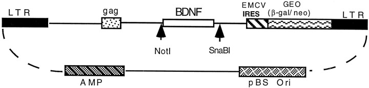
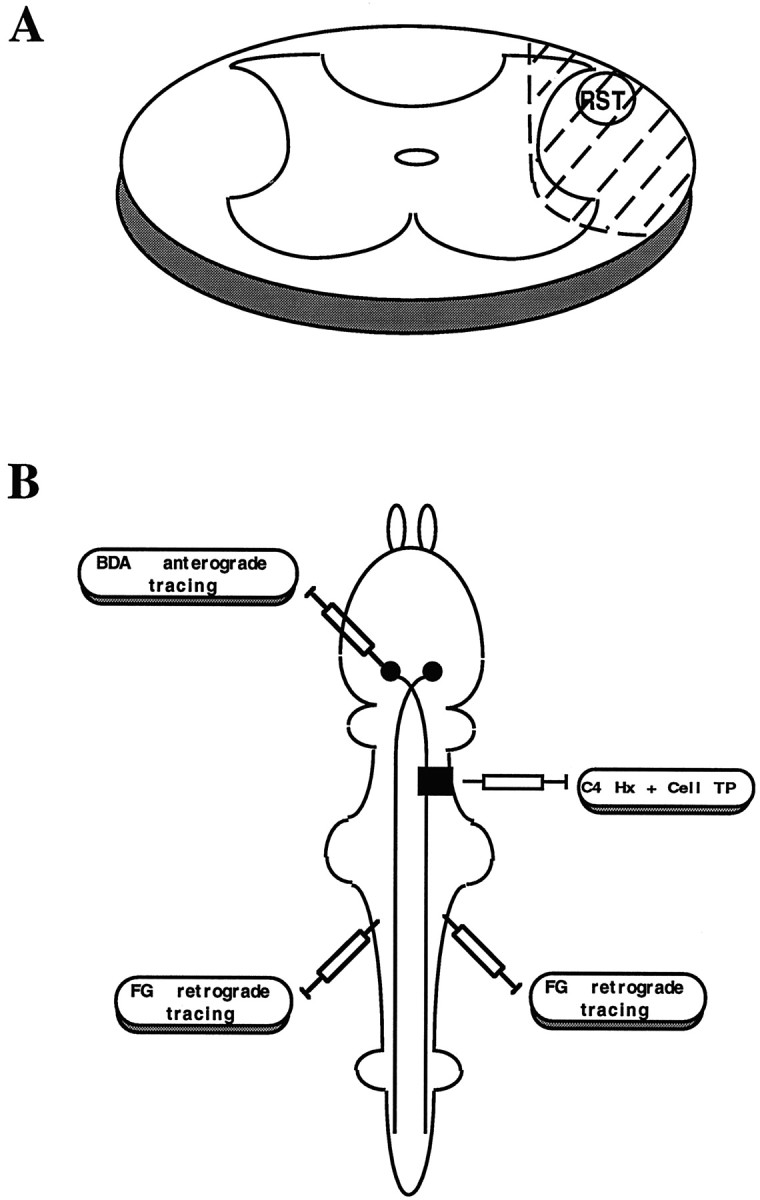
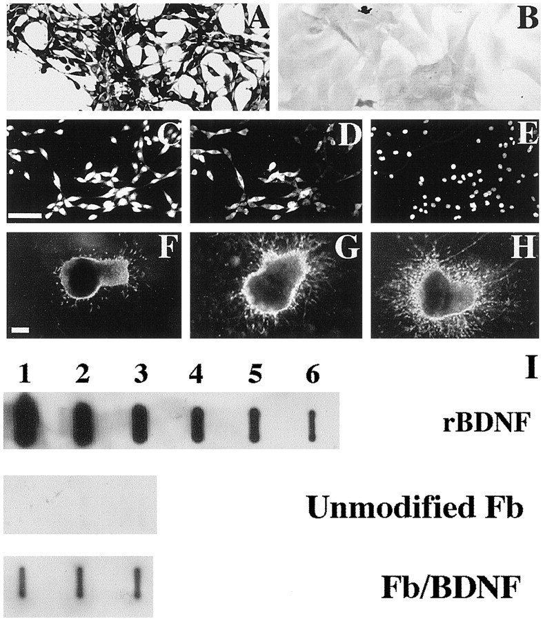
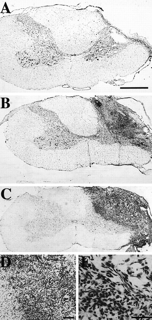
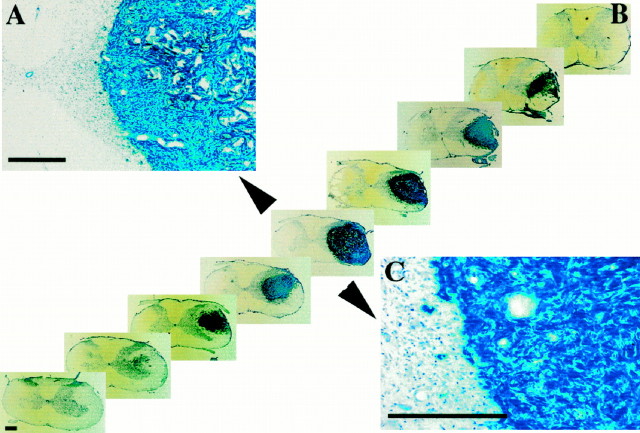
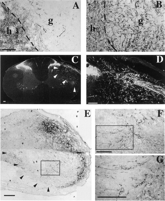
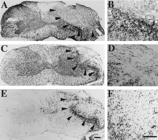
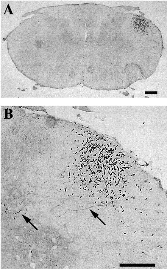
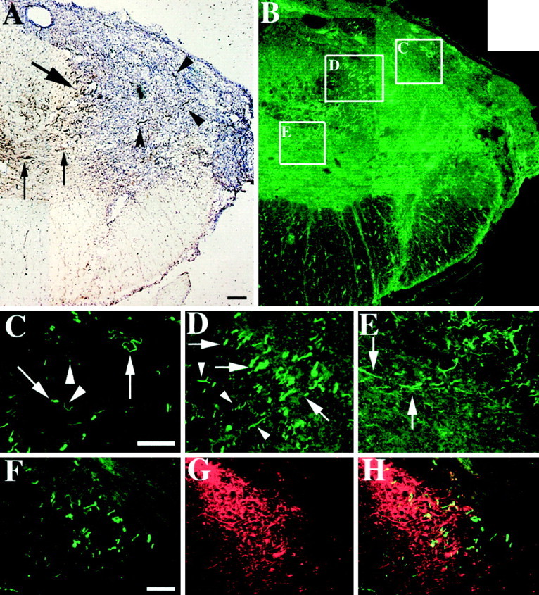
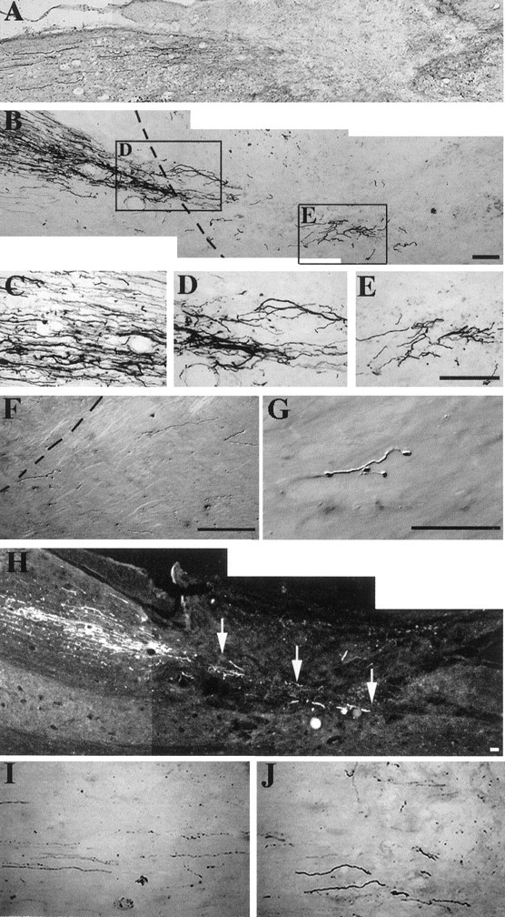
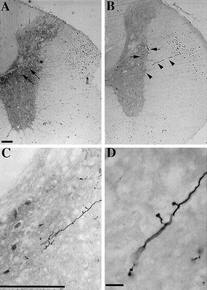
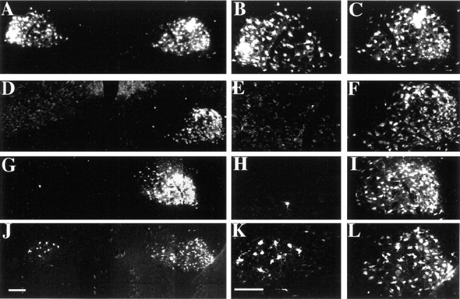
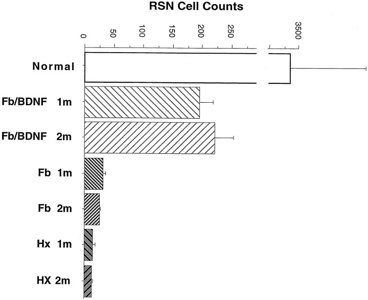
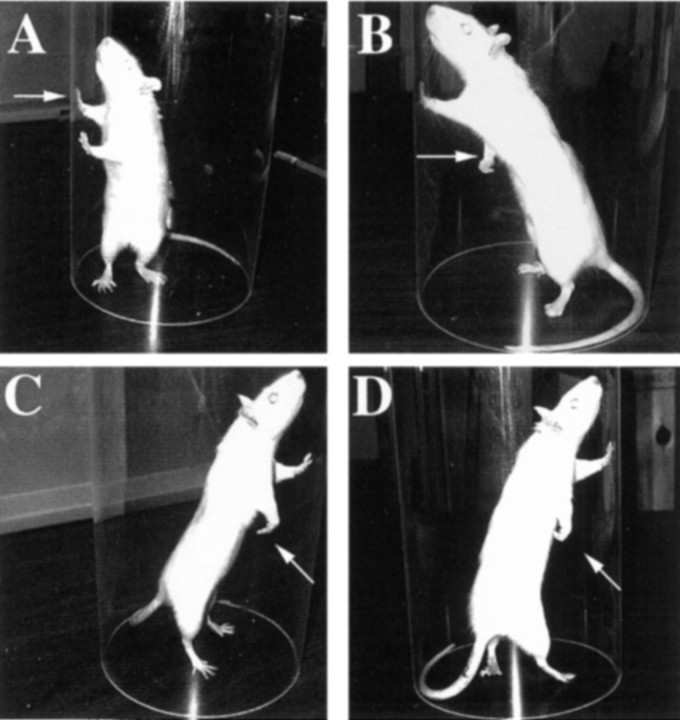
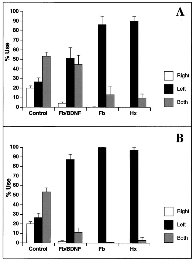
Adult mammalian CNS neurons do not normally regenerate their severed axons. This failure has been attributed to scar tissue and inhibitory molecules at the injury site that block the regenerating axons, a lack of trophic support for the axotomized neurons, and intrinsic neuronal changes that follow axotomy, including cell atrophy and death. We studied whether transplants of fibroblasts genetically engineered to produce brain-derived neurotrophic factor (BDNF) would promote rubrospinal tract (RST) regeneration in adult rats. Primary fibroblasts were modified by retroviral-mediated transfer of a DNA construct encoding the human BDNF gene, an internal ribosomal entry site, and a fusion gene of lacZ and neomycin resistance genes. The modified fibroblasts produce biologically active BDNF in vitro. These cells were grafted into a partial cervical hemisection cavity that completely interrupted one RST. One and two months after lesion and transplantation, RST regeneration was demonstrated with retrograde and anterograde tracing techniques. Retrograde tracing with fluorogold showed that approximately 7% of RST neurons regenerated axons at least three to four segments caudal to the transplants. Anterograde tracing with biotinylated dextran amine revealed that the RST axons regenerated through and around the transplants, grew for long distances within white matter caudal to the transplant, and terminated in spinal cord gray matter regions that are the normal targets of RST axons. Transplants of unmodified primary fibroblasts or Gelfoam alone did not elicit regeneration. Behavioral tests demonstrated that recipients of BDNF-producing fibroblasts showed significant recovery of forelimb usage, which was abolished by a second lesion that transected the regenerated axons.


Figures

Fig. 1.
Fig. 1.
The LIG/BDNF retrovirus. The virus encodes the full-length human BDNF cDNA and GEO, which is a fusion gene of β-gal and neomycin resistance genes. The entire BDNF-IRES-GEO sequence is driven by the RSV LTR promoter and is transcribed into a polycistron mRNA. The EMCV IRES located between BDNF and GEO allows cap-independent initiation of translation of the polycistron mRNA.
Fig. 2.
Fig. 2.
Schematic diagram of the experimental paradigm. Animals received a right C3–4 partial hemisection that disrupted the axons from the left RN. A, Drawing of a spinal cord cross section; the lesion and transplant are represented by theshaded area. Immediately after the spinal cord lesion, Gelfoam, Fb, or Fb/BDNF cells were grafted into the lesion cavity. RST regeneration was studied using either BDA anterograde tracing or FG retrograde tracing (B).
Fig. 3.
Fig. 3.
Analysis of in vitro transgene expression. Cultured Fb/BDNF (A, C–E) or Fb (B) cells were stained with an anti-BDNF antibody (A, B) or double-labeled with anti-BDNF (C) and anti-β-gal antibody (D). E, From the same visual field as C and D; cells are labeled with a nuclear dye (4′,6-diamidino-2-phenylindole) to reveal the entire cell population in the culture. Conditioned media from Fb (F) or Fb/BDNF (G) cells were analyzed for production of biologically active BDNF using an E8 chicken DRG explant assay. Recombinant human BDNF was used as a positive control (H). The rate that Fb/BDNF cells secrete BDNF was calculated relative to control recombinant BDNF using a slot blot assay (I). Recombinant BDNF (rBDNF) was loaded onto a slot blot apparatus at 2000, 400, 80, 16, 3.2, and 0.064 ng (lanes 1–6, respectively) and compared with 20 μl conditioned media from Fb and Fb/BDNF (5000 cells in triplicates). Fb/BDNF cells secrete BDNF at a rate of 12.8 ng/106 cells per 24 hr, whereas Fb cells do not secrete detectable levels of BDNF. Scale bars, 100 μm.
Fig. 4.
Fig. 4.
Photomicrographs of cervical spinal cord sections showing lesion and transplant in different animal groups. Animals received a right C3–4 partial hemisection and a transplant of Gelfoam (A), Fb (B), or Fb/BDNF (C). The animals were killed 1 month after surgery. Spinal cord tissue was cut into cross sections and stained with cresyl violet. D, E, High-power images ofC showing the host–graft interface (D) and the characteristic morphology of fibroblasts in the transplants (E). Scale bars:A–C, 500 μm; D, E, 100 μm.
Fig. 5.
Fig. 5.
Photomicrographs of cervical spinal cord sections showing transgene expression in Fb/BDNF transplants. Animals received a right C3–4 partial hemisection and a transplant of Fb/BDNF cells. Serial sections of spinal cord tissue were stained with X-gal histochemistry and lightly counterstained with cresyl violet. InB, the sections (spaced by 500 μm) were serially reconstructed to show the extent of the lesion and the transplant.A, C, High-power images fromB (arrowheads). A, Host–graft interface and numerous blood vessels in the transplant.C, Host–graft integration and the intense X-gal staining, suggesting high levels of transgene expression. One week survival. Scale bars: A, C, 200 μm; B, 500 μm.
Fig. 6.
Fig. 6.
Photomicrographs of cervical spinal cord sections showing host axon growth into Fb/BDNF or Fb transplants. Spinal cord cross sections from animals receiving Fb (A) or Fb/BDNF (B) transplants were stained with the RT-97 antibody and show cell grafts (g), the host gray matter (h), and the host–graft interface (dashed lines). Numerous axons are present within the Fb/BDNF transplant and at the host–graft interface (B), whereas host axons penetrate Fb transplant sparsely and superficially (A). One month survival. Scale bars, 100 μm. In C andD, a spinal cord cross section from a Fb/BDNF recipient was stained with an anti-CGRP antibody. A dorsal root had regenerated into the transplant and elongated toward the dorsal horn, which was partially disrupted by the transplantation procedures, as intended.Arrowheads outline the graft. D, Higher-power view of axons that had reached the dorsal horn. Two month survival. Scale bars: C, 200 μm; D, 100 μm. In E–G, a spinal cord cross section from an Fb/BDNF recipient was stained with an anti-serotonin antibody. Numerous serotonin-immunoreactive fibers are present throughout the transplant (E). At higher power (F, G), the axons show the characteristic “beads on a string” morphology. InE arrowheads outline the graft–host interface. One month survival. Scale bars, 100 μm.
Fig. 7.
Fig. 7.
Photomicrographs of cervical spinal cord sections showing host immune response. Spinal cord cross sections from animals receiving Fb/BDNF transplants were immunostained with GFAP (A, B), OX-42 (C, D), and ED-1 (E, F) antibodies. A mild astrocytic activation is visible along the graft–host interface, but few if any astrocytes are in the graft (A, B). Macrophages and activated microglia accumulate at the graft–host interface, but few are in the transplants (C–F). One month survival. Scale bars, 100 μm.
Fig. 8.
Fig. 8.
Photomicrographs of cervical spinal cord sections showing the distribution of the RST in normal animals after BDA anterograde tracing. BDA was injected into the maganocellular portion of the left RN. Cervical spinal cord sections were stained with DAB and demonstrate the discrete location of RST in the superficial dorsolateral quadrant (A). B, Higher-power view of the morphology and organization of RST axons. InB arrows point to axon branches in the gray matter. Scale bars, 100 μm.
Fig. 9.
Fig. 9.
Photomicrographs of cervical spinal cord sections showing BDA anterograde tracing of RST axons in the lesion–transplant site. The animal received a right cervical hemisection and an Fb/BDNF transplant. The left RN was anterogradely traced with BDA 15 d before killing. Two month survival after transplantation.A, Section through the transplant 1000 μm from its rostral pole. The section was stained with DAB as chromagen for BDA-labeled fibers and counterstained with cresyl violet. Numerous BDA-labeled axons are present in the transplant (arrowheads) and at the graft–host interface (large arrow) and send off branches into the gray matter (small arrows). However, most of the BDA-labeled axons are obscured by the counterstain. B, Adjacent section stained with FITC to identify BDA-labeled fibers, no counterstain.C–E, Higher magnification of the corresponding regions in B. C, Numerous BDA-labeled axons in the transplant. Most are cut transversely. Arrowheadspoint to smaller-caliber axons, and arrows point to larger-caliber axons. In D, many transversely sectioned larger-caliber axons are labeled at the graft–host interface (arrows) and send off branches perpendicular to the main stem toward the gray matter (arrowheads).E, Higher-power view of axon branches that enter the gray matter (arrows). F–H, Section double-labeled with FITC–avidin (F) and an anti-GFAP antibody (G). H, Merged image of F and G. Numerous RST axons are intermingled with processes of activated astrocytes. Scale bars:A, 200 μm; C, F, 100 μm. Scale bar inA applies to B; scale bar inC applies to D and E; scale bar in F applies to G andH.
Fig. 10.
Fig. 10.
Photomicrographs of cervical spinal cord sections showing regeneration of RST axons through Fb/BDNF transplants. Animals received a right cervical hemisection and Fb (A) or Fb/BDNF transplants (B–J). The left RN was anterogradely traced with BDA 15 d before killing. One month survival after transplantation. Spinal cord tissue was cut into sagittal (A–H) or horizontal (I, J) sections. For all sections left is rostral, and right is caudal. A, All BDA-labeled RST axons are interrupted by the lesion–transplant and failed to enter an Fb transplant. B, Some RST axons regenerated into an Fb/BDNF transplant; the dashed lineindicates the rostral graft–host interface. C, Region rostral to the transplant. Numerous axon branches are evident, suggesting sprouting induced by the transplant. D,E, Higher magnifications of regions fromB. D, BDA-labeled axons that have penetrated the rostral graft–host interface. E, RST axons deeply within the transplant. F, Region in the host white matter immediately caudal to the transplant. Thedashed line indicates the caudal graft–host interface. BDA-labeled axons exit the transplant and elongate caudally. Some axons bear varicosities resembling terminal boutons. G, Terminal bouton-like structure at higher power. H, BDA-labeled axons stained by FITC. Regenerated axons (arrows) pass through the rostral graft–host interface and continue for several millimeters through the transplant and the caudal graft–host interface. I, J, Higher-power views of many smaller-caliber and some larger-caliber RST axons in a rostrocaudal direction in an Fb/BDNF transplant. Scale bars, 100 μm. The scale bar in E applies to C, D, I, and J.
Fig. 11.
Fig. 11.
Photomicrographs of upper-thoracic (A, B) and mid-thoracic (C, D) spinal cord showing BDA-labeled RST axons. A, Cross section from a normal animal demonstrating the normal RST location and organization.Arrows point to axon branches in the gray matter.B, Cross section from an animal with an Fb/BDNF transplant. One month survival. Numerous BDA-labeled axons are present in the lateral funiculus, but their location is aberrant and more diffuse than normal. A few transversely sectioned BDA-labeled axons are also present in the gray matter (arrows). One axon branch arises perpendicular to the main stem and enters lamina VII (arrowheads). C, Axon branches in the gray matter with varicosities resembling terminal boutons.D, Terminal bouton-like structures at higher power. Scale bars: A–C, 100 μm; D, 25 μm. Scale bar in A also applies to B.
Fig. 12.
Fig. 12.
Photomicrographs of midbrain showing FG retrograde tracing of RN neurons. Neurons in both RNs were retrogradely traced by injection of FG into both sides of the spinal cord in normal animals (A–C) or in recipients of Gelfoam (D–F), Fb (G–I), or Fb/BDNF (J–L) transplants. Survival after transplantation was 1 month. All sections were taken ∼480 μm from the caudal pole of RN. B, E, H, K, Higher-power views of the left RN regions corresponding to A, D, G, andJ. C, F, I, L, Higher-power views of the right RN regions corresponding to A, D, G, andJ. In normal animals both RNs are equally labeled (A–C). In Gelfoam recipients virtually no RN neurons are labeled by FG on the left (D, E), whereas labeling on the right is normal (D, F). In Fb transplant recipients, very few RN neurons are labeled in the left RN (G, H), whereas the right RN is normally labeled (G, I). In recipients of Fb/BDNF transplants, numerous RN neurons are labeled in the left RN (J, K), and labeling is normal in the right RN (J, L). Scale bars, 100 μm. The scale bar in Japplies to A, D, G, and J; the scale bar in K applies to B, C, E, F, H, I, K, andL.
Fig. 13.
Fig. 13.
Bar graph comparing numbers of FG-labeled RN neurons among animal groups. The FG-labeled RN neurons in normal animals and animals that had received Fb/BDNF, Fb, or Gelfoam transplants were counted and compared by one-way ANOVA, followed by Fisher’s post hoc test 1 or 2 months after transplantation. Significantly more RN neurons (∼7%) were labeled contralateral to surgery in animals receiving Fb/BDNF transplants than in those receiving Fb or Gelfoam (<1%). p < 0.00001; n = 3 for normal; n = 5 for Fb/BDNF 1m, Hx 1m, and Hx 2m; n = 6 for Fb/BDNF 2m, Fb 1m, and Fb 2m.
Fig. 14.
Fig. 14.
Photograph comparing forelimb use. Animals were analyzed in a cylinder test to study preferred forelimb use.A, C, Seven weeks after transplantation, Fb/BDNF recipients used their forelimb ipsilateral to the lesion to explore the environment (A), whereas Fb and Gelfoam recipients rarely did so (C). The forepaw posture in animals with Fb/BDNF transplants was nearly normal (A), but Fb or Gelfoam recipients kept the forepaw ipsilateral to the lesion strongly flexed (C). B, D, Seven weeks after the relesion at C2, animals with Fb/BDNF transplants lost the normal forelimb posture and the ability to use the forelimb ipsilateral to the lesion (B). In contrast, the relesion had little effect on forelimb posture and forelimb use in animals with Fb transplants (D).A, B, Photographs of the same animal with an Fb/BDNF graft taken before and after the relesion. C, D, From the same animal with an Fb graft before and after the relesion. Arrows in A–D point to the forelimb ipsilateral to the lesion.
Fig. 15.
Fig. 15.
Behavioral analysis of forelimb use. Three weeks after transplantation, Fb/BDNF recipients showed significant recovery of use of the injured limb alone or together with the uninjured limb, whereas Fb or Gelfoam recipients showed negligible use of the injured limb alone (A). The relesion abolished most of the forelimb use in Fb/BDNF recipients but had little effect on the Fb or Gelfoam recipients (B). n= 6 for Fb/BDNF and Fb, n = 5 for Hx.

Similar articles

Cited by

References

    1. Bernstein-Goral H, Bregman BS. Spinal cord transplants support the regeneration of axotomized neurons after spinal cord lesions at birth: a quantitative double-labeling study. Exp Neurol. 1993;123:118–132. - PubMed
    1. Bregman BS, Kunkel-Bagden E, Schnell L, Dai HN, Gao D, Schwab ME. Recovery from spinal cord injury mediated by antibodies to neurite growth inhibitors. Nature. 1995;378:498–501. - PubMed
    1. Bregman BS, McAtee M, Dai HN, Kuhn PL. Neurotrophic factors increase axonal growth after spinal cord injury and transplantation in the adult rat. Exp Neurol. 1997;148:475–494. - PubMed
    1. Brosamle C, Schwab ME. Cells of origin, course, and termination patterns of the ventral, uncrossed component of the mature rat corticospinal tract. J Comp Neurol. 1997;386:293–303. - PubMed
    1. Brown LT. Rubrospinal projections in the rat. J Comp Neurol. 1974;154:169–187. - PubMed

Publication types

MeSH terms

Substances

LinkOut - more resources