. 1999 Jun 1;19(11):4437-48.
doi: 10.1523/JNEUROSCI.19-11-04437.1999.

Collapsin-1/semaphorin-III/D is regulated developmentally in Purkinje cells and collapses pontocerebellar mossy fiber neuronal growth cones

Affiliations

Collapsin-1/semaphorin-III/D is regulated developmentally in Purkinje cells and collapses pontocerebellar mossy fiber neuronal growth cones

S A Rabacchi et al. J Neurosci. .

Abstract

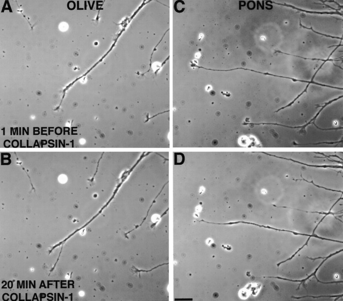
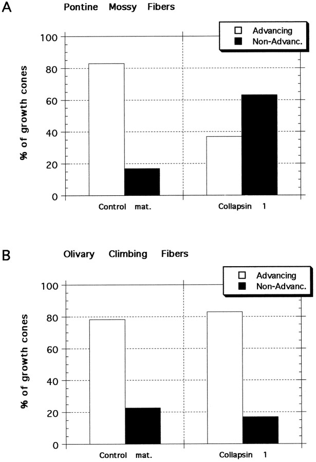
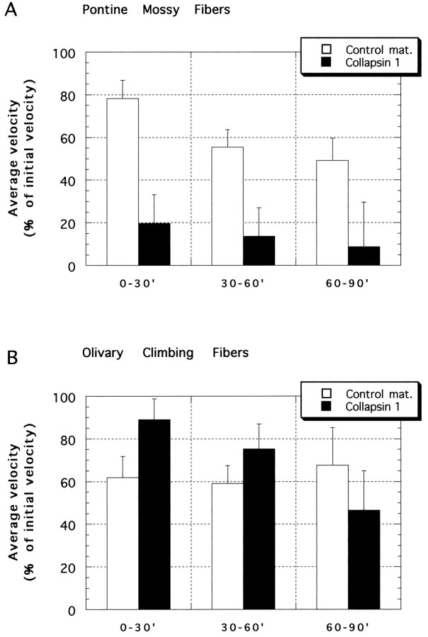
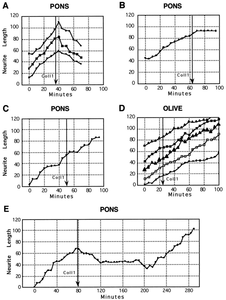
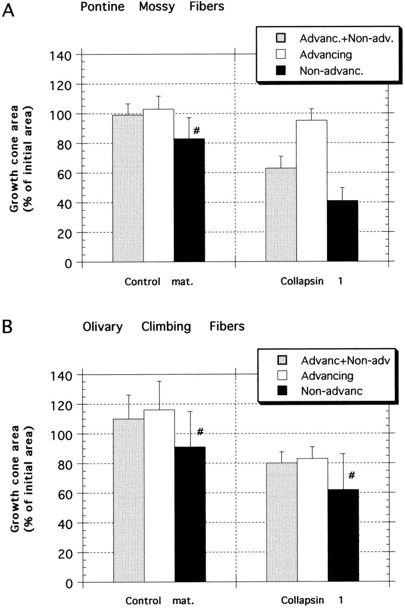
Most axons in the CNS innervate specific subregions or layers of their target regions and form contacts with specific types of target neurons, but the molecular basis of this process is not well understood. To determine whether collapsin-1/semaphorin-III/D, a molecule known to repel specific axons, might guide afferent axons within their cerebellar targets, we characterized its expression by in situ hybridization and observed its effects on mossy and climbing fiber extension and growth cone size in vitro. In newborn mice sema-D is expressed by cerebellar Purkinje cells in parasagittal bands located medially and in some cells of the cerebellar nuclei. Later, sema-D expression in Purkinje cells broadens such that banded expression is no longer prominent, and expression is detected in progressively more lateral regions. By postnatal day 16, expression is observed throughout the cerebellar mediolateral axis. Collapsin-1 protein, the chick ortholog of sema-D, did not inhibit the extension of neurites from explants of inferior olivary nuclei, the source of climbing fibers that innervate Purkinje cells. In contrast, when it was applied to axons extending from basilar pontine explants, a source of mossy fiber afferents of granule cells, collapsin-1 caused most pontine growth cones to collapse, as evidenced by a reduction in growth cone size of up to 59%. Moreover, 63% of pontine growth cones arrested their extension or retracted. Its effects on mossy fiber extension and its distribution suggest that sema-D prevents mossy fibers from innervating inappropriate cerebellar target regions and cell types.


Figures

Fig. 1.
Fig. 1.
In situ hybridization of sema-D in postnatal cerebellum. A, Sema-D expression at P0 is prominent in four bands of cells flanking the midline in the developing Purkinje cell layer (arrowheads). Expression is absent at the midline itself and also is absent or very low lateral to the expressing cells in the developing cerebellar hemispheres. Expression also is present in cells of the developing cerebellar nuclei (asterisks) and in or near the developing hypoglossal nuclei of the brainstem beneath the cerebellum (arrows).B, Cerebellar section adjacent to A, immunolabeled for the Purkinje cell marker calbindin-D28k. Calbindin-positive cells are found in areas of sema-D expression (arrowheads, compare with A) and in areas with little or no expression (between arrowheads), including the midline and much of the developing hemispheres (lateral to arrowheads).Asterisk in B indicates labeling of calbindin-positive Purkinje cell axons. C–H, Sema-D expression at P10 (C–E) and P16 (F–H). C, F, Purkinje cells of developing vermis show high expression at P10 (C) and P16 (F). D, G, Low magnification of P10 (D) and P16 (G). Hybrids are indicated by thepurple product of alkaline phosphatase histochemistry.Brown areas are the external germinal layer (EGL) seen, for example, at the edges of the sections and the larger developing internal granule layer (IGL), seen adjacent to the purplesema-D-expressing Purkinje cells. The brown coloration was nonspecific and also was present in sense controls (data not shown). A double-headed arrow indicates the approximate location of the midline. The arrowheads betweenV and H indicate the approximate boundary between vermis (V) and hemisphere (H). Arrows indicate examples of sema-D expression in the Purkinje cell layer. At P10 (D) expression in the Purkinje layer is nonuniform, with areas of low expression interspersed with areas of higher expression. At P16 (G) expression in the vermal Purkinje layer is more homogeneous, but vermal expression is still higher than in the hemisphere. An asteriskindicates expression in cerebellar nuclei, which are only visible in the P16 section (G). Boxed areasat left indicated medial locations shown at higher magnification in C and F. Boxed areas at right indicate lateral locations shown at higher magnification in E and H.E, H, Purkinje cells of developing hemisphere show little expression at P10 (E) and higher expression at P16 (F).I–L, Early postnatal sema-D expression in the lateral cerebellar hemispheres is confined to the region of the developing flocculus. I, At birth the developing Purkinje cell layer is revealed by calbindin immunolabeling of a very anterior frontal section. J, A section neighboringI. High levels of sema-D hybridization (arrow) are seen in the region containing the most ventral Purkinje cells, located within the developing flocculus.K, At P6 a low-magnification image is shown for orientation. L, The boxed area inK is shown at higher magnification to reveal hybridization in the Purkinje cell layer of the most ventral lobule of this very anterior frontal section. This region is part of the developing flocculus. Little or no hybridization is present in the neighboring lobules. Scale bars: A, B, 270 μm; C, E, F,H, 40 μm; D, G, 160 μm; I, 110 μm; J, 100 μm;K, 200 μm; L, 100 μm.
Fig. 2.
Fig. 2.
Effect of collapsin-1 on growth cones from inferior olivary nuclei (A, B,OLIVE) and basilar pontine nuclei (C,D, PONS). Most olivary growth cones maintained their lamellipodia and continued to advance after exposure to collapsin-1 (∼50 ng/ml) for 20 min (compare A andB). Most pontine growth cones collapsed within 20 min after collapsin-1 exposure (∼20 ng/ml) and became neurites with tapered endings that did not extend further or retracted slightly (compare C and D). Scale bar, 20 μm.
Fig. 3.
Fig. 3.
Response of pontine and olivary growth cones to collapsin-1 and control material. Neurites were scored as either advancing or nonadvancing on the basis of their rate of extension between 15 and 30 min after treatment. A, Pontine mossy fibers. Most growth cones from basilar pontine neurites continue to advance after the addition of control material, and few growth cones are seen to arrest their extension or to retract. The growth cones that either arrest their extension or retract are referred to asNon-Advanc. When collapsin-1 is applied to pontine growth cones, 63% collapse and then arrest their extension or retract. χ2 analysis indicates that the frequency of responses of pontine neurites to collapsin-1 is significantly different (p < 0.003) from their response to control material and significantly different from the responses of olivary neurites to either collapsin-1 or control material. B, Olivary climbing fibers. The responses of neurites extending in collapsin-1 or control material were similar; ∼80% of neurites continued to advance.
Fig. 4.
Fig. 4.
Mean rates of neurite extension from pontine and inferior olivary explants after the addition of collapsin-1 or control material. 100% = mean rate of neurite extension during the 30 min before the addition of collapsin or control material. Collapsin-1 (20 or 50 ng/ml final) was added to the cultures at time = 0.A, Pontine mossy fibers. By 30 min after collapsin-1 (filled bars) application, growth cone velocities declined to <20% of their initial velocity. By 90 min after collapsin-1 application the mean rate of neurite extension was reduced to <10% of the mean initial rate. During the 90 min after control material (open bars) application, pontine neurites slowly reduced their rate of extension to ∼50% of their initial rate. The reduced extension rate of pontine neurites in collapsin-1 was highly significant (p < 0.001) as compared with extension in control material or with olivary neurites with either collapsin-1 or control material. B, Olivary climbing fibers. Between 0 and 30 min after treatment, neurite extension rates were significantly higher in collapsin-1 than in control material (p < 0.015, unpaired ttest). Between 30 and 60 min and then between 60 and 90 min after treatment there was no significant difference between extension rates of collapsin- and control-treated neurites. Numbers of growth cones: pontine + collapsin-1 = 27; pontine + control material = 23; olivary + collapsin-1 = 24; olivary + control material = 23.
Fig. 5.
Fig. 5.
Neurite length versus time for individual pontine (A–C, E) and olivary growth cones (D) after the addition of collapsin-1 (50 ng/ml). Each trace represents the extension of a single growth cone. Each graph represents growth cones observed in a separate culture. A, Pontine neurite extension is initially steady, but neurites rapidly retract after collapsin-1 addition (arrow); ∼40% of pontine growth cones exhibit this response to collapsin-1. B, Pontine neurite extends and then rapidly arrests after collapsin-1 addition; ∼20% of pontine growth cones exhibit this type of response to collapsin-1.C, Pontine growth cone does not alter its advance after collapsin-1 addition; ∼40% of pontine growth cones continue to advance in collapsin-1. D, Examples of the ∼80% of growth cones from inferior olivary explants that do not alter their extension rates after the application of collapsin-1. E, Long-term observation of a pontine growth cone. Approximately 2 hr after collapsin-1-induced neurite retraction the growth cone recovered and advanced at a rate comparable to that before collapsin addition (arrow).
Fig. 6.
Fig. 6.
Growth cone areas 30 min after collapsin-1 or control treatment. Areas are expressed as the mean percentage of the area 5 min before treatment with either collapsin-1 or control material. A, Pontine mossy fibers. Control material did not significantly affect the area of growth cones from explants of basilar pontine nuclei (n = 21), even considering only those few growth cones that did not advance in control material (n = 4 of 21). Collapsin-1, however, reduced growth cone areas to 64% of their pretreatment area (n = 29) and to 41% of the pretreatment area if only growth cones that did not advance after collapsin-1 treatment are considered (n = 12 of 29). B, Olivary climbing fibers. Control material did not significantly affect the area of growth cones from explants of inferior olivary nuclei (n = 23), even considering only those few growth cones that did not advance in control material (n = 5 of 23). Growth cone areas after collapsin-1 treatment were 83% of their areas 5 min before collapsin treatment (n = 29), which was not a significant reduction as compared with areas of control material-treated growth cones, but it was a significant reduction when compared with their precollapsin areas. The areas of those few olivary growth cones (n = 4 of 29) that did not advance in collapsin-1 were reduced to 62% of their pretreatment value, not a significant reduction when compared with growth cones that did not advance in control material. #, Low n (≤5).

Similar articles

Cited by

References

    1. Adams RH, Betz H, Püschel AW. A novel class of murine semaphorins with homology to thrombospondin is differentially expressed during early embryogenesis. Mech Dev. 1996;57:33–45. - PubMed
    1. Altman J. Postnatal development of the cerebellar cortex in the rat. III. Maturation of the components of the granule layer. J Comp Neurol. 1972;145:465–514. - PubMed
    1. Altman J, Bayer SA. Development of the precerebellar nuclei in the rat. IV. The anterior precerebellar extramural migratory stream and the nucleus reticularis tegmenti pontis and the basal pontine gray. J Comp Neurol. 1987;257:529–552. - PubMed
    1. Altman J, Bayer SA. Development of the cerebellar system: in relation to its evolution, structure and functions. CRC; New York: 1996.
    1. Azizi SA, Mihailoff GA, Burne RA, Woodward DJ. The pontocerebellar system in the rat: an HRP study. I. Posterior vermis. J Comp Neurol. 1981;197:543–558. - PubMed

Publication types

MeSH terms

LinkOut - more resources