. 1999 Jun 1;19(11):4520-32.
doi: 10.1523/JNEUROSCI.19-11-04520.1999.

Odor coding in a model olfactory organ: the Drosophila maxillary palp

Affiliations

Odor coding in a model olfactory organ: the Drosophila maxillary palp

M de Bruyne et al. J Neurosci. .

Abstract

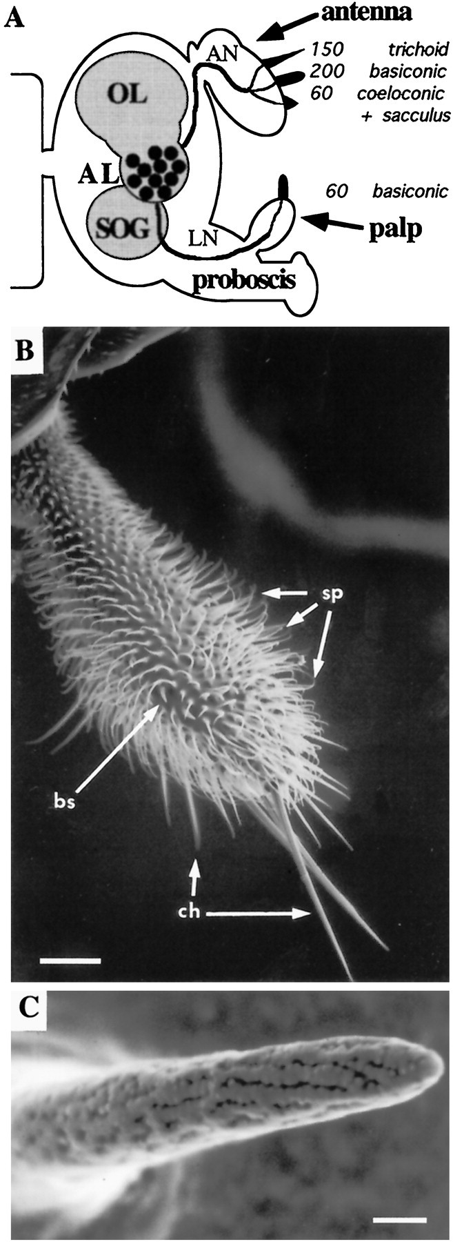
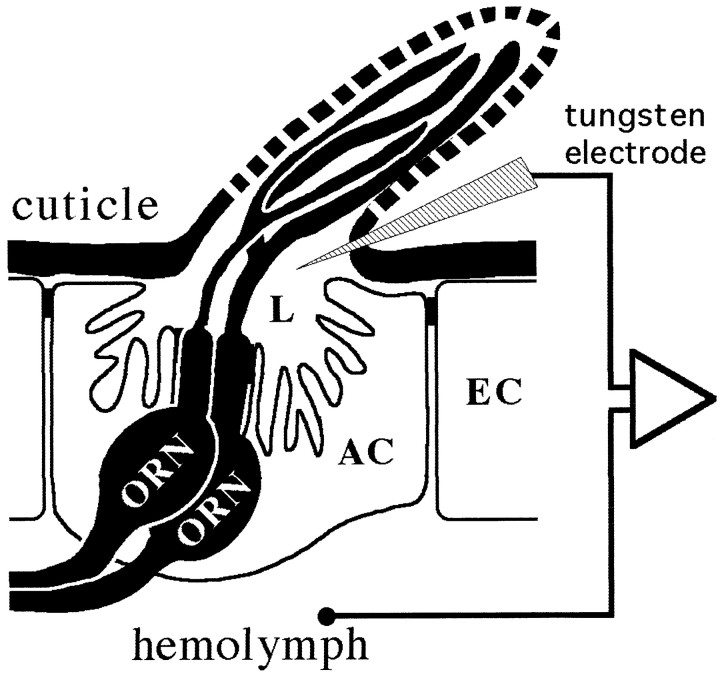
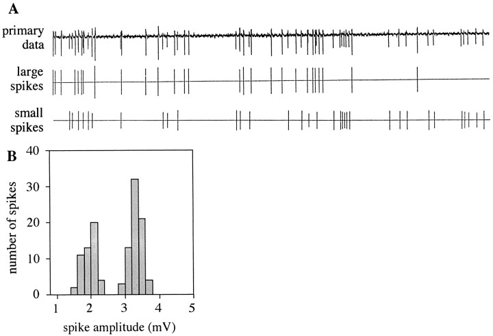
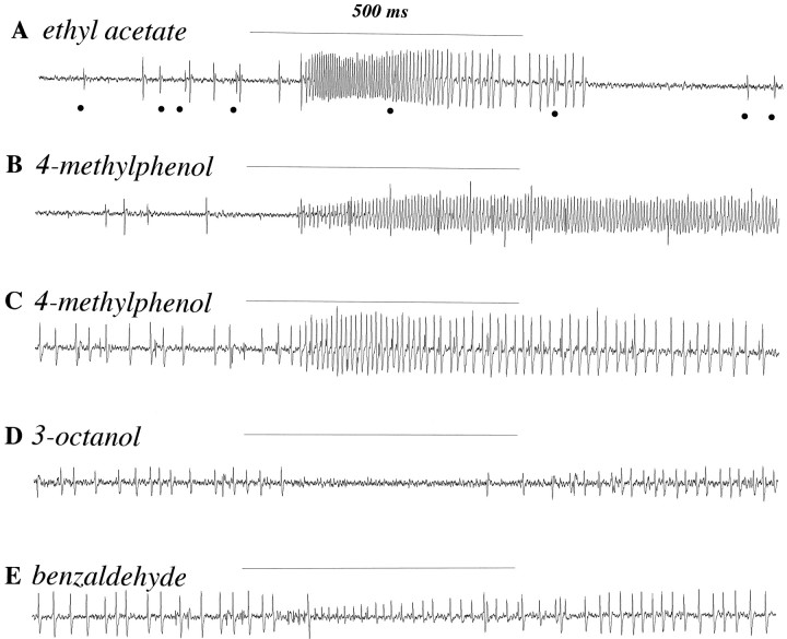
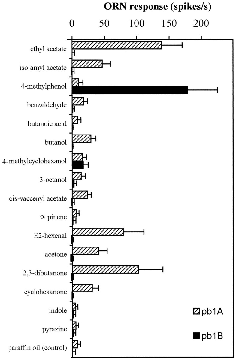
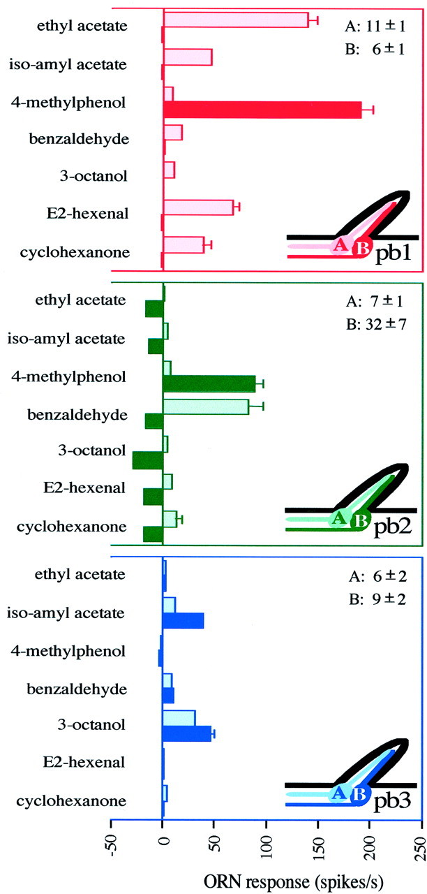
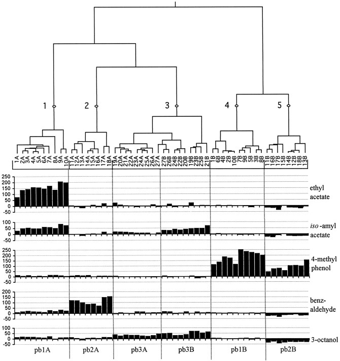
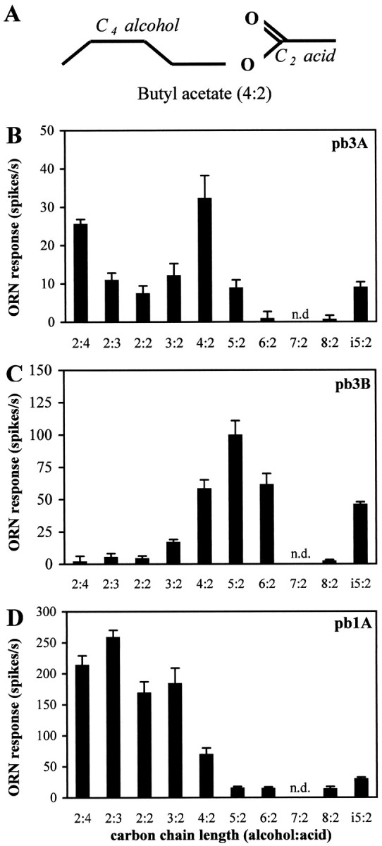
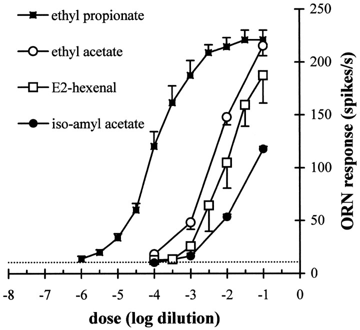
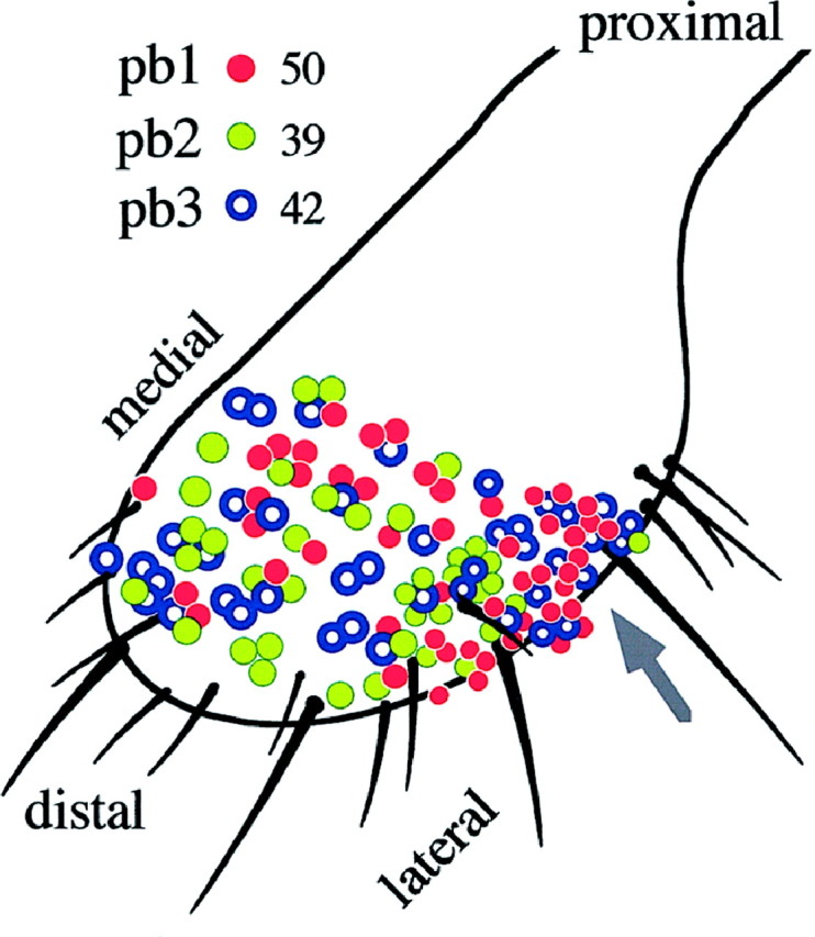
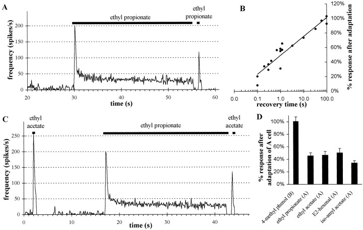
Odor coding relies on the activity of different classes of receptor neurons, each with distinct response characteristics. We have examined odor coding in a model olfactory organ, the maxillary palp of Drosophila. This organ contains only 120 olfactory receptor neurons, compartmentalized in sensory hairs called sensilla, and provides an opportunity to characterize all neurons in an entire olfactory organ. Extensive extracellular recordings from single sensilla reveal that the neurons fall into six functional classes. Each of the 60 sensilla houses two neurons, which observe a pairing rule: each sensillum combines neurons of two particular classes, thereby yielding three sensillum types. The sensillum types are intermingled on the surface of the palp, but their distribution is not random. The neurons exhibit diverse response characteristics, providing the basis for an olfactory code. A particular odor can excite one neuron and inhibit another, and a particular neuron can be excited by one odor and inhibited by another. Some excitatory responses continue beyond the end of odor delivery, but responses to most odors terminate abruptly after the end of odor delivery, with some followed by a period of poststimulus quiescence. The specificity of odor response is examined in detail for the neurons of one sensillum, which were found to differ in their relative responses to a homologous series of esters. Adaptation and cross-adaptation are documented, and cross-adaptation experiments demonstrate that the two neurons within one type of sensillum can function independently. The analysis of all neuronal types in this model olfactory organ is discussed in terms of its functional organization and the mechanisms by which it encodes olfactory information.


Figures

Fig. 1.
Fig. 1.
The maxillary palps carry olfactory sensilla.A, Schematic overview of the Drosophilaolfactory system showing three structurally different sensillum types and their numbers on two olfactory organs as well as their projections to the CNS. Sensilla in the sacculus, a multichambered sensory pit, are not enumerated. AL, Antennal lobe; AN, antennal nerve; LN, labiomaxillary nerve;OL, optic lobe; SOG, subesophageal ganglion. B, Scanning electron micrograph of the maxillary palp showing three types of cuticular hairs: olfactory sensilla (bs, sensilla basiconica), mechanosensory setae (ch, sensilla chaetica), and uninnervated hairs (sp, spinules). Scale bar, 25 μm. C, Scanning electron micrograph detail of a basiconic sensillum showing a multitude of pores through which odorants may pass. Scale bar, 1 μm.B and C are reprinted with permission from Riesgo-Escovar et al. (1997).
Fig. 2.
Fig. 2.
Schematic overview of single-unit recordings from ORNs in a basiconic sensillum, showing electrode positions for the extracellular recording of voltage differences between the sensillum lymph (L) and the hemolymph. AC, Accessory cells; EC, epidermal cells.
Fig. 3.
Fig. 3.
Single-unit recordings from palpal basiconic sensilla confirm the presence of two olfactory receptor neurons.A, Differences in spike amplitudes allow separate analysis of firing rates of the two neurons in a single sensillum.Top, Primary data from a 3 sec period of spontaneous activity from a sensillum are shown. Middle, bottom, The extracted data are presented with large spikes shown separately from small spikes. B, The distribution of amplitudes of individual action potentials (measured from peak-to-trough) is bimodal. Data shown are for 123 spikes from 6 sec of spontaneous activity of the recording in A.
Fig. 4.
Fig. 4.
Sensilla house receptor neurons with a variety of different response characteristics to different odors.A–E, Five 1500 msec traces of recordings from two different sensilla showing excitatory and inhibitory responses of the two cells to 500 msec stimulations (horizontal lines) with different odorants. For odor stimulation, air was expelled from 5 ml syringes over filter paper laden with 20 μl of odorant. The odorants were diluted 10−2 in paraffin oil. We do not know the exact concentrations of the odor present in the air reaching the preparation. A, B, Recordings from one sensillum, later classified as pb1. Large action potentials, from the A neuron, increase their frequency in response to ethyl acetate. Dots indicate smaller action potentials from the B neuron, which is not excited by ethyl acetate but which responds to 4-methylphenol. C–E, Recordings from another sensillum, later classified as pb2. Large spikes are from the B neuron, which is excited by 4-methylphenol and inhibited by other odors. Smaller spikes are from the A neuron, excited by benzaldehyde.
Fig. 5.
Fig. 5.
Responses of the two pb1 neurons to a set of 16 odorants (error bars indicate SD; n = 13). pb1B responds strongly only to one of the tested odors. Responses of pb1A to several odorants are significantly larger than the control response to the paraffin oil diluent alone. The indicated ORN response is measured as the increase in spikes per second over the spontaneous frequency.
Fig. 6.
Fig. 6.
Six classes of olfactory receptor neurons are found in characteristic pairs (A and B neurons) in three functional types of sensilla on the Drosophila maxillary palp. Response profiles of ORNs are shown for the three sensillum types: pb1 (n = 17), pb2 (n = 15), and pb3 (n = 14). The spontaneous frequencies of the two neurons (± SD) are indicated in the top right corner for each type. The ORN response is measured as the increase (or decrease) in spikes per second over the spontaneous frequency. Error bars indicate SEM and are too small to be seen in some cases.
Fig. 7.
Fig. 7.
Dendrogram of a hierarchical cluster analysis of 54 ORNs in 27 sensilla, comparing their responses to five odors. Branch length is proportional to distance (see Materials and Methods), and five clusters are indicated by the numbered points. Individual ORNs are indicated as the A or B cell of a single sensillum, and their responses, measured as the increase in spikes per second over the spontaneous frequency (y-axis), are shown. Six classes of neurons are indicated at the bottom of the figure. Clusters 1, 2, 4, and 5 correspond to pb1A, pb2A, pb1B, and pb2B, respectively; cluster 3 includes two cell types within the pb3 sensillum, neurons pb3A and pb3B, that can be clearly distinguished by their response to other odors (see Fig. 8) and by differences in spike amplitude (data not shown).
Fig. 8.
Fig. 8.
Responses of olfactory receptor neurons on theDrosophila maxillary palp to aliphatic esters confirm and characterize the distinct identities of pb3A and pb3B.A, Nomenclature illustrated with the structure of butyl acetate. i5:2 is isoamyl acetate, a branched analog of pentyl acetate (5:2). BD, Responses of pb3A, pb3B, and pb1A, respectively, to a set of esters with varying chain lengths and structures. The indicated ORN response is measured as the increase in spikes per second over the spontaneous frequency (error bars indicate SEM; n = 8). n.d., Not done.
Fig. 9.
Fig. 9.
Dose–response relations for pb1A to four odorants. Stimuli were presented as dilutions in 20 μl of paraffin oil on filter paper as described in Figure 4. The indicated ORN response is measured as the increase in spikes per second over the spontaneous frequency (error bars indicate SEM; n = 13). The dotted horizontal line indicates the mean response of pb1A to the paraffin oil diluent alone.
Fig. 10.
Fig. 10.
The distribution of the three sensillum types, all of which reside on the dorsal (top) and lateral surfaces of the maxillary palp. Each colored circle represents one recording from a basiconic sensillum. Clusters ofcircles indicate recordings made from sensilla in corresponding positions on different flies. The total number of recordings from each sensillum type are indicated. The arrow points to a zone where pb2 sensilla are virtually absent.
Fig. 11.
Fig. 11.
Adaptation and cross-adaptation in pb1 sensilla show convergence of olfactory information in pb1A neurons but independence of two neurons in a sensillum. A, Frequency of action potentials (averaged over 120 msec intervals) during prolonged exposure of pb1A to ethyl propionate is shown. The long stimulation (25 sec) was generated using the flask flow method (10−2 dilution), whereas the ensuing short stimulus (500 msec) was delivered with the syringe puff method (10−3.5 dilution; see Materials and Methods). Both stimulations elicit the same spike frequency during the first 500 msec from unadapted neurons. Note that frequencies over 500 msec are not the same as peak responses averaged over 120 msec. B, The response to the second stimulation with ethyl propionate increases, approximately following a linear function of the logarithm of the recovery time after adaptation. Responses are presented as a percentage of the response to an identical 500 msec stimulus given before adaptation. C, The pb1A neuron cross-adapts to ethyl acetate, after stimulation with ethyl propionate. A 10−2 dilution of ethyl acetate and a 10−3.5 dilution of ethyl propionate were used, both using the syringe puff method, because they elicit the same response from pb1A (see Fig. 9). D, The pb1A neuron was adapted to ethyl propionate as in A. Then the response was measured from pb1B to 4-methylphenol (10−2dilution; n = 13) or from pb1A to ethyl propionate (10−3.5 dilution; n = 10), ethyl acetate (10−2 dilution; n= 8), E2-hexenal (10−1.5 dilution;n = 8), or isoamyl acetate (10−1 dilution; n = 7). All the doses used were determined from Figure 9 to give approximately equal responses before adaptation. Error bars indicate SEM. Cross-adaptation is observed for all odorants exciting pb1A but not for 4-methylphenol, which stimulates pb1B.

Similar articles

Cited by

References

    1. Ache BW, Zhainazarov A. Dual second-messenger pathways in olfactory transduction. Curr Opin Neurobiol. 1995;5:461–466. - PubMed
    1. Almaas TJ, Christensen TA, Mustaparta H. Chemical communication in heliothine moths. I. Antennal receptor neurons encode several features of intra- and interspecific odorants in the male corn earworm moth Helicoverpa zea. J Comp Physiol [A] 1991;169:249–258.
    1. Altner H, Prillinger L. Ultrastructure of invertebrate chemo-, thermo- and hygroreceptors and its functional significance. Int Rev Cytol. 1980;67:69–139.
    1. Anderberg MR. Cluster analysis for applications. Academic; New York: 1973.
    1. Anderson P, Hansson BS, Löfqvist J. Plant-odour-specific receptor neurones on the antennae of female and male Spodoptera littoralis. Physiol Entomol. 1995;20:189–198.

Publication types

LinkOut - more resources