. 1999 Jun 1;19(11):4544-58.
doi: 10.1523/JNEUROSCI.19-11-04544.1999.

Presynaptically located CB1 cannabinoid receptors regulate GABA release from axon terminals of specific hippocampal interneurons

Affiliations

Presynaptically located CB1 cannabinoid receptors regulate GABA release from axon terminals of specific hippocampal interneurons

I Katona et al. J Neurosci. .

Abstract

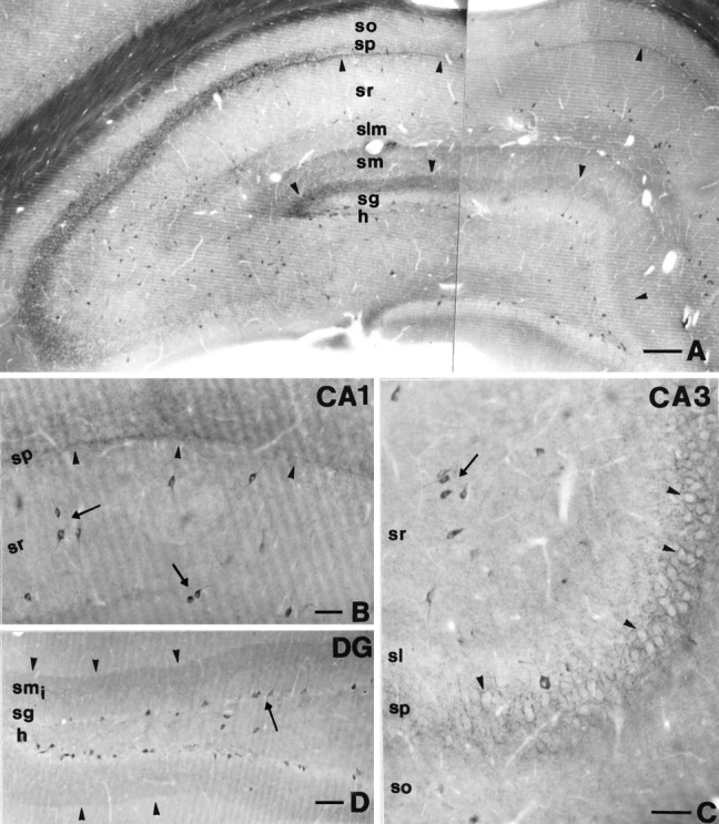
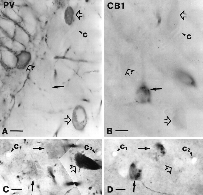
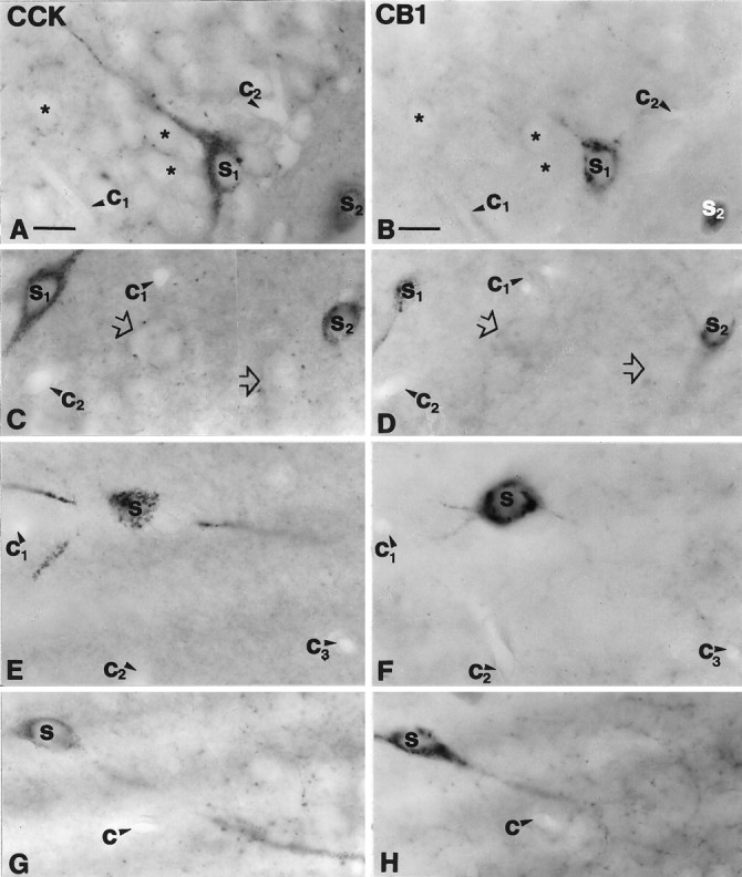
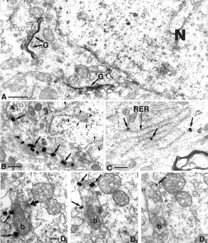
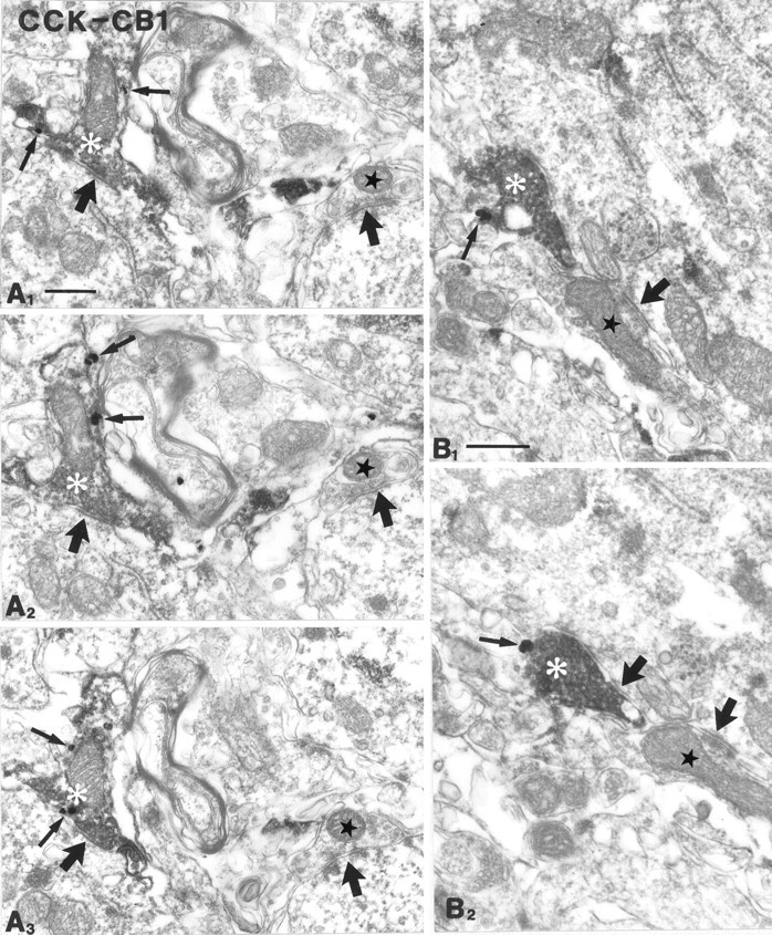
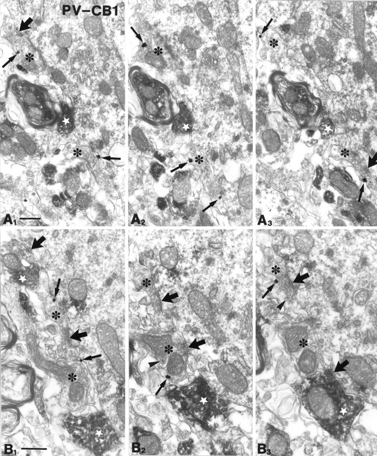
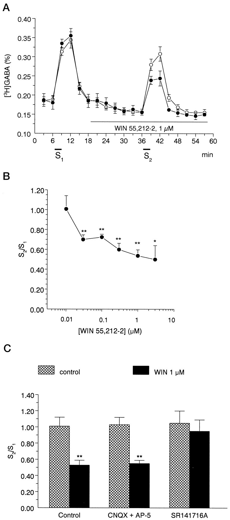
To understand the functional significance and mechanisms of action in the CNS of endogenous and exogenous cannabinoids, it is crucial to identify the neural elements that serve as the structural substrate of these actions. We used a recently developed antibody against the CB1 cannabinoid receptor to study this question in hippocampal networks. Interneurons with features typical of basket cells showed a selective, intense staining for CB1 in all hippocampal subfields and layers. Most of them (85.6%) contained cholecystokinin (CCK), which corresponded to 96.9% of all CCK-positive interneurons, whereas only 4.6% of the parvalbumin (PV)-containing basket cells expressed CB1. Accordingly, electron microscopy revealed that CB1-immunoreactive axon terminals of CCK-containing basket cells surrounded the somata and proximal dendrites of pyramidal neurons, whereas PV-positive basket cell terminals in similar locations were negative for CB1. The synthetic cannabinoid agonist WIN 55,212-2 (0.01-3 microM) reduced dose-dependently the electrical field stimulation-induced [3H]GABA release from superfused hippocampal slices, with an EC50 value of 0. 041 microM. Inhibition of GABA release by WIN 55,212-2 was not mediated by inhibition of glutamatergic transmission because the WIN 55,212-2 effect was not reduced by the glutamate blockers AP5 and CNQX. In contrast, the CB1 cannabinoid receptor antagonist SR 141716A (1 microM) prevented this effect, whereas by itself it did not change the outflow of [3H]GABA. These results suggest that cannabinoid-mediated modulation of hippocampal interneuron networks operate largely via presynaptic receptors on CCK-immunoreactive basket cell terminals. Reduction of GABA release from these terminals is the likely mechanism by which both endogenous and exogenous CB1 ligands interfere with hippocampal network oscillations and associated cognitive functions.


Figures

Fig. 1.
Fig. 1.
A, Low-power light micrograph showing CB1-immunostaining in the dorsal hippocampus. Arrowheadsindicate characteristic CB1-immunopositive bands in the inner third of stratum moleculare and at the border of strata pyramidale and radiatum in the CA1 subfield. Note that most CB1-immunoreactive cell bodies resembling interneurons are distributed in all subfields and layers of the hippocampus. B, In the CA1 subfield, arrowsdepict typical CB1-positive interneurons with multipolar, thin proximal dendrites located at the border of strata radiatum and lacunosum-moleculare, and interneurons with a bitufted dendritic tree in the middle part of stratum radiatum. Although immunostained axon terminals covered the entire stratum pyramidale, an even denser band of axonal staining was observed at the border of strata pyramidale and radiatum (arrowheads). C, In the CA3 subfield, this band was absent in stratum lucidum, whereas a dense meshwork of CB1-immunostained basket-like axons surrounded the immunonegative somata of pyramidal cells (arrowheads) as in CA1. Most of the CB1-positive cell bodies were found in stratum radiatum (arrow). D, In the dentate gyrus, most of the CB1-immunoreactive cell bodies were located at the border of the hilus and stratum granulosum (arrow). The apical dendrites of these cells crossed stratum granulosum without branching.Arrowheads indicate dense punctate immunostaining in the inner third of stratum moleculare. DG, Dentate gyrus;h, hilus; sg, stratum granulosum; smi, inner third of stratum moleculare; sm, stratum moleculare;slm, stratum lacunosum-moleculare; sr, stratum radiatum; sp, stratum pyramidale; so, stratum oriens; sl, stratum lucidum. Scale bars: A, 200 μm; B, C, 60 μm; D, 100 μm.
Fig. 2.
Fig. 2.
Parvalbumin-positive cells represent a subpopulation of perisomatic inhibitory interneurons that does not contain CB1-immunoreactivity. A, B, Parvalbumin-positive neurons (open arrows in A) in the dentate gyrus never showed CB1-immunoreactivity. The other halves of the somata, negative for CB1, are labeled by open arrows inB. Filled arrows depict a CB1-immunoreactive cell body in B and its PV-negative half in A. C, D, Parvalbumin-positive neurons (open arrow inC) proved to be CB1-negative (open arrow onD) in stratum pyramidale of the CA1 subfield. Filled arrows depict CB1-immunoreactive but parvalbumin-negative somata. Capillaries labeled by c1–2 served as landmarks and confirmed the localization of the halved cell bodies.PV, Parvalbumin; CB1, CB1 cannabinoid receptor. Scale bars, 10 μm.
Fig. 3.
Fig. 3.
Cholecystokinin-containing interneurons express CB1 cannabinoid receptor in the hippocampus. A, B, A CCK-immunoreactive pyramidal-like basket cell (S1) in the dentate gyrus contains CB1-immunoreactivity. Granule cells (*) were negative for both markers.C, D, In the CA3 subfield large somata with thick proximal dendrites (S1) and smaller multipolar cells (S2) were positive for both CCK and CB1.Open arrows indicate double-negative cell bodies.E–H, These two morphological types colocalized CCK and CB1 in the CA1 subfield as well. A multipolar CCK-positive cell (S in E) in stratum radiatum is cut in half on the surface of the section. The same cell (S inF) shows CB1-immunoreactivity in the adjacent section. Three primary dendrites are also seen to continue in the adjacent section. G, H, A large bitufted neuron at the border of strata oriens and pyramidale is shown to be double-labeled. Capillaries labeled by c1–3 serve as landmarks to confirm precise alignment. CCK, Cholecystokinin;CB1, CB1 cannabinoid receptor. Scale bars (shown inA and B for A–H): 15 μm.
Fig. 4.
Fig. 4.
A, Low-power electron micrograph of a CB1-immunoreactive cell body. In this experiment, DAB was used as chromogen. Note the rather selective localization of the DAB precipitate in the Golgi apparatus (G). B, C, Immunogold labeling confirmed at a higher resolution that CB1 is localized in the Golgi apparatus (arrows in B) and in the rough endoplasmic reticulum (RER,arrows in C). Arrowheads inB label the invaginated nucleus, which further confirms that CB1 is expressed by interneurons. D1–3, In stratum pyramidale, gold particles representing CB1-immunoreactivity were found on the plasma membrane of axon terminals (arrows), on the side facing the extracellular space. This confirms that the antibody used in this study was raised against the N-terminal domain of CB1, which is located extracellularly. Moreover, this figure shows that CB1 is localized presynaptically on boutons of inhibitory neurons (b), because these boutons formed exclusively symmetrical synapses with their targets (thick arrow). N, Nucleus; G, Golgi apparatus;RER, rough endoplasmic reticulum. Scale bars:A–C, 0.5 μm; D1–3, 0.1 μm.
Fig. 5.
Fig. 5.
CB1 cannabinoid receptors are localized presynaptically on cholecystokinin-immunoreactive axon terminals. Most of the CCK-positive boutons (white asterisks; diffuse DAB end-product) were found to be positive for CB1 (gold particles labeled by thin arrows). The serial sections inA1–3 were taken from the CA1 subfield; those inB1–2 derive from CA3. These terminals formed symmetrical synapses mainly on somata and proximal dendrites of their targets. Adjacent boutons (stars), which were negative for both markers, also formed symmetrical synapses. Thick arrowsindicate symmetrical, probably GABAergic synapses. CCK, Cholecystokinin; CB1, CB1 cannabinoid receptor. Scale bars:A, B, 0.4 μm.
Fig. 6.
Fig. 6.
Parvalbumin-immunoreactive axon terminals are negative for CB1 cannabinoid receptors. Parvalbumin-immunoreactive boutons (diffuse DAB precipitate; white stars) are shown from stratum pyramidale of the CA1 (A1–3) and CA3 (B1–3) subfields, forming symmetrical synapses with their targets. Several CB1-positive (gold particles labeled by arrows) axon terminals (*) were found nearby, but CB1 and PV did not colocalize to the same boutons. These CB1-positive boutons contained several dense-core vesicles (arrowheads), which probably contain the neuropeptide cholecystokinin. These findings further confirm that the two basket cell populations are distinct regarding their presynaptic receptors. Thick arrows label symmetrical synapses. Thin arrows depict gold particles representing CB1 receptor immunoreactivity. PV, Parvalbumin;CB1, CB1 cannabinoid receptor. Scale bars: A, B, 0.4 μm.
Fig. 7.
Fig. 7.
A, A cannabinoid agonist, 1 μmWIN55,212-2, inhibits electrically evoked [3H]GABA release from rat hippocampal slices. The slices were superfused for 60 min at a rate of 0.7 ml/min. After the preperfusion period, 3 min samples were collected and assayed for radioactivity. [3H]GABA release was significantly increased in response to electrical field stimulation (S1, S2). Open circles represent control experiments; filled circles show experiments when WIN 55,212-2 was present during the second stimulation period (S2). The release of [3H]GABA was expressed as fractional release (%; for calculation see Materials and Methods). The values show the mean ± SEM of 7–12 identical experiments. B, Concentration dependence of the effect of WIN55,212-2 on stimulation-induced [3H]GABA release from rat hippocampal slices. The cannabinoid receptor agonist WIN 55,212-2 was applied to the slices by perfusion according to the protocol shown in Ain different concentrations ranging from 0.01 to 3 μm, and its effect on stimulation-evoked [3H]GABA outflow was expressed as S2/S1 ratios (for calculation, see Materials and Methods). The values show the mean ± SEM of 7–12 identical experiments. Asterisksrepresent significant differences from the control S2/S1 ratio, measured in the absence of drugs (1.01 ± 0.11, n = 12), calculated by ANOVA followed by the Dunnett test (*p < 0.05, **p < 0.01). C, Interaction of the effect of WIN55,212-2 on stimulation-induced [3H]GABA release with glutamate receptor antagonists AP-5 and CNQX and with the CB1 receptor antagonist SR141716A in rat hippocampal slices. The effect of drugs on stimulation-induced release of [3H]GABA was expressed as S2/S1 ratio, measured in the absence (cross-hatched bars) and presence of WIN55,212-2 (black bars). The perfusion with WIN55,212-2 (1 μm) started 18 min before S2 and continued thereafter, whereas AP-5 (10 μm), CNQX (10 μm), and SR141716A (1 μm) were perfused from 15 min before S1. Data show the mean ± SEM of 6–12 identical experiments. Asterisks represent significant difference from respective controls (**p < 0.01 calculated by Student’s t test).

Similar articles

Cited by

References

    1. Abood ME, Martin BR. Neurobiology of marijuana abuse. Trends Pharmacol Sci. 1992;13:201–206. - PubMed
    1. Acsády L, Arabadzisz D, Freund TF. Correlated morphological and neurochemical features identify different subsets of vasoactive intestinal polypeptide-immunoreactive interneurons in rat hippocampus. Neuroscience. 1996a;73:299–315. - PubMed
    1. Acsády L, Görcs TJ, Freund TF. Different populations of vasoactive intestinal polypeptide-immunoreactive interneurons are specialized to control pyramidal cells or interneurons in the hippocampus. Neuroscience. 1996b;73:317–334. - PubMed
    1. Acsády L, Katona I, Gulyás AI, Shigemoto R, Freund TF. Immunostaining for substance P receptor labels GABAergic cells with distinct termination patterns in the hippocampus. J Comp Neurol. 1997;378:320–336. - PubMed
    1. Baimbridge KG, Miller JJ. Immunohistochemical localization of calcium-binding protein in the cerebellum, hippocampal formation and olfactory bulb of the rat. Brain Res. 1982;245:223–229. - PubMed

Publication types