. 1999 Feb;58(2):72-8.
doi: 10.1136/ard.58.2.72.

The shape of the distal femur: a palaeopathological comparison of eburnated and non-eburnated femora

Affiliations

The shape of the distal femur: a palaeopathological comparison of eburnated and non-eburnated femora

L Shepstone et al. Ann Rheum Dis. 1999 Feb.

Abstract

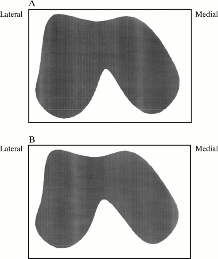
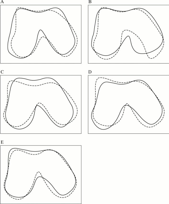
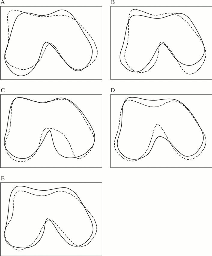
Objectives: To determine the difference in shape of the distal femur, viewed axially in two dimensions, between eburnated and non-eburnated femora.

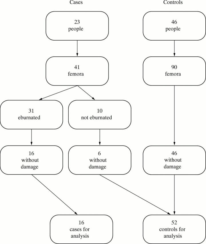
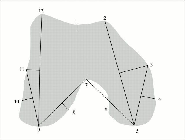
Methods: A comparison of 52 non-eburnated and 16 eburnated femora drawn from a large archeological skeletal population. Eburnation was taken to indicate late stage osteoarthritis. Shape variability, based on landmarks, was quantified using a principal components analysis after a Procrustes alignment.

Results: A statistically significant difference was found between the two groups. This was with respect to the patellar groove and the shape of the medial condyle. The latter difference is consistent with bone remodelling as a knee stabilising mechanism.

Conclusions: Anatomical shape can be quantified using an uncomplicated statistical technique. It was used to quantify the shape of the distal femur and demonstrate shape differences associated with osteoarthritis of the knee.


Figures

Figure 1
Figure 1
Illustration of selection of study material.
Figure 2
Figure 2
Alignment of femora to camera: femora are allowed to rest naturally upon the trochanters and most posterior points of the condyles.
Figure 3
Figure 3
Example of bitmap digitised from video image.
Figure 4
Figure 4
Location of 12 landmarks used for analysis. Landmarks 1, 2, 5, 7, 9, and 12 were located by hand. The remaining six landmarks were defined relative to these.
Figure 5
Figure 5
Mean configurations. Periodic splines were used to produce outlines for the 12 landmark mean configurations. The mean shape of the 16 eburnated femora is shown (A) compared with that of the 52 non-eburnated (B).
Figure 6
Figure 6
Effect of principal components on the overall mean. The mean configuration plus (solid line) or minus (dotted line) three standard deviations of the first five principal components (A)-(E). A significant difference was found between the two groups with respect to the second principal component scores. The eburnated femora tended to have higher second principal component scores.
Figure 7
Figure 7
Effect of varimax rotated principal components on the overall mean. The mean configuration plus (solid line) or minus (dotted line) three standard deviations of the rotated first five principal components (A)-(E). A significant difference was found between the two groups with respect to the second and third principal component scores. The eburnated femora tended to have higher scores for both of these rotated principal components.

References

    1. J Bone Joint Surg Am. 1971 Dec;53(8):1561-70 - PubMed
    1. Ann Rheum Dis. 1957 Dec;16(4):494-502 - PubMed
    1. Clin Orthop Relat Res. 1978 May;(132):225-32 - PubMed
    1. Clin Orthop Relat Res. 1981 May;(156):61-6 - PubMed
    1. Br J Rheumatol. 1984 Aug;23(3):166-9 - PubMed

Publication types