Clinical Trial
. 1999 Jan;58(1):20-6.
doi: 10.1136/ard.58.1.20.

Bone scintigraphy in chronic knee pain: comparison with magnetic resonance imaging

Affiliations
Clinical Trial

Bone scintigraphy in chronic knee pain: comparison with magnetic resonance imaging

T Boegård et al. Ann Rheum Dis. 1999 Jan.

Abstract

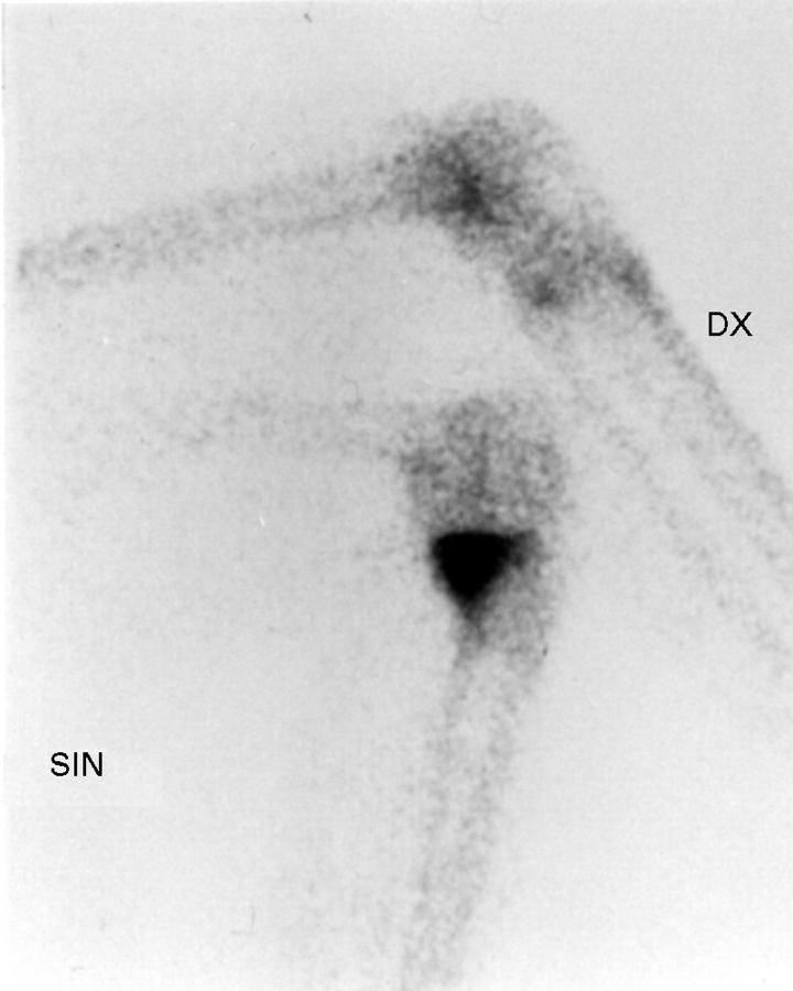
Objective: To compare increased bone uptake of 99Tcm-MDP and magnetic resonance (MR) detected subchondral lesions, osteophytes, and cartilage defects in the knee in middle aged people with long-standing knee pain.

Methods: Fifty eight people (aged 41-58 years, mean 50) with chronic knee pain, with or without radiographic knee osteoarthritis, were examined with bone scintigraphy. The pattern and the grade of increased bone uptake was assessed. On the same day, a MR examination on a 1.0 T imager was performed. The presence and the grade of subchondral lesions, osteophytes, and cartilage defects were registered.

Results: The kappa values describing the correlation between increased bone uptake and MR detected subchondral lesions varied between 0.79 and 0.49, and between increased bone uptake and MR detected osteophytes or cartilage defects the values were < 0.54. The kappa values describing the correlation between the grade of bone uptake and the grade of the different MR findings was < 0.57.

Conclusions: Good agreement was found between increased bone uptake and MR detected subchondral lesion. The agreement between increased bone uptake and osteophytes or cartilage defects was in general poor as well as the agreement between the grade of bone uptake and the grade of the MR findings.


Figures

Figure 1
Figure 1
Bone scintigram of the knees in right medial and left lateral projection in a 51 year old woman, showing an increased bone uptake with a point-like pattern of grade 2 in the medial femoral condyle of the right knee (the signal knee). The left knee has normal uptake.
Figure 2
Figure 2
Bone scintigram of the knees in right medial and left lateral projection in a 58 year old woman, demonstrating an increased bone uptake with a tramline pattern of grade 1 in the lateral tibial condyle of the left knee (arrow) (the signal knee) as well as in the medial tibial condyle of the right knee (double arrow).
Figure 3
Figure 3
Bone scintigram of the knees in right lateral and left medial projections in a 42 year old woman, showing an increased bone uptake with an extended pattern in the medial tibial condyle of the left knee (the signal knee). The right knee has normal uptake.
Figure 4
Figure 4
(A) A sagittal T2 weighted STIR+ MR image of the right knee in a 43 year old man demonstrating a subchondral lesion of grade 2 dorsal in the lateral femoral condyle as well as in the lateral tibial condyle (arrows). (B) In a corresponding sagittal proton density weighted MR image is shown adjacent cartilage defects of grade 2 in both locations (arrows).
Figure 5
Figure 5
A coronal proton density weighted MR image of the right knee in a 57 year old woman demonstrating a peripheral osteophyte of grade 1, at the medial femoral condyle (white arrow). Note the cartilage defect of grade 2 in the same condyle (black arrow).

Similar articles

Cited by

References

    1. Ann Rheum Dis. 1986 Aug;45(8):617-21 - PubMed
    1. Orthop Clin North Am. 1986 Apr;17(2):249-62 - PubMed
    1. Br J Rheumatol. 1988;27 Suppl 2:23-8 - PubMed
    1. Acta Radiol. 1988 Jul-Aug;29(4):451-5 - PubMed
    1. Radiology. 1990 Aug;176(2):479-83 - PubMed

Publication types

Substances