

doi: 10.1101/gad.13.10.1240.
Loss of transcription factor IRF-1 affects tumor susceptibility in mice carrying the Ha-ras transgene or nullizygosity for p53
Affiliations
- PMID: 10346812
- PMCID: PMC316726
- DOI: 10.1101/gad.13.10.1240
Item in Clipboard
Loss of transcription factor IRF-1 affects tumor susceptibility in mice carrying the Ha-ras transgene or nullizygosity for p53
Genes Dev.
.
Abstract
The transcription factor IRF-1 has been implicated in tumor suppression: IRF-1 suppresses cell transformation and mediates apoptosis in vitro. Here we show that the loss of IRF-1 alleles per se has no effect on spontaneous tumor development in the mouse but dramatically exacerbates previous tumor predispositions caused by the c-Ha-ras transgene or by nullizygosity for p53. Grossly altered tumor spectrum, as compared to p53-null mice, was also observed in mice lacking both IRF-1 and p53, and cells from these mice show significantly higher mutation rate. Our results suggest that IRF-1 is a new member of the tumor susceptibility genes.
Figures
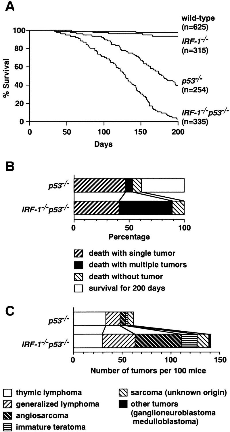
Survival rate, cause of death, and histology of tumors in subject mice. (A) Survival curves of wild-type, IRF-1−/−, p53−/−, and IRF-1−/−p53−/− mice followed up to 200 days by the Kaplan–Meier method. Mice were sacrificed upon becoming apparently moribund. (n) Total number of mice in each genotype. At 200 days, 2% of IRF-1−/− mice, 56% of p53−/− mice, and 96% of IRF-1−/−p53−/− mice developed tumors; 0.2% of wild-type mice died before 200 days, but no tumors were found. (B) The first 100 mice of each genotype were monitored for up to 200 days and autopsied to assess for the presence and number of tumors. (C) Raw numbers of histological types of tumors in p53−/− and IRF-1−/−p53−/− mice. Data shown are from the same cohort of 100 mice as in B. The bar patterns for tumor types are shown in B and C.
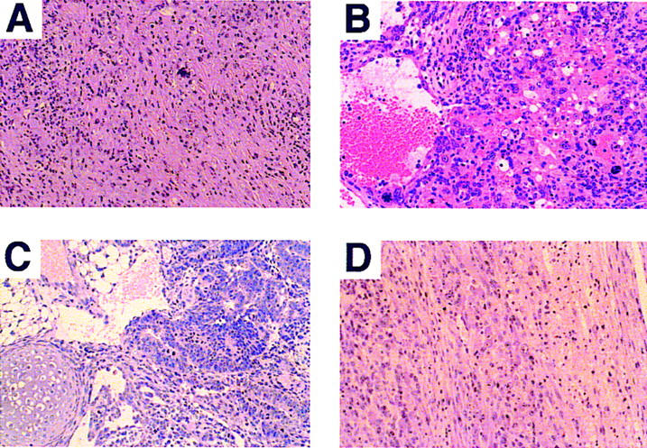
Histological analysis of representative tumors obtained from IRF-1−/− and IRF-1−/−p53−/− mice (hematoxylin and eosin staining). (A) Malignant fibrous histiocytoma-like sarcoma characteristically found in IRF-1−/− mice. Tumor chiefly consists of atypical spindle-shaped cells. (B) Angiosarcoma in an IRF-1−/−p53−/− mouse. Atypical tumor cells surround individual or groups of erythrocytes and also form neoplastic blood vessels. (C) Immature teratoma in testis of a male IRF-1−/−p53−/− mouse. Tumor consists of immature adenomatous, adipose-like, and chondroid tissue components. (D) Ganglioneuroblastoma in an IRF-1−/−p53−/− mouse. The spinal cord (right) is invaded by neuron-like tumor cells with enlarged nuclei and prominent nucleoli (left). This type of tumor was not observed in singly null mice. Original magnifications: (A,C,D) 500×; (B) 600×.
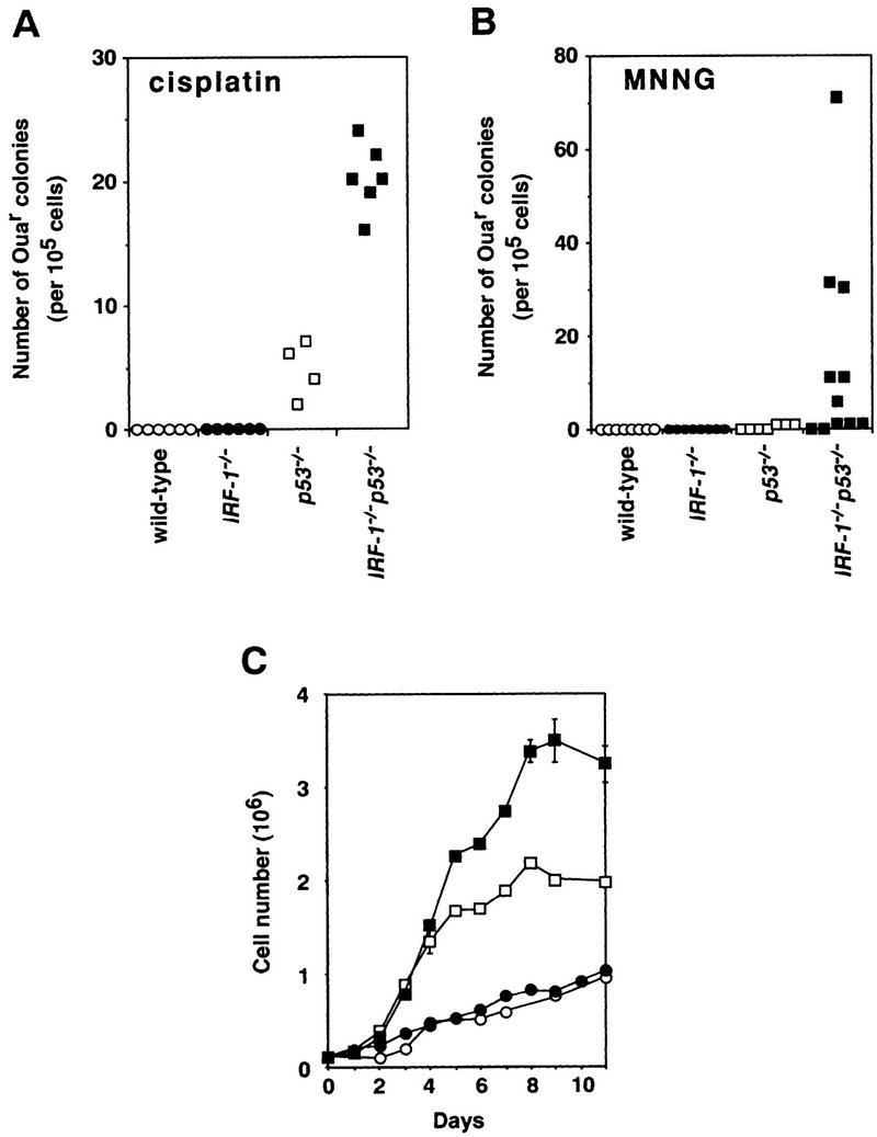
Cellular abnormalities in IRF-1−/−p53−/− MEFs. (A,B) Mutation frequency in MEFs treated with cisplatin (0.05 μg/ml for 72 hr; A) or MNNG (5 μm for 3 hr; B). The numbers of Ouar colonies/105 MEF cells were plotted. Plating efficiencies of p53−/− and IRF-1−/−p53−/− MEFs were similar (∼90%). (C) Representative growth curves of MEFs. Cells were plated at a density of 105 cells/35-mm dish at passage 4, and cell number counted (bars indicate s.d. ). Experiments performed on at least four clones of each genotype showed the results to be essentially reproducible. Mean doubling times in log phase for wild-type and IRF-1−/− MEFs were similar (66.6 ± 39.8 and 56.9 ± 18.6 hr, respectively). p53−/− and IRF-1−/−p53−/− MEFs showed similar growth rates (mean doubling time, 22.9 ± 2.5 and 25.0 ± 4.3 hr, respectively). Saturation density of MEFs of each genotype was 11.0 ± 2.5 × 10⁵ (wild-type, ○), 8.6 ± 1.9 × 10⁵ (IRF-1−/−, ●), 26.8 ± 4.4 × 10⁵ (p53−/−, □), and 35.4 ± 3.4 × 10⁵ cells (IRF-1−/− p53−/−, █).
Similar articles
-
Altered DNA repair and dysregulation of p53 in IRF-1 null hepatocytes.FASEB J. 1998 Feb;12(2):181-8. doi: 10.1096/fasebj.12.2.181. FASEB J. 1998. PMID: 9472983
-
IRF-1 reverts the transformed phenotype of oncogenically transformed cells in vitro and in vivo.Oncogene. 2003 Feb 20;22(7):1045-56. doi: 10.1038/sj.onc.1206260. Oncogene. 2003. PMID: 12592391
-
Cellular commitment to oncogene-induced transformation or apoptosis is dependent on the transcription factor IRF-1.Cell. 1994 Jun 17;77(6):829-39. doi: 10.1016/0092-8674(94)90132-5. Cell. 1994. PMID: 8004672
-
IRF-1 as a negative regulator of cell proliferation.J Interferon Cytokine Res. 2002 Jan;22(1):39-47. doi: 10.1089/107999002753452647. J Interferon Cytokine Res. 2002. PMID: 11846974 Review.
-
Transcription factors IRF-1 and IRF-2: linking the immune responses and tumor suppression.J Cell Physiol. 1997 Nov;173(2):128-30. doi: 10.1002/(SICI)1097-4652(199711)173:2<128::AID-JCP8>3.0.CO;2-P. J Cell Physiol. 1997. PMID: 9365509 Review. No abstract available.
Cited by
-
Diverse mechanisms evolved by DNA viruses to inhibit early host defenses.Crit Rev Biochem Mol Biol. 2016 Nov/Dec;51(6):452-481. doi: 10.1080/10409238.2016.1226250. Epub 2016 Sep 21. Crit Rev Biochem Mol Biol. 2016. PMID: 27650455 Free PMC article. Review.
-
IFN-zeta/ limitin: a member of type I IFN with mild lympho-myelosuppression.J Cell Mol Med. 2005 Apr-Jun;9(2):244-54. doi: 10.1111/j.1582-4934.2005.tb00353.x. J Cell Mol Med. 2005. PMID: 15963247 Free PMC article. Review.
-
Genetic Programs Driving Oncogenic Transformation: Lessons from in Vitro Models.Int J Mol Sci. 2019 Dec 12;20(24):6283. doi: 10.3390/ijms20246283. Int J Mol Sci. 2019. PMID: 31842516 Free PMC article.
-
Frequent loss of heterozygosity at the interferon regulatory factor-1 gene locus in breast cancer.Breast Cancer Res Treat. 2010 May;121(1):227-31. doi: 10.1007/s10549-009-0509-8. Epub 2009 Aug 21. Breast Cancer Res Treat. 2010. PMID: 19697121 Free PMC article.
-
Tumor suppressor IRF-1 mediates retinoid and interferon anticancer signaling to death ligand TRAIL.EMBO J. 2004 Aug 4;23(15):3051-60. doi: 10.1038/sj.emboj.7600302. Epub 2004 Jul 8. EMBO J. 2004. PMID: 15241475 Free PMC article.
References
-
- Barlow C, Hirotsune S, Paylor R, Liyanage M, Eckhaus M, Collins F, Shiloh Y, Crawley JN, Ried T, Tagle D, Wynshaw-Boris A. Atm-deficient mice: A paradigm of ataxia telangiectasia. Cell. 1996;86:159–171. - PubMed
-
- Buckwalter MS, Lossie AC, Scarlett LM, Camper SA. Localization of the human chromosome 5q genes Gabra-1, Gabrg-2, Il-4, Il-5, and Irf-1 on mouse chromosome 11. Mamm Genome. 1992;3:604–607. - PubMed
-
- Demant P. Genetic resolution of susceptibility to cancer—New perspectives. Semin Cancer Biol. 1992;3:159–166. - PubMed
-
- Dietrich WF, Lander ES, Smith JS, Moser AR, Gould KA, Luongo C, Borenstein N, Dove W. Genetic identification of Mom-1, a major modifier locus affecting Min-induced intestinal neoplasia in the mouse. Cell. 1993;75:631–639. - PubMed
-
- Donehower LA, Harvey M, Slagle BL, McArthur MJ, Montgomery CA, Jr, Butel JS, Bradley A. Mice deficient for p53 are developmentally normal but susceptible to spontaneous tumours. Nature. 1992;356:215–221. - PubMed
Publication types
MeSH terms
Substances
LinkOut - more resources
Full Text Sources
Molecular Biology Databases
Research Materials
Miscellaneous