. 1999 May 15;13(10):1263-75.
doi: 10.1101/gad.13.10.1263.

Crystal structure of the human Pax6 paired domain-DNA complex reveals specific roles for the linker region and carboxy-terminal subdomain in DNA binding

Affiliations

Crystal structure of the human Pax6 paired domain-DNA complex reveals specific roles for the linker region and carboxy-terminal subdomain in DNA binding

H E Xu et al. Genes Dev. .

Abstract

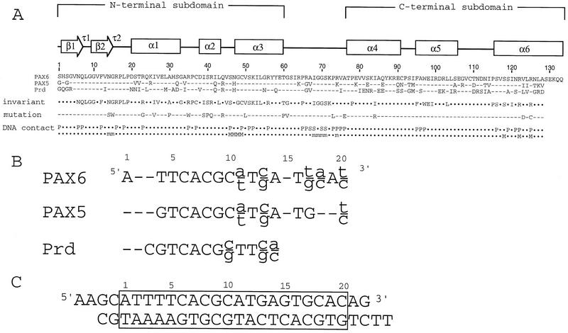
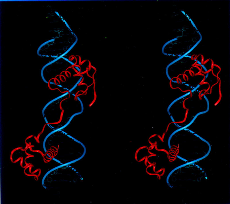
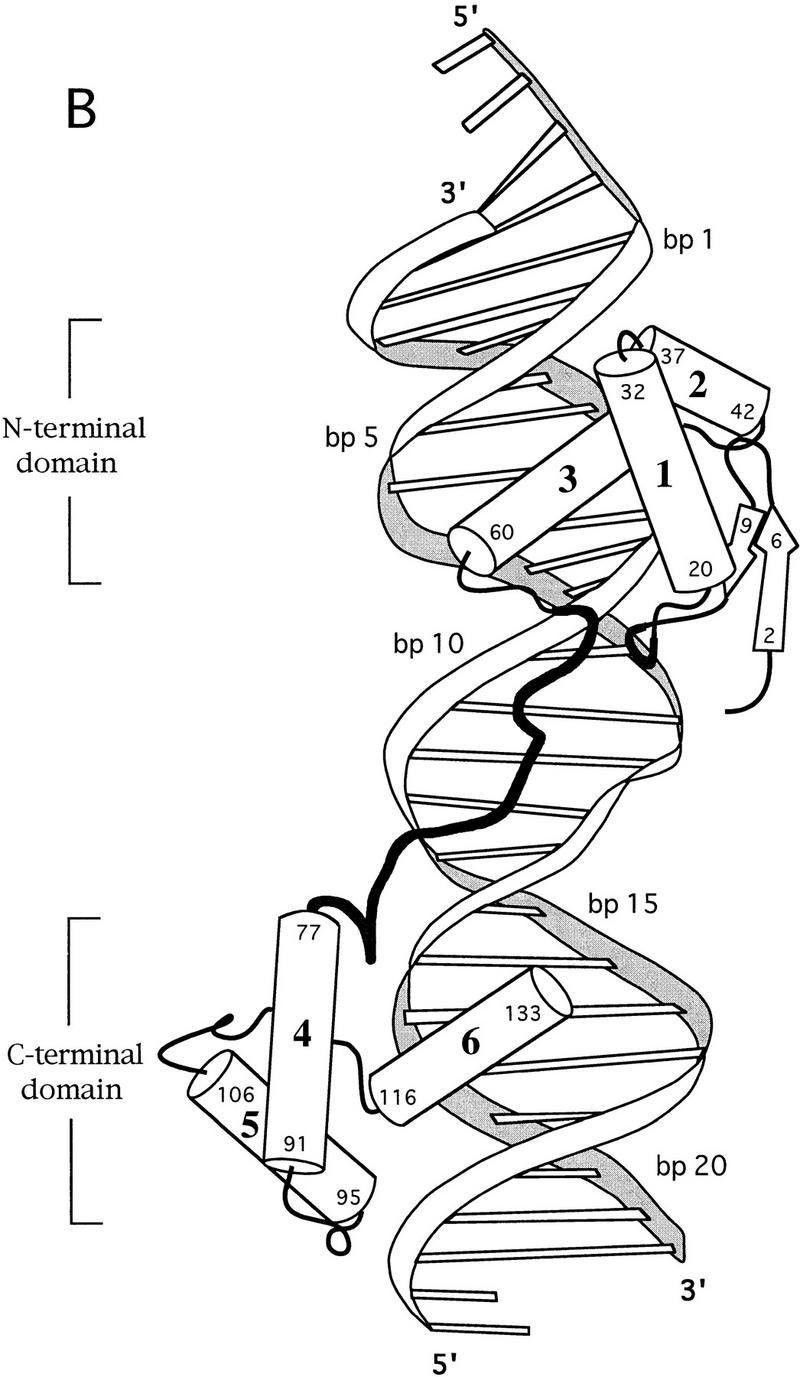
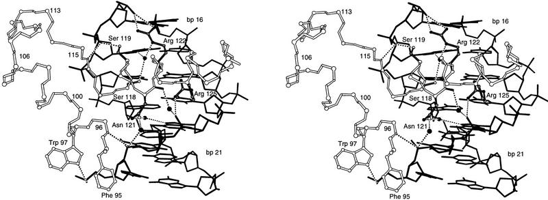
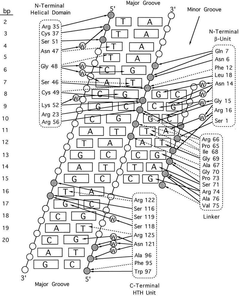
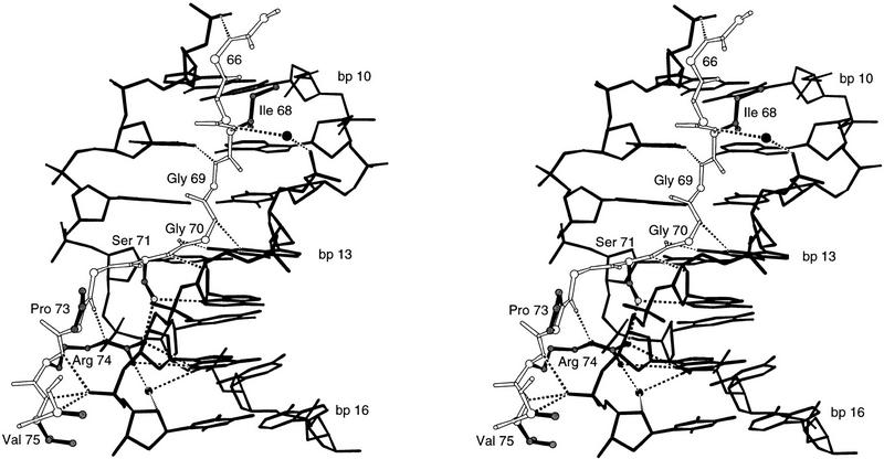
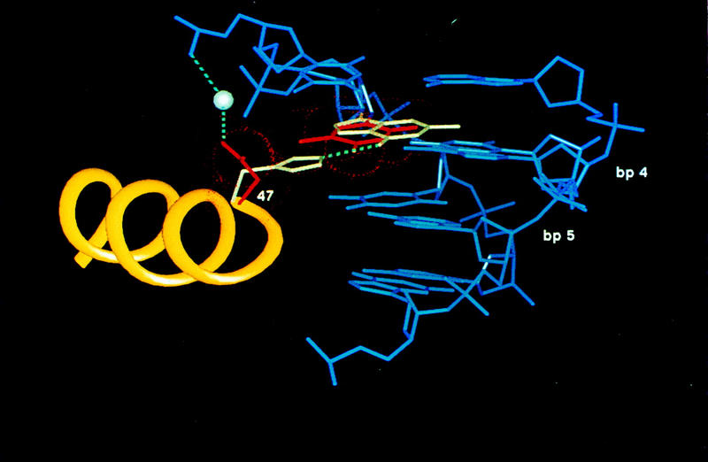
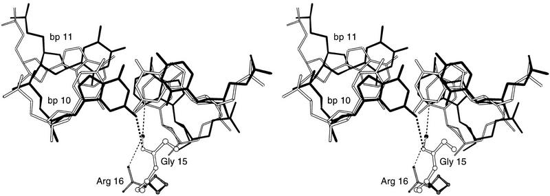
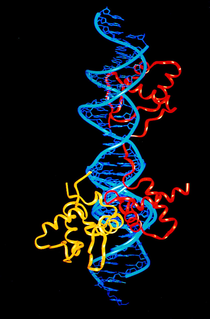
Pax6, a transcription factor containing the bipartite paired DNA-binding domain, has critical roles in development of the eye, nose, pancreas, and central nervous system. The 2.5 A structure of the human Pax6 paired domain with its optimal 26-bp site reveals extensive DNA contacts from the amino-terminal subdomain, the linker region, and the carboxy-terminal subdomain. The Pax6 structure not only confirms the docking arrangement of the amino-terminal subdomain as seen in cocrystals of the Drosophila Prd Pax protein, but also reveals some interesting differences in this region and helps explain the sequence specificity of paired domain-DNA recognition. In addition, this structure gives the first detailed information about how the paired linker region and carboxy-terminal subdomain contact DNA. The extended linker makes minor groove contacts over an 8-bp region, and the carboxy-terminal helix-turn-helix unit makes base contacts in the major groove. The structure and docking arrangement of the carboxy-terminal subdomain of Pax6 is remarkably similar to that of the amino-terminal subdomain, and there is an approximate twofold symmetry axis relating the polypeptide backbones of these two helix-turn-helix units. Our structure of the Pax6 paired domain-DNA complex provides a framework for understanding paired domain-DNA interactions, for analyzing mutations that map in the linker and carboxy-terminal regions of the paired domain, and for modeling protein-protein interactions of the Pax family proteins.


Figures

Figure 1
Figure 1
Sequences of paired domains and binding sites. (A) The sequence and secondary structure of the Pax6 paired domain, with sequences of paired domains from Pax5 and the Drosophila Prd protein, are shown (Treisman et al. 1991; Adams et al. 1992; Glaser et al. 1992). The protein contains the (conventional) 128-residue paired domain and five subsequent residues (SEKQQ) from Pax6. Lines below these sequences indicate residues that are conserved in almost all paired domains and also show missense mutations in the Pax6 paired domain (Azuma et al. 1996, ; Tang et al. 1997; Prosser and van Heyningen 1998 and http://www.mrc.hgu.ac.uk/Softdata/Pax6/ cited therein; Wolf et al. 1998; Grønskov et al. 1999; Hanson et al. 1999; T. Glaser, pers. comm.). Note that both the N17S and I29V missense mutations were identified in the same allele along with a 12-bp insertion in intron 5; hence, the functional significance of each charge by itself is uncertain. DNA contacts from the Pax6 crystals are summarized in the last two lines: One line indicates contacts with the sugar (S) phosphate (P) backbone; the other indicates base contacts [(M) major groove; (m) minor groove]. Note that the Pax6 nomenclature differs by three residues from that used in this paper because the Pax6 paired domain begins at residue 4 of the Pax6 protein. (B) DNA-binding sites for the Pax6 and Pax5 paired domains are longer than that for the Prd paired domain. Consensus binding sites for the Prd and Pax6 paired domains were determined from in vitro selections (Epstein et al. 1994a; Jun and Desplan 1996); the binding site for Pax5 paired domain was deduced from a combination of in vitro selection (Czerny and Busslinger 1995) and alignments of functional promoter sequences (Czerny et al. 1993). The extended sites recognized by Pax6 and Pax5 reflect binding of the C subdomain. (C) DNA oligonucleotide used in cocrystallization, with a box marking the Pax6 binding site. (D) The density-modified MI map shows clear electron density for the protein and the DNA. The map is contoured at 2.0 ς; this section shows the interface of the carboxy-terminal HTH motif (red) with the DNA (yellow). Several residues are labeled.
Figure 1
Figure 1
Sequences of paired domains and binding sites. (A) The sequence and secondary structure of the Pax6 paired domain, with sequences of paired domains from Pax5 and the Drosophila Prd protein, are shown (Treisman et al. 1991; Adams et al. 1992; Glaser et al. 1992). The protein contains the (conventional) 128-residue paired domain and five subsequent residues (SEKQQ) from Pax6. Lines below these sequences indicate residues that are conserved in almost all paired domains and also show missense mutations in the Pax6 paired domain (Azuma et al. 1996, ; Tang et al. 1997; Prosser and van Heyningen 1998 and http://www.mrc.hgu.ac.uk/Softdata/Pax6/ cited therein; Wolf et al. 1998; Grønskov et al. 1999; Hanson et al. 1999; T. Glaser, pers. comm.). Note that both the N17S and I29V missense mutations were identified in the same allele along with a 12-bp insertion in intron 5; hence, the functional significance of each charge by itself is uncertain. DNA contacts from the Pax6 crystals are summarized in the last two lines: One line indicates contacts with the sugar (S) phosphate (P) backbone; the other indicates base contacts [(M) major groove; (m) minor groove]. Note that the Pax6 nomenclature differs by three residues from that used in this paper because the Pax6 paired domain begins at residue 4 of the Pax6 protein. (B) DNA-binding sites for the Pax6 and Pax5 paired domains are longer than that for the Prd paired domain. Consensus binding sites for the Prd and Pax6 paired domains were determined from in vitro selections (Epstein et al. 1994a; Jun and Desplan 1996); the binding site for Pax5 paired domain was deduced from a combination of in vitro selection (Czerny and Busslinger 1995) and alignments of functional promoter sequences (Czerny et al. 1993). The extended sites recognized by Pax6 and Pax5 reflect binding of the C subdomain. (C) DNA oligonucleotide used in cocrystallization, with a box marking the Pax6 binding site. (D) The density-modified MI map shows clear electron density for the protein and the DNA. The map is contoured at 2.0 ς; this section shows the interface of the carboxy-terminal HTH motif (red) with the DNA (yellow). Several residues are labeled.
Figure 2
Figure 2
Overview of the Pax6 paired domain–DNA complex. (A) Stereo view with ribbons drawn through the Cα atoms of the protein (red) and through the phosphate atoms of the DNA backbone (blue). The N subdomain is at the top. (B) Sketch of the Pax6 paired domain–DNA complex in a similar orientation. Cylinders represent α helices; arrows represent β strands. Helices 1–6 are labeled; residue numbers indicate termini of the corresponding secondary structure elements.
Figure 2
Figure 2
Overview of the Pax6 paired domain–DNA complex. (A) Stereo view with ribbons drawn through the Cα atoms of the protein (red) and through the phosphate atoms of the DNA backbone (blue). The N subdomain is at the top. (B) Sketch of the Pax6 paired domain–DNA complex in a similar orientation. Cylinders represent α helices; arrows represent β strands. Helices 1–6 are labeled; residue numbers indicate termini of the corresponding secondary structure elements.
Figure 3
Figure 3
Stereo view of the interface between the C domain and the DNA. The orientation of the complex is similar to that in Fig. 2, A and B. DNA is represented by solid sticks; the protein backbone is represented with open sticks. Side chains of key residues that contact the DNA are shown (Phe-95 and Trp-97 with open sticks; Ser-118, Ser-119, Asn-121, Arg-122, and Arg-125 with solid sticks). (●) Water molecules; (broken lines) hydrogen bonds. Corresponding superpositions between the C subdomain (residues 80–128) and the three helices of Engrailed (residues 10–58) give an rms distance of 1.71 Å; superpositions with the three helices of the Hin recombinase (residues 148–180) give rms distances of 1.86 Å.
Figure 4
Figure 4
Diagram of DNA contacts in the Pax6 paired domain–DNA complex. DNA is represented as a cylindrical projection. Circles labeled W denote water molecules; other circles represent phosphates; shaded circles mark sites where Pax6 contacts the DNA backbone. All contacts made by Pax6 are indicated with arrows. (Solid arrows) Hydrogen bonds; (broken arrows) van der Waals contacts.
Figure 5
Figure 5
Stereo view of the interface between the linker and the DNA. The orientation of the complex is similar to that in Figs. 2, A and B. DNA is represented by solid sticks; the protein backbone is represented by open sticks. Side chains of key residues (Ile-68, Ser-71, Pro-73, Arg-74, and Val-75) that contact DNA are in black. (●) Water molecules; (broken lines) hydrogen bonds.
Figure 6
Figure 6
Key differences in DNA contacts made by the Pax6 and Prd N subdomains. (A) Comparison of the role of residue 47 in Pax6 and Prd. Complexes were aligned by superimposing the amino-terminal HTH motifs of Prd and Pax6. Helix 3 is yellow; neighboring regions of the DNA are blue. His-47 of Prd (white) makes a hydrogen bond (broken line) with the guanine (white) at base pair 4 of the Prd site; Asn-47 of Pax6 (shown in red) makes van der Waals contacts (dotted red spheres) with the thymine (red) at base pair 4 of the Pax6 site and makes a water-mediated contact with a phosphate. (B) Stereo view of contacts made by Gly-15 and Arg-16 where the β turn of Pax6 fits into the minor groove. (Broken lines) Hydrogen bonds with the Pax6 site (bases shown in black); (●) critical water molecule. Bases from the corresponding region of paired are shown with open lines. (Complexes were superimposed by superimposing the β turns.) In Pax6, the carbonyl oxygen of Gly-15 contacts the N2 of a guanine at base pair 10; Prd has a contact at essentially the same position in space but it involves the N2 of a guanine on the opposite strand of the DNA. In Pax6, the critical water molecule contacts the N3 of the adenine at base pair 11; Prd has a water molecule at essentially the same position in space, but it contacts the O2 of a thymine, which occurs at base pair 11 of the Prd site.
Figure 6
Figure 6
Key differences in DNA contacts made by the Pax6 and Prd N subdomains. (A) Comparison of the role of residue 47 in Pax6 and Prd. Complexes were aligned by superimposing the amino-terminal HTH motifs of Prd and Pax6. Helix 3 is yellow; neighboring regions of the DNA are blue. His-47 of Prd (white) makes a hydrogen bond (broken line) with the guanine (white) at base pair 4 of the Prd site; Asn-47 of Pax6 (shown in red) makes van der Waals contacts (dotted red spheres) with the thymine (red) at base pair 4 of the Pax6 site and makes a water-mediated contact with a phosphate. (B) Stereo view of contacts made by Gly-15 and Arg-16 where the β turn of Pax6 fits into the minor groove. (Broken lines) Hydrogen bonds with the Pax6 site (bases shown in black); (●) critical water molecule. Bases from the corresponding region of paired are shown with open lines. (Complexes were superimposed by superimposing the β turns.) In Pax6, the carbonyl oxygen of Gly-15 contacts the N2 of a guanine at base pair 10; Prd has a contact at essentially the same position in space but it involves the N2 of a guanine on the opposite strand of the DNA. In Pax6, the critical water molecule contacts the N3 of the adenine at base pair 11; Prd has a water molecule at essentially the same position in space, but it contacts the O2 of a thymine, which occurs at base pair 11 of the Prd site.
Figure 7
Figure 7
Proposed model for cooperative DNA binding of Pax6 (red) with the Ets domain (yellow). In this model, protein–protein interactions are mediated by the second and third helices of the Pax6 C subdomain with the third helix and the following hairpin region of Ets domain. The model was generated by superimposing phosphates of the Pax and Ets (Liang et al. 1994; Kodandapani et al. 1996) complexes in a way that reflects the relative spacing of the binding sites (Fitzsimmons et al. 1996). (The N subdomain is at the top, but the complex has been rotated, relative to Fig. 2, around a vertical axis so that the proposed contacts between Pax6 and Ets are easier to see.)

Similar articles

Cited by

References

    1. Adams B, Dörfler P, Aguzzi A, Kozmik Z, Urbánek P, Maurer-Fogy I, Busslinger M. Pax-5 encodes the transcription factor BSAP and is expressed in B lymphocytes, the developing CNS, and adult testis. Genes & Dev. 1992;6:1589–1607. - PubMed
    1. Azuma N, Nishina S, Yanagisawa H, Okuyama T, Yamada M. PAX6 missense mutation in isolated foveal hypoplasia. Nat Genet. 1996;18:141–142. - PubMed
    1. Azuma N, Hotta Y, Tanaka H, Yamada M. Missense mutations in the PAX6 gene in Aniridia. Invest Ophthalmol Vis Sci. 1998;39:2524–2528. - PubMed
    1. Baldwin CT, Hoth CF, Macina RA, Milunsky A. Mutations in Pax3 that cause Waardenburg syndrome type I: Ten new mutations and review of the literature. Am J Med Genet. 1995;58:115–122. - PubMed
    1. Barr FG. Chromosomal translocations involving paired box transcription factors in human cancer. Int J Biochem Cell Biol. 1997;29:1449–1461. - PubMed

Publication types

Associated data

LinkOut - more resources