. 1999 May 15;13(10):1322-8.
doi: 10.1101/gad.13.10.1322.

IKK1-deficient mice exhibit abnormal development of skin and skeleton

Affiliations

IKK1-deficient mice exhibit abnormal development of skin and skeleton

Q Li et al. Genes Dev. .

Abstract

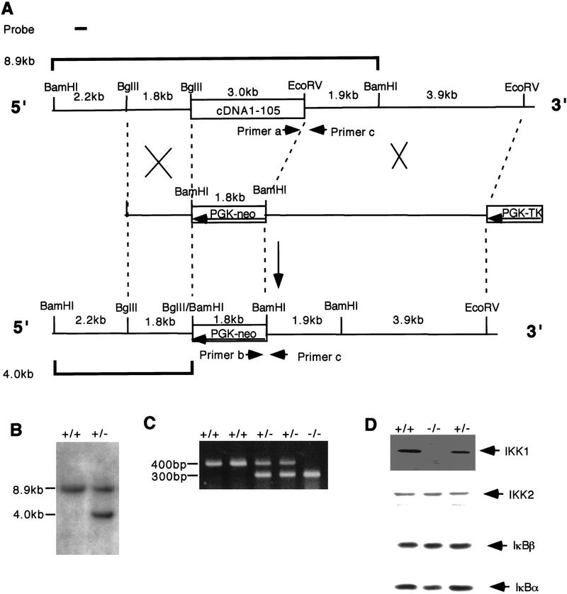
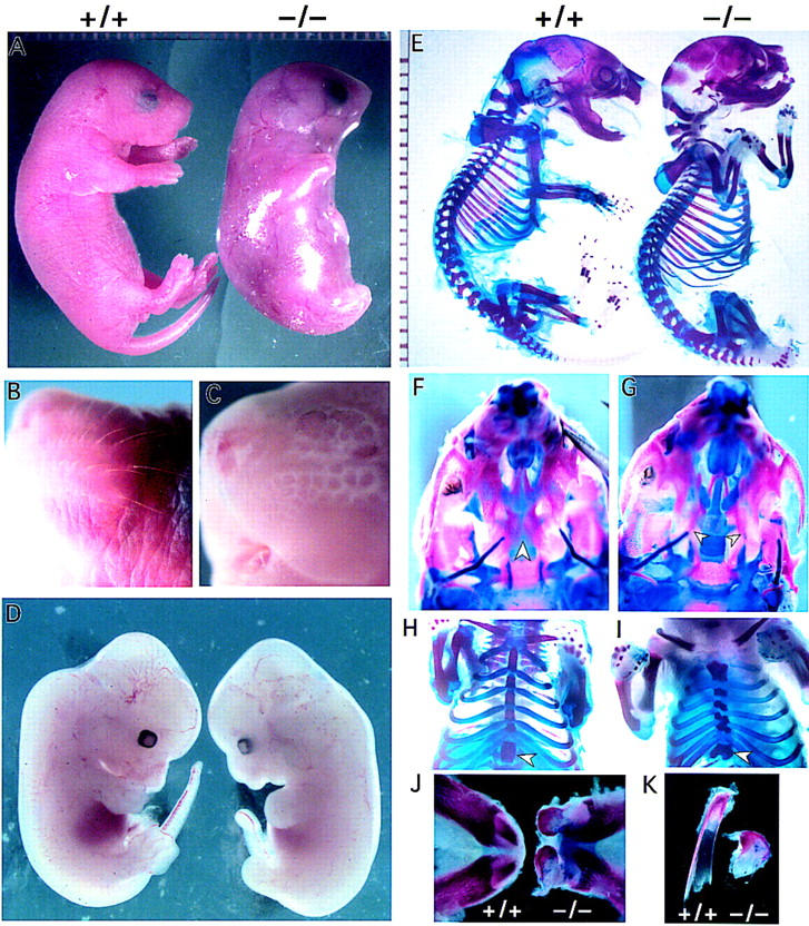
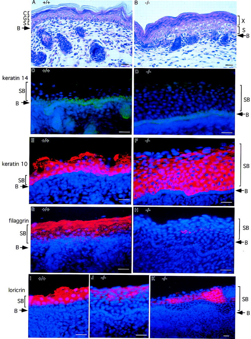
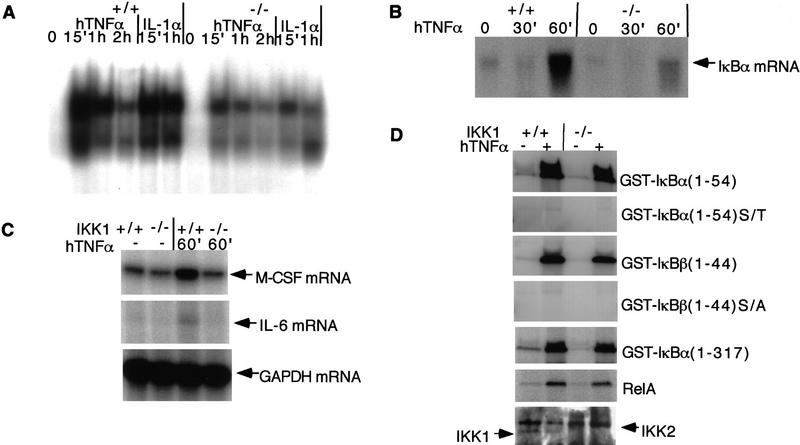
IkappaB kinases (IKKs) IKK1 and IKK2 are two putative IkappaBalpha kinases involved in NF-kappaB activation. To examine the in vivo functions of IKK1, we generated IKK1-deficient mice. The mutant mice are perinatally lethal and exhibit a wide range of developmental defects. Newborn mutant mice have shiny, taut, and sticky skin without whiskers. Histological analysis shows thicker epidermis, which is unable to differentiate. Limbs and tail are wrapped inside the skin and do not extend properly out of the body trunk. Skeleton staining reveals a cleft secondary palate, split sternebra 6, and deformed incisors. NF-kappaB activation mediated by TNFalpha and IL-1 is diminished in IKK1-deficient mouse embryonic fibroblast (MEF) cells. The IKK complex in the absence of IKK1 is capable of phosphorylating IkappaBalpha and IkappaBbeta in vitro. Our results support a role for IKK1 in NF-kappaB activation and uncover its involvement in skin and skeleton development. We conclude further that the two related kinases IKK1 and IKK2 have distinct functions and can not be substituted for each other's functions.


Figures

Figure 1
Figure 1
Targeting of the IKK1 gene in mice. (A) Strategy for targeting the IKK1 allele. Simplified restriction maps of the wild-type IKK1 allele, the targeting construct, and the mutated allele are shown. A 3-kb genomic fragment is deleted and replaced by a PGK–neo cassette in an antisense orientation. (B) Southern blot analysis of an ES clone showing the correct insertion of the targeting construct. DNA was digested with BamHI and hybridized to the probe shown in A. The wild-type allele yields an 8.9-kb fragment whereas the mutant allele yields a 4-kb fragment. (C) PCR detection of mouse genotypes. (D) Western blot analysis confirmed the absence of IKK1 in IKK1−/− MEFs, whereas the expression of IKK2, IκBα, and IκBβ was not changed. Forty micrograms of whole-cell protein lysates from IKK2+/+, IKK2+/−, and IKK2−/− MEFs were loaded and immunoblotted with IKK1, IKK2, IκBα, and IκBβ antibodies.
Figure 2
Figure 2
Gross phenotypes of IKK1−/−newborn mice. (A) Wild-type newborn mouse (left) and an IKK1−/− homozygous (right) littermate are shown. The mutant embryos exhibit severe alterations in the overall morphology. Limbs and tail are barely protruding and seem to be shortened. The skin appears translucent and tense. (B,C) A close-up view of a wild-type (B) and mutant (C) head. The mutant embryos lack whiskers, compared with the wild-type embryos. (D) Comparison of E12.5 wild-type (left) and IKK1 (right) mutant embryos. The mutant shows mild phenotypical alterations in morphology of the limb buds, tail and craniofacial region. (E) Bone staining of the embryos shown in A. The mutant skeleton (right) exhibits a few gross alterations. The craniofacial bone structure shows malformation, the tail appears to be shorter, and the bending of the distal limb region is abnormal. However, in contrast to the appearance of a newborn embryo, all long bones of the limbs are formed. (F,G) Ventral view of wild-type (F) and mutant (G) skulls. The skull exhibits a nearly complete fusion of the palate (F; arrowhead), whereas the mutant bilateral palate shelves (arrowhead) remains unfused, allowing the more dorsal lying vomer and presphenoid to be visible (G). (H,I) A ventral view of the ribcage and the sternum of a wild-type skeleton (H) and an IKK1−/− skeleton (F). The mutant embryo displays a broader sternum and the ribs exhibit a kinky fusion to the sternum and split sternebra 6 (arrowhead). (J) Lower jaw and lower incisors of wild-type (left) and mutant (right) embryos. The morphology of the protruding incisors in mutant jaw appears malformed. (K) A detailed view of the incisors (left, +/+; right , −/−) shows a reduced and distorted mutant incisor.
Figure 3
Figure 3
Lack of epidermal differentiation in the skin from IKK1−/− mice. Staining was performed on skin from newborn wild-type (+/+) or mutant mice (−/−). (A,B) Hematoxylin and eosin staining of skin. In the superficial layer of mutant skin, an unusual thick layer of flatten cells (layer X in B) was present instead of cornified layer. (C,D) Keratin 14 immunostaining (green) shows normal staining pattern of positive cells in the stratum basal layer. Suprabasal layer above basal layer was much thicker in mutant skin than in wild-type skin. (E,F) Anti-keratin 10 antibody stains the cells (red) in suprabasal layers in normal epidermis. The number of keratin 10 positive cells (red) in mutant skin increased dramatically. (G,H) Filaggrin is expressed in the granule and cornified layers of normal skin; however, its expression is diminished and limited to small patches of cells (red) in the mutant skin. (I–K) Expression of loricrin (red) is also reduced dramatically in the mutant skin. Nuclei (blue) were labeled by DAPI in C–K. Abbreviations: (B) Basal layer of epidermis; (S) spinous layer; (G) granular layer; (C) cornified layer; (SB) suprabasal layer. Bar size, 24 μm.
Figure 4
Figure 4
NF-κB activity in IKK1−/− MEFs. (A) NF-κB binding activity is reduced in IKK1−/− MEF. Gel mobility shift analysis was conducted on 5 μg of nuclear extract from IKK1+/+ and IKK1−/− MEFs with or without 10 ng/ml TNFα induction for the indicated time. (B) TNFα-induced IκBα RNA synthesis is reduced in MEFs lacking IKK1. Northern blot analysis was performed on 10 μg of total RNA from each sample, using probe from a full-length IκBα cDNA. (C) RNase protection shows that TNFα-induced mRNA expression of M-CSF and IL-6 are diminished in IKK1−/− MEF. (D) Phosphorylation of IκBα, IκBβ, and p65 by NEMO immunocomplexes from IKK1−/− MEFs is not affected. Cells from three 15-cm plates were lysed and immunoprecipitated with anti-NEMO serum. Equal amounts of NEMO immunoprecipitates were incubated with the substrates indicated for the in vitro kinase assay. NEMO immunoprecipitation samples were loaded on a 10% SDS–polyacrylamide gel for anti-IKK1 and anti-IKK2 immunoblotting (D, bottom).

References

    1. Attar RM, Caamano J, Carrasco D, Iotsova V, Ishikawa H, Ryseck RP, Weih F, Bravo R. Genetic approaches to study Rel/NF-κB/I κB function in mice. Semin Cancer Biol. 1997;8:93–101. - PubMed
    1. Baeuerle PA, Baichwal VR. NF-κ B as a frequent target for immunosuppressive and anti-inflammatory molecules. Adv Immunol. 1997;65:111–137. - PubMed
    1. Beg AA, Baltimore D. An essential role for NF-κ in preventing TNF-alpha-induced cell death. Science. 1996;274:782–784. - PubMed
    1. Beg AA, Sha WC, Bronson RT, Ghosh S, Baltimore D. Embryonic lethality and liver degeneration in mice lacking the RelA component of NF-κ B. Nature. 1995;376:167–170. - PubMed
    1. Bushdid PB, Brantley DM, Yull FE, Blaeuer GL, Hoffman LH, Niswander L, Kerr LD. Inhibition of NF-κB activity results in disruption of the apical ectodermal ridge and aberrant limb morphogenesis. Nature. 1998;392:615–618. - PubMed

Publication types

LinkOut - more resources