. 1999 May 15;13(10):1329-41.
doi: 10.1101/gad.13.10.1329.

Mice devoid of all known thyroid hormone receptors are viable but exhibit disorders of the pituitary-thyroid axis, growth, and bone maturation

Affiliations

Mice devoid of all known thyroid hormone receptors are viable but exhibit disorders of the pituitary-thyroid axis, growth, and bone maturation

S Göthe et al. Genes Dev. .

Abstract

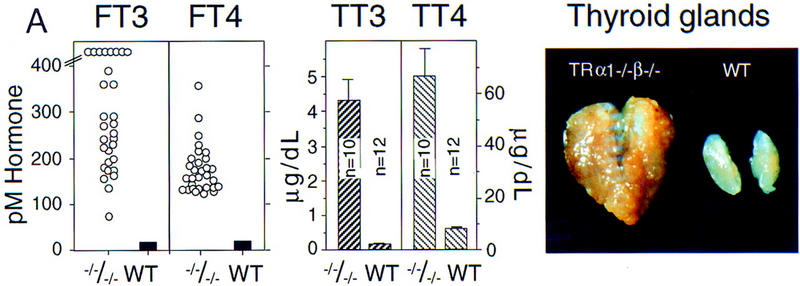
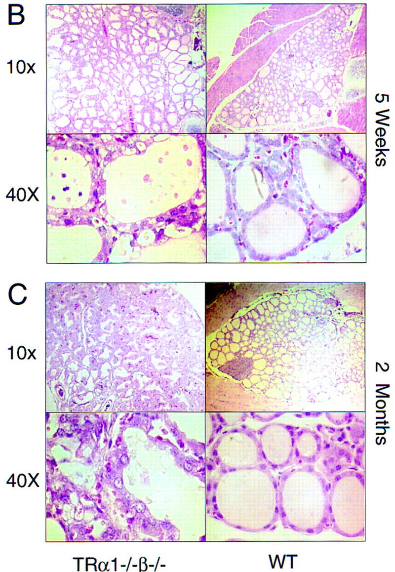
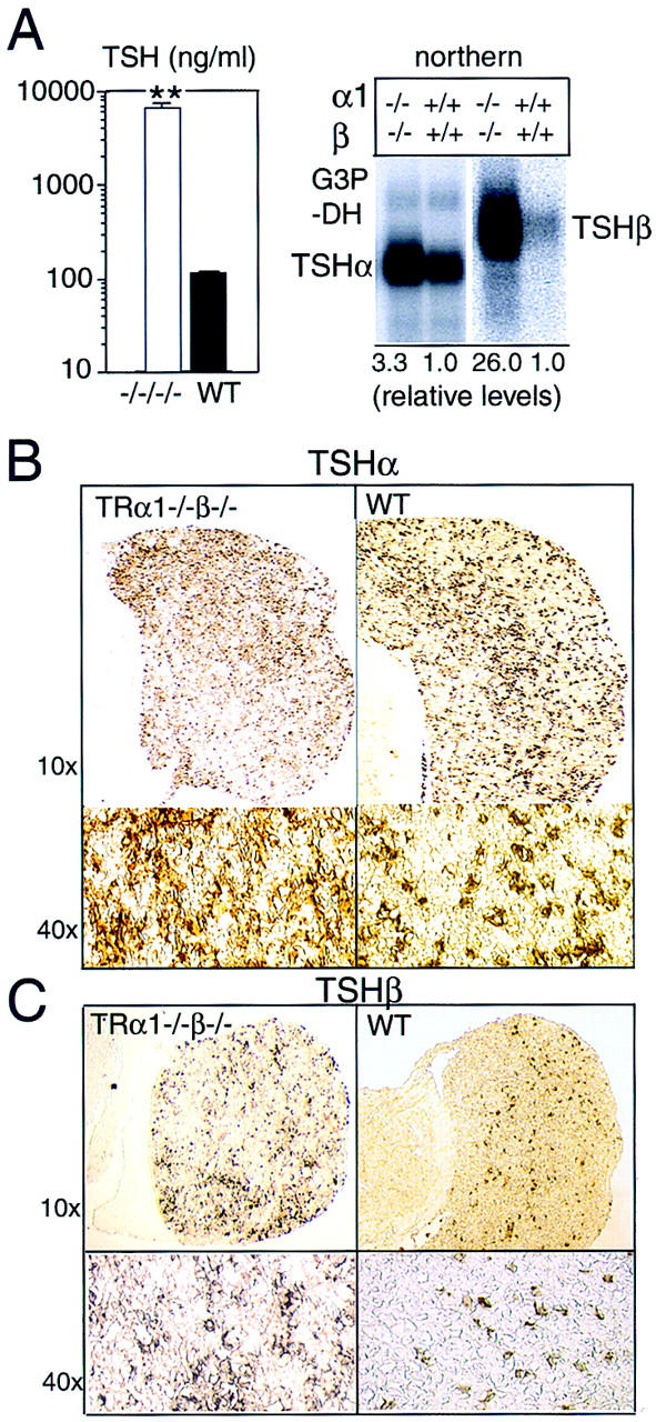
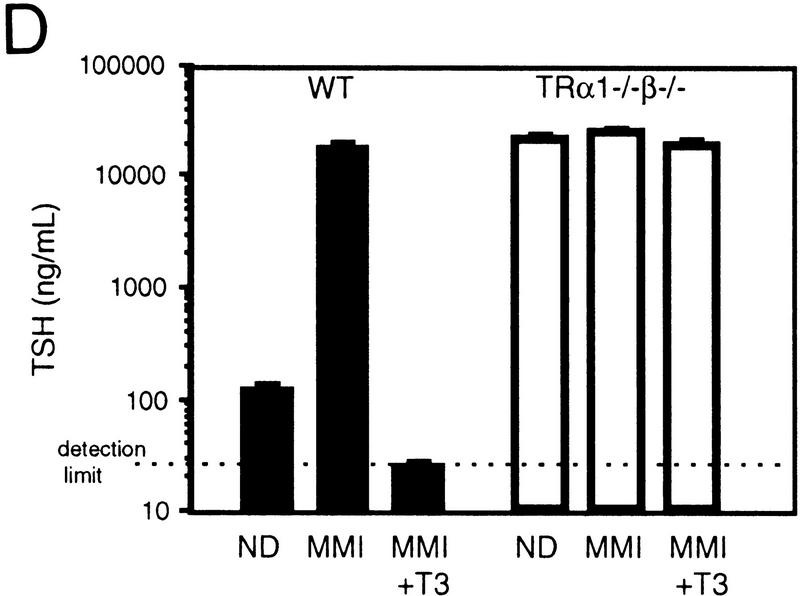
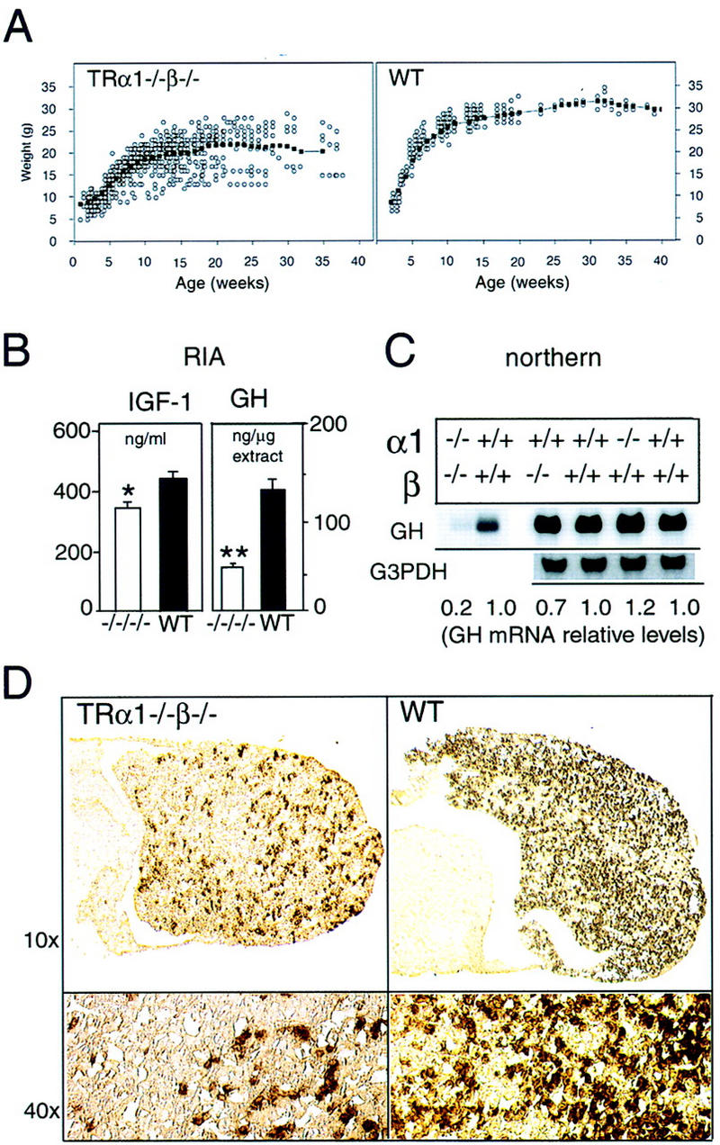
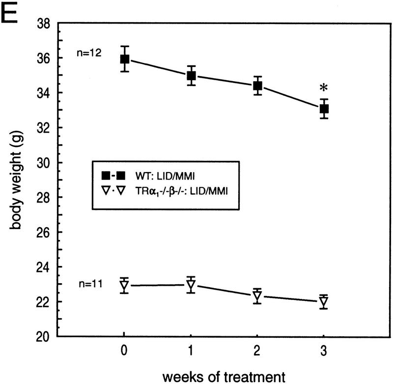
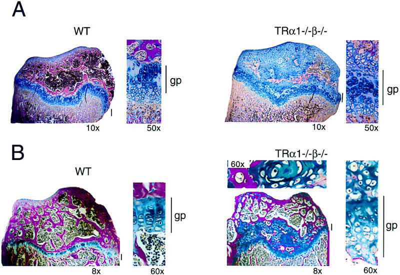
Thyroid hormone (T3) has widespread functions in development and homeostasis, although the receptor pathways by which this diversity arises are unclear. Deletion of the T3 receptors TRalpha1 or TRbeta individually reveals only a small proportion of the phenotypes that arise in hypothyroidism, implying that additional pathways must exist. Here, we demonstrate that mice lacking both TRalpha1 and TRbeta (TRalpha1(-/-)beta-/-) display a novel array of phenotypes not found in single receptor-deficient mice, including an extremely hyperactive pituitary-thyroid axis, poor female fertility and retarded growth and bone maturation. These results establish that major T3 actions are mediated by common pathways in which TRalpha1 and TRbeta cooperate with or substitute for each other. Thus, varying the balance of use of TRalpha1 and TRbeta individually or in combination facilitates control of an extended spectrum of T3 actions. There was no evidence for any previously unidentified T3 receptors in TRalpha1(-/-)beta-/- mouse tissues. Compared to the debilitating symptoms of severe hypothyroidism, the milder overall phenotype of TRalpha1(-/-)beta-/- mice, lacking all known T3 receptors, indicates divergent consequences for hormone versus receptor deficiency. These distinctions suggest that T3-independent actions of T3 receptors, demonstrated previously in vitro, may be a significant function in vivo.


Figures

Figure 1
Figure 1
Absence of detectable T3 receptors in nuclear extracts from TRα1−/−β−/− mouse tissues. (A,B) Gel mobility shift experiments showing absence of TR proteins that can bind the F2 T3 response element in nuclear extracts from liver (A) and lung (B) of TRα1−/−β−/− mice. Specific TR-containing bands were identified by addition of antibodies against TR (anti-TR) or RXR (anti-RXR); (No ab) No antibodies added. Parallel analysis of samples with a β-fibrinogen probe demonstrated the integrity of other DNA-binding proteins in TR-deficient samples. (C) Absence of a specific T3 binding capacity in nuclear extracts from brain and liver of TRα1−/−β−/− mice. The 125I-labeled T3 binding capacity of TRα1−/−β−/− and wild-type samples is shown after subtraction of the binding that was not competed by a 1000-fold excess of cold T3 (Torresani and DeGroot 1975). (D) Absence of a saturable, specific T3 binding capacity in nuclear extracts from TRα1−/−β−/− mice. Nuclear extracts were prepared from mice that had been made hypothyroid to exclude the possibility that residual T3 binding sites were present but were masked by prior saturation by the excessive T3 levels in TRα1−/−β−/− mice (see Fig. 2A).
Figure 1
Figure 1
Absence of detectable T3 receptors in nuclear extracts from TRα1−/−β−/− mouse tissues. (A,B) Gel mobility shift experiments showing absence of TR proteins that can bind the F2 T3 response element in nuclear extracts from liver (A) and lung (B) of TRα1−/−β−/− mice. Specific TR-containing bands were identified by addition of antibodies against TR (anti-TR) or RXR (anti-RXR); (No ab) No antibodies added. Parallel analysis of samples with a β-fibrinogen probe demonstrated the integrity of other DNA-binding proteins in TR-deficient samples. (C) Absence of a specific T3 binding capacity in nuclear extracts from brain and liver of TRα1−/−β−/− mice. The 125I-labeled T3 binding capacity of TRα1−/−β−/− and wild-type samples is shown after subtraction of the binding that was not competed by a 1000-fold excess of cold T3 (Torresani and DeGroot 1975). (D) Absence of a saturable, specific T3 binding capacity in nuclear extracts from TRα1−/−β−/− mice. Nuclear extracts were prepared from mice that had been made hypothyroid to exclude the possibility that residual T3 binding sites were present but were masked by prior saturation by the excessive T3 levels in TRα1−/−β−/− mice (see Fig. 2A).
Figure 2
Figure 2
Hyperactive goiter in TRα1−/−β−/− mice. (A) Elevated serum levels of thyroid hormones and goiter in adult TRα1−/−β−/− mice. Free (FT3, FT4) and total (TT3, TT4) levels of thyroid hormones were elevated in TRα1−/−β−/− mice. Values are shown for males; females gave similar results. (Right) The grossly enlarged thyroid gland dissected from an 8-week-old TRα1−/−β−/− mouse. (B,C) Progressive pathological changes in the thyroid glands of TRα1−/−β−/− mice (left) compared to wild-type mice (right) at 5 (B) and 8 (C) weeks. Sections were stained with hematoxylin and eosin. The enlarged follicles often contained degenerating cellular material in the colloid. Hyperproliferation progressed considerably by 8 weeks.
Figure 2
Figure 2
Hyperactive goiter in TRα1−/−β−/− mice. (A) Elevated serum levels of thyroid hormones and goiter in adult TRα1−/−β−/− mice. Free (FT3, FT4) and total (TT3, TT4) levels of thyroid hormones were elevated in TRα1−/−β−/− mice. Values are shown for males; females gave similar results. (Right) The grossly enlarged thyroid gland dissected from an 8-week-old TRα1−/−β−/− mouse. (B,C) Progressive pathological changes in the thyroid glands of TRα1−/−β−/− mice (left) compared to wild-type mice (right) at 5 (B) and 8 (C) weeks. Sections were stained with hematoxylin and eosin. The enlarged follicles often contained degenerating cellular material in the colloid. Hyperproliferation progressed considerably by 8 weeks.
Figure 3
Figure 3
Overproduction of TSH in TRα1−/−β−/− mice. (A) Serum TSH levels were 60-fold elevated (left) in 5-month-old adult male mice, and the pituitary glands contained increased levels of TSHα and TSHβ subunit mRNAs (right). As a control for the amount and integrity of RNA loaded, the same Northern blot was hybridized with a probe for G3PDH, as shown in the TSHα lanes. (B,C) Immunostaining showed increased numbers of TSHα-positive cells (1.9-fold; B) and TSHβ-positive cells (2.8-fold; C) in the anterior lobe of the pituitary gland. Cell counts were determined for n = 2 wild-type and n = 3 TRα1−/−β−/− female mice at 5 weeks of age. (D) Resistance of TSH levels to change under MMI-induced hypothyroid conditions. Serum TSH levels were sequentially determined in groups of n = 4 wild-type or TRα1−/−β−/− male mice (4 weeks old) provided with normal diet (ND), low iodine diet with MMI/perchlorate in drinking water for 4 weeks (MMI), and finally under MMI with the addition of 5.0 μg/ml L-T3 in the drinking water for 8 days (MMI+T3). MMI resulted in hypothyroidism (T4 < 0.4 μg/dl) in both groups. MMI+T3 resulted in serum total T3 levels of 9440 ± 2110 and 14,937 ± 1,347 ng/dl, respectively in wild-type and TRα1−/−β−/− mice (116- and 5.5-fold elevated above the respective levels under normal diet). One wild-type mouse died under MMI+T3 conditions. TSH detection limit, 25 ng/ml.
Figure 3
Figure 3
Overproduction of TSH in TRα1−/−β−/− mice. (A) Serum TSH levels were 60-fold elevated (left) in 5-month-old adult male mice, and the pituitary glands contained increased levels of TSHα and TSHβ subunit mRNAs (right). As a control for the amount and integrity of RNA loaded, the same Northern blot was hybridized with a probe for G3PDH, as shown in the TSHα lanes. (B,C) Immunostaining showed increased numbers of TSHα-positive cells (1.9-fold; B) and TSHβ-positive cells (2.8-fold; C) in the anterior lobe of the pituitary gland. Cell counts were determined for n = 2 wild-type and n = 3 TRα1−/−β−/− female mice at 5 weeks of age. (D) Resistance of TSH levels to change under MMI-induced hypothyroid conditions. Serum TSH levels were sequentially determined in groups of n = 4 wild-type or TRα1−/−β−/− male mice (4 weeks old) provided with normal diet (ND), low iodine diet with MMI/perchlorate in drinking water for 4 weeks (MMI), and finally under MMI with the addition of 5.0 μg/ml L-T3 in the drinking water for 8 days (MMI+T3). MMI resulted in hypothyroidism (T4 < 0.4 μg/dl) in both groups. MMI+T3 resulted in serum total T3 levels of 9440 ± 2110 and 14,937 ± 1,347 ng/dl, respectively in wild-type and TRα1−/−β−/− mice (116- and 5.5-fold elevated above the respective levels under normal diet). One wild-type mouse died under MMI+T3 conditions. TSH detection limit, 25 ng/ml.
Figure 4
Figure 4
Retarded growth and reduced expression of IGF-I and GH in TRα1−/−β−/− mice. (A) Weight gain of TRα1−/−β−/− and wild-type mice measured over the first 40 postnatal weeks. The average weight gain curve, determined as a moving three-point average, is shown by the linked, black points. (B) Reduced serum levels of IGF-I and of pituitary GH content in TRα1−/−β−/− mice, determined by radioimmunoassay. (C) Northern blot analysis of GH mRNA in TRα1−/−β−/−, TRα1−/−, and TRβ−/− mice. Adult male samples are shown alongside wild-type samples of the corresponding genetic background. Quantitation was determined by PhosphorImager with values normalized to G3PDH signals; for TRα1−/−β−/− mice, the same Northern blot was used as in Fig. 3A; thus, normalization was to the G3PDH bands shown in Fig. 3A. (D) Immunostaining of pituitary gland sections showed 4.5-fold reduced numbers of GH-positive cells in the anterior lobe, determined on the same pituitaries as for TSH immunostaining in Fig. 3. (E) Groups of adult male mice were pretreated on low iodine diet for 3 weeks and were then supplemented with MMI/perchlorate (LID/MMI) in the drinking water for 3 weeks, starting at 0 weeks. Over 3 weeks, weight decreased significantly in wild-type but not TRα1−/−β−/− mice. (*) P < 0.01 vs. weight at time 0 weeks. MMI treatment resulted in free T3 levels ≤ 2.6 pM and free T4 ≤ 3.5 pM in both groups (normal wild-type levels are 6–9 pM for FT3 and 12–16 pM for FT4).
Figure 4
Figure 4
Retarded growth and reduced expression of IGF-I and GH in TRα1−/−β−/− mice. (A) Weight gain of TRα1−/−β−/− and wild-type mice measured over the first 40 postnatal weeks. The average weight gain curve, determined as a moving three-point average, is shown by the linked, black points. (B) Reduced serum levels of IGF-I and of pituitary GH content in TRα1−/−β−/− mice, determined by radioimmunoassay. (C) Northern blot analysis of GH mRNA in TRα1−/−β−/−, TRα1−/−, and TRβ−/− mice. Adult male samples are shown alongside wild-type samples of the corresponding genetic background. Quantitation was determined by PhosphorImager with values normalized to G3PDH signals; for TRα1−/−β−/− mice, the same Northern blot was used as in Fig. 3A; thus, normalization was to the G3PDH bands shown in Fig. 3A. (D) Immunostaining of pituitary gland sections showed 4.5-fold reduced numbers of GH-positive cells in the anterior lobe, determined on the same pituitaries as for TSH immunostaining in Fig. 3. (E) Groups of adult male mice were pretreated on low iodine diet for 3 weeks and were then supplemented with MMI/perchlorate (LID/MMI) in the drinking water for 3 weeks, starting at 0 weeks. Over 3 weeks, weight decreased significantly in wild-type but not TRα1−/−β−/− mice. (*) P < 0.01 vs. weight at time 0 weeks. MMI treatment resulted in free T3 levels ≤ 2.6 pM and free T4 ≤ 3.5 pM in both groups (normal wild-type levels are 6–9 pM for FT3 and 12–16 pM for FT4).
Figure 5
Figure 5
Retarded bone growth and maturation in TRα1−/−β−/− mice. (A) Histological section showing disorganized growth plates of the proximal tibia in pubertal (5-week-old) TRα1−/−β−/− and wild-type mice. (B) Histological section of the proximal tibia in adult (4-month-old) TRα1−/−β−/− and wild-type mice. For the TRα1−/−β−/− sample, the top shows a higher magnification of a cartilage area from the epiphysis. Note the extensive areas of remaining cartilage (blue) in the epiphysis above the growth plate in both pubertal and adult TRα1−/−β−/−mice. (gp) Growth plate (also indicated by vertical bars). Sections were stained with alcian blue/Van Gieson (cartilage area, blue; bone area, red). Magnifications are indicated.

Similar articles

Cited by

References

    1. Aguanno A, Afar R, Albert V. Tissue-specific expression of the non-neuronal promoter of the aromatic l-amino acid decarboxylase gene is regulated by hepatocyte nuclear factor 1. J Biol Chem. 1996;271:4528–4538. - PubMed
    1. Beamer W, Eicher E, Maltais L, Southard J. Inherited primary hypothyroidism in mice. Science. 1981;212:61–63. - PubMed
    1. Beamer W, Maltais L, DeBaets M, Eicher E. Inherited congenital goiter in mice. Endocrinology. 1987;120:838–840. - PubMed
    1. Bradley DJ, Towle HC, Young WS., III Spatial and temporal expression of α- and β-thyroid hormone receptor mRNAs, including the β2-subtype, in the developing mammalian nervous system. J Neurosci. 1992;12:2288–2302. - PMC - PubMed
    1. Brent G, Moore D, Larsen P. Thyroid hormone regulation of gene expression. Annu Rev Physiol. 1991;53:17–35. - PubMed

Publication types

MeSH terms

Substances