. 1999 Jun;181(11):3438-44.
doi: 10.1128/JB.181.11.3438-3444.1999.

Mapping an interface of SecY (PrlA) and SecE (PrlG) by using synthetic phenotypes and in vivo cross-linking

Affiliations

Mapping an interface of SecY (PrlA) and SecE (PrlG) by using synthetic phenotypes and in vivo cross-linking

C R Harris et al. J Bacteriol. 1999 Jun.

Abstract

SecY and SecE are integral cytoplasmic membrane proteins that form an essential part of the protein translocation machinery in Escherichia coli. Sites of direct contact between these two proteins have been suggested by the allele-specific synthetic phenotypes exhibited by pairwise combinations of prlA and prlG signal sequence suppressor mutations in these genes. We have introduced cysteine residues within the first periplasmic loop of SecY and the second periplasmic loop of SecE, at a specific pair of positions identified by this genetic interaction. The expression of the cysteine mutant pair results in a dominant lethal phenotype that requires the presence of DsbA, which catalyzes the formation of disulfide bonds. A reducible SecY-SecE complex is also observed, demonstrating that these amino acids must be sufficiently proximal to form a disulfide bond. The use of cysteine-scanning mutagenesis enabled a second contact site to be discovered. Together, these two points of contact allow the modeling of a limited region of quaternary structure, establishing the first characterized site of interaction between these two proteins. This study proves that actual points of protein-protein contact can be identified by using synthetic phenotypes.


Figures
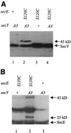
FIG. 1
FIG. 1
Disulfide bonding of PrlA3 and SecE(S120C). Alleles are shown, including the wild type (+), the prlA3 gene, which codes for an F67C point mutation (A3), and secE(S120C) (S120C). (A) Total protein from strains expressing the secE and secY alleles indicated was TCA precipitated in the presence of iodoacetamide and then electrophoresed in the oxidized state (lanes 1 to 3) or reduced with DTT (lane 4). SecY antisera were used to visualize SecY and SecY protein complexes. (B) Total protein was precipitated in the presence of iodoacetamide and then electrophoresed in the oxidized state. SecE antisera were used to visualize SecE and SecE protein complexes.
FIG. 2
FIG. 2
Toxicity of secE(S120C) expression in prlA3 strain. At 0 min, arabinose was added to secE(S120C) prlA3 (squares), secE(S120C) secY+ (diamonds), and secE+ prlA3 (circles) strains. Aliquots were removed at various times postinduction, and CFU were measured. Also shown are CFU of the secE(S120C) prlA3 strain culture in the absence of arabinose (triangles).
FIG. 3
FIG. 3
Protein translocation in prlA3 secE(S120C) cells. (A) prlA3 secE(S120C) cells were induced for secE(S120C) expression with arabinose (lanes 1 and 2) for 2 h or were left uninduced (lanes 3 and 4). Cultures were pulse labeled for 30 s with [35S]methionine and chased for 30 s (lanes 1 and 3) or 4 min (lanes 2 and 4), and then the protein was TCA precipitated. β-Lactamase was collected by immunoprecipitation and separated by SDS-PAGE. The positions of precursor (p) and mature (m) β-lactamase are shown. (B) prlA3 secE(S120C) cells were induced for secE(S120C) expression with arabinose (lanes 3 and 4) for 2 h or were left uninduced (lanes 1 and 2). Cultures were pulse labeled for 30 s with [35S]methionine and chased for 30 s (lanes 1 and 3) or 4 min (lanes 2 and 4), and then the protein was TCA precipitated. Lipoprotein was collected by immunoprecipitation and separated by SDS-PAGE. Only mature lipoprotein was present. The lipoprotein precursor could not be detected.
FIG. 4
FIG. 4
PrlA3 and SecE(S120C) cross-linking inhibits the incorporation of the radioactive methionine into polypeptide. At 0 min, arabinose was added to the strains with the following genotypes: secE(S120C) prlA3 (squares), secE(S120C) secY+ (diamonds), and secE+ prlA3 (circles). At various time points samples were removed, and the ability of cells to incorporate [35S]methionine into polypeptide was measured by pulse labeling followed by TCA precipitation and radioactivity measurement as described in Materials and Methods.
FIG. 5
FIG. 5
Molecular modeling of the regions subjected to cysteine scanning mutagenesis. The second periplasmic loop of the SecE protein and the first periplasmic loop of the SecY protein are depicted as antiparallel α-helices. The positions of the amino acids subjected to cysteine scanning are shown as numbers within circles. We have linked the amino acids at points sufficiently proximal to form disulfide bonds when replaced by cysteines.

Similar articles

Cited by

References

    1. Baba T, Taura T, Shimoike T, Akiyama Y, Yoshihisa T, Ito K. A cytoplasmic domain is important for the formation of a SecY-SecE translocator complex. Proc Natl Acad Sci USA. 1994;91:4539–4543. - PMC - PubMed
    1. Bardwell J C, Lee J O, Jander G, Martin N, Belin D, Beckwith J. A pathway for disulfide bond formation in vivo. Proc Natl Acad Sci USA. 1993;90:1038–1042. - PMC - PubMed
    1. Bardwell J C A, McGovern K, Beckwith J. Identification of a protein required for disulfide bond formation in vivo. Cell. 1991;67:581–589. - PubMed
    1. Bieker K L, Phillips G J, Silhavy T J. The sec and prl genes of Escherichia coli. J Bioenerg Biomembr. 1990;22:291–310. - PubMed
    1. Bieker K L, Silhavy T J. PrlA (SecY) and PrlG (SecE) interact directly and function sequentially during protein translocation in E. coli. Cell. 1990;61:833–842. - PubMed

Publication types

MeSH terms

LinkOut - more resources