. 1999 Jun;181(11):3452-61.
doi: 10.1128/JB.181.11.3452-3461.1999.

A functional 4-hydroxysalicylate/hydroxyquinol degradative pathway gene cluster is linked to the initial dibenzo-p-dioxin pathway genes in Sphingomonas sp. strain RW1

Affiliations

A functional 4-hydroxysalicylate/hydroxyquinol degradative pathway gene cluster is linked to the initial dibenzo-p-dioxin pathway genes in Sphingomonas sp. strain RW1

J Armengaud et al. J Bacteriol. 1999 Jun.

Abstract

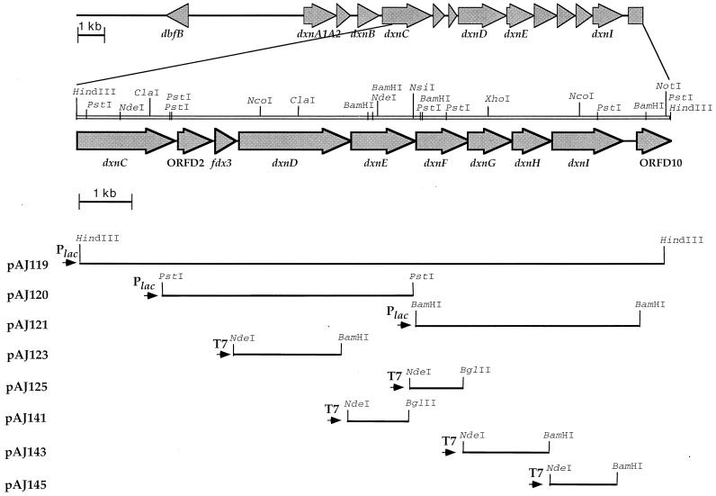
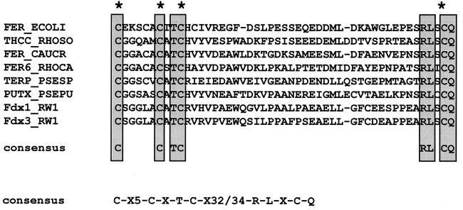
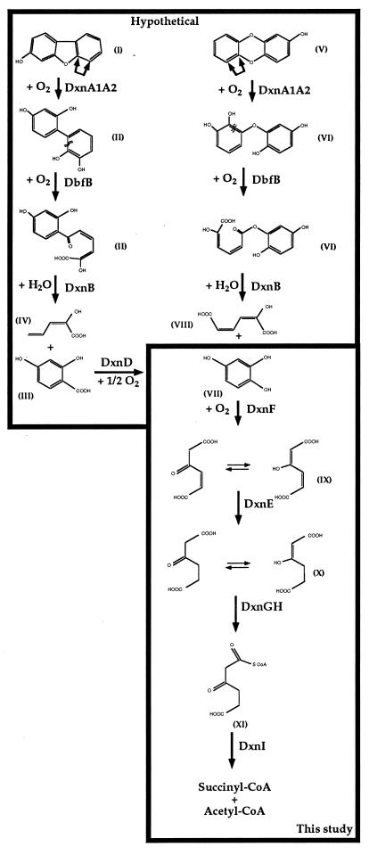
The bacterium Sphingomonas sp. strain RW1 is able to use dibenzo-p-dioxin, dibenzofuran, and several hydroxylated derivatives as sole sources of carbon and energy. We have determined and analyzed the nucleic acid sequence of a 9,997-bp HindIII fragment downstream of cistrons dxnA1A2, which encode the dioxygenase component of the initial dioxygenase system of the corresponding catabolic pathways. This fragment contains 10 colinear open reading frames (ORFs), apparently organized in one compact operon. The enzymatic activities of some proteins encoded by these genes were analyzed in the strain RW1 and, after hyperexpression, in Escherichia coli. The first three ORFs of the locus, designated dxnC, ORF2, and fdx3, specify a protein with a low homology to bacterial siderophore receptors, a polypeptide representing no significant homology to known proteins, and a putative ferredoxin, respectively. dxnD encodes a 69-kDa phenol monooxygenase-like protein with activity for the turnover of 4-hydroxysalicylate, and dxnE codes for a 37-kDa protein whose sequence and activity are similar to those of known maleylacetate reductases. The following gene, dxnF, encodes a 33-kDa intradiol dioxygenase which efficiently cleaves hydroxyquinol, yielding maleylacetate, the ketoform of 3-hydroxy-cis,cis-muconate. The heteromeric protein encoded by dxnGH is a 3-oxoadipate succinyl coenzyme A (succinyl-CoA) transferase, whereas dxnI specifies a protein exhibiting marked homology to acetyl-CoA acetyltransferases (thiolases). The last ORF of the sequenced fragment codes for a putative transposase. DxnD, DxnF, DxnE, DxnGH, and DxnI (the activities of most of them have also been detected in strain RW1) thus form a complete 4-hydroxysalicylate/hydroxyquinol degradative pathway. A route for the mineralization of the growth substrates 3-hydroxydibenzofuran and 2-hydroxydibenzo-p-dioxin in Sphingomonas sp. strain RW1 thus suggests itself.


Figures

FIG. 1
FIG. 1
Genetic organization of the 10-kb HindIII fragment of plasmid pAJ119 and subcloning strategies. Cosmid pAJ114 was previously identified in a pLAFR3 library of Sphingomonas sp. strain RW1 genomic DNA by hybridization with a probe specific for the dxnA1A2 cistrons. A 9,997-bp HindIII fragment was subcloned from this cosmid into pBluescript, yielding pAJ119. The positions and orientations of the different ORFs detected within the locus are shown by large arrows on the top of the figure. The orientation of the vector promoter in each construct designed for hyperexpression is shown by small black arrows. Relevant restriction sites present on the fragment are indicated, as well as sites artificially introduced by PCR.
FIG. 2
FIG. 2
Conserved sequences that characterize putidaredoxin-type [2Fe-2S] ferredoxins. Well-conserved fingerprint sequence regions in putidaredoxin-type ferredoxins are shown. Cysteines implicated in binding the [2Fe-2S] cluster are indicated with an asterisk. A consensus sequence for the whole family of this type of ferredoxin is also indicated. The sequences were compiled and aligned by using GeneWorks software (version 2.5N) from IntelliGenetics. The proteins are labeled by trivial abbreviations. Their accession codes in the GenBank/EMBL/DDBJ databases and their origins are as follows: P25528 for ferredoxin from E. coli (FER_ECOLI), P43493 for ferredoxin from Rhodococcus erythropolis sp. strain NI86/21 (THCC_RHOSO), P37098 for ferredoxin from Caulobacter crescentus (FER_CAUCR), P80306 for ferredoxin FdVI from Rhodobacter capsulatus (FER6_RHOCA), P33007 for ferredoxin from Pseudomonas sp. (TERP_PSESP), P00259 for ferredoxin from P. putida (PUTX_PSEPU), Y13118 for ferredoxin Fdx1 from Sphingomonas sp. RW1 (Fdx1_RW1), and X72850 for ferredoxin Fdx3 from Sphingomonas sp. RW1 (Fdx3_RW1).
FIG. 3
FIG. 3
SDS-PAGE analysis of the hyperexpression products of dxnD, dxnE, dxnF, dxnGH, and dxnI. E. coli cells were treated with lysis buffer and subjected to electrophoresis on a 10% (left gel) or 15% (right gel) glycine polyacrylamide gel, and the gels were subsequently stained with Coomassie blue. Protein standards (lysozyme 14.4 kDa; trypsin inhibitor, 21.5 kDa; carbonic anhydrase, 31 kDa; ovalbumin, 45 kDa; serum albumin, 66.2 kDa; and phosphorylase B, 97.4 kDa) (lanes M) and induced whole cells of E. coli BL21(DE3)(pLysS) containing plasmids pAJ125, pAJ123, pAJ111, pAJ141, pAJ143, and pAJ145 (lanes 2 to 6, respectively) were loaded on the gels. Construct pAJ111, designed for the hyperexpression of reductase RedA2 from RW1 (25), was used as a control. The hyperproduced polypeptides are indicated by arrows: DxnF (f), DxnD (d), RedA2 (r), DxnE (e), DxnG (g), and DxnH (h).
FIG. 4
FIG. 4
Phylogenetic tree obtained by alignment of DxnF with related dioxygenases. The sequences were compiled by using the software mentioned in the legend to Fig. 2; the multiple alignment analysis was performed with the Phylips package programs. The phylogenetic unrooted tree was drawn by using TreeView. The horizontal bar indicates the percent divergence (distance). The numbers on some of the branches refer to the confidence (percent) estimated by bootstrap analysis (100 replications). The proteins are labeled by trivial abbreviations. Their accession codes in the GenBank/EMBL/DDBJ databases and their origins are as follows: D86544 for hydroxyquinol 1,2-dioxygenase from Ralstonia pickettii strain DTP0602 (HadC-DTP0602), U19883 for hydroxyquinol 1,2-dioxygenase from B. cepacia AC1100 (TftH-AC1100), AF003948 for chlorocatechol 1,2-dioxygenase from Rhodococcus opacus 1CP (ClcA-1CP), AF044314 for chlorocatechol 1,2-dioxygenase from Variovorax paradoxus TV1 (TfdC-TV1), U32188 for the so-called 3,5-dichlorocatechol 1,2-dioxygenase from P. putida EST4011 (TfdC-EST4011), M57629 for chlorocatechol 1,2-dioxygenase II from Pseudomonas sp. strain P51 (TcbC-P51), M35097 for chlorocatechol 1,2-dioxygenase I from plasmid pJP4 of Ralstonia eutropha JMP134 (TfdC-pJP4), M36279 for chlorocatechol 1,2-dioxygenase II from plasmid pJP4 of R. eutropha JMP134 (TfdCII-pJP4), M16964 for chlorocatechol 1,2-dioxygenase from plasmid pAC27 (ClcA-pAC27), AJ006307 for chlorocatechol 1,2-dioxygenase from Ralstonia sp. strain JS705 (ClcA-JS705), D16356 for the so-called 3,5-dichlorocatechol 1,2-dioxygenase from B. cepacia CSV90 (TfdC-CSV90), AF003947 for the α-subunit of protocatechuate 3,4-dioxygenase from R. opacus 1CP (PcaG-1CP), P15110 for the β-subunit of protocatechuate 3,4-dioxygenase from B. cepacia (PcxB-cepacia), L14836 for the β-subunit of protocatechuate 3,4-dioxygenase from P. putida (PcaH-putida), L23213 for the α-subunit of protocatechuate 3,4-dioxygenase from P. putida (PcaG-putida), L05770 for the β-subunit of protocatechuate 3,4-dioxygenase from Acinetobacter sp. ADP1 (PcaH-ADP1), P20372 for the β-subunit of protocatechuate 3,4-dioxygenase from Acinetobacter calcoaceticus (PcaH-acine), X99622 for the dioxygenase-like enzyme from R. opacus (Dle-1CP), U77658 for catechol 1,2-dioxygenase I from Acinetobacter lwoffii K24 (CatA1-K24), Z36909 for catechol 1,2-dioxygenase from A. calcoaceticus NCIB8250 (ORF7-NCBI8250), U77659 for catechol 1,2-dioxygenase II from Acinetobacter lwoffii K24 (CatA2-K24), P07773 for catechol 1,2-dioxygenase from A. calcoaceticus BD413 (CatA-BD413), U12557 for catechol 1,2-dioxygenase from P. putida PRS1 (CatA-PRS1), D37783 for catechol 1,2-dioxygenase from P. putida C-1 (CatA-C1), D37782 for catechol 1,2-dioxygenase from P. putida mt2 (CatA-mt2), P31019 for catechol 1,2-dioxygenase from Pseudomonas sp. strain EST1001 (PheB-EST1001), M57500 for catechol 1,2-dioxygenase from plasmid pEST1226 of Pseudomonas sp. strain EST1001 (PheB-pEST1226), M94318 for catechol 1,2-dioxygenase from Arthrobacter sp. strain mA3 (CatA-mA3), X99622 for catechol 1,2-dioxygenase from R. opacus (CatA-1CP), D83237 for catechol 1,2-dioxygenase from R. erythropolis AN-13 (CatA-AN13), and AF043741 for catechol 1,2-dioxygenase from Rhodococcus rhodochrous NCIMB 13259 (CatA-NCIMB13259).
FIG. 5
FIG. 5
Proposed converging pathways for the degradation of 3-hydroxydibenzofuran and 2-hydroxydibenzo-p-dioxin through 4-hydroxysalicylate and hydroxyquinol in Sphingomonas sp. strain RW1. The unstable compounds (hemiacetals) formed by angular dioxygenation by DnxA1A2, which spontaneously decay, are not indicated in order to simplify the pathway. The double-arrows indicate the position of possible dioxygenolytic attack by the initial dioxin dioxygenase. Chemical designations: I, 3-hydroxydibenzofuran; II, 2,2′,3,4′-tetrahydroxybiphenyl; III, 2-hydroxy-6-oxo-6-(2,4-dihydroxyphenyl)-hexa-2,4-dienoic acid; IV, 2-hydroxypenta-2,4-dienoic acid; V, 2-hydroxydibenzo-p-dioxin; VI, 2,2′,3,5′-tetrahydroxydiphenyl ether; VII, 6-(2,5-dihydroxyphenyl)-ester of 2-hydroxy-cis,cis-muconic acid; VIII, 2-hydroxy-cis,cis-muconic acid; IX, 4-hydroxysalicylic acid; X, hydroxyquinol; XI, 3-hydroxy-cis,cis-muconic acid (maleylacetic acid); XII, 3-oxoadipic acid (and its enol); XIII, 3-oxoadipyl-CoA.

Similar articles

Cited by

References

    1. Arfmann H-A, Timmis K N, Wittich R-M. Mineralization of 4-chlorodibenzofuran by a consortium consisting of Sphingomonas sp. strain RW1 and Burkholderia sp. strain JWS. Appl Environ Microbiol. 1997;63:3458–3462. - PMC - PubMed
    1. Armengaud J, Gaillard J, Forest E, Jouanneau Y. Characterization of a 2[4Fe-4S] ferredoxin obtained by chemical insertion of the Fe-S clusters into the apoferredoxin II from Rhodobacter capsulatus. Eur J Biochem. 1995;231:396–404. - PubMed
    1. Armengaud J, Happe B, Timmis K N. Genetic analysis of dioxin dioxygenase of Sphingomonas sp. strain RW1: catabolic genes dispersed on the genome. J Bacteriol. 1998;180:3954–3966. - PMC - PubMed
    1. Armengaud J, Meyer C, Jouanneau Y. Recombinant expression of the fdxD gene of Rhodobacter capsulatus and characterization of its product, a [2Fe-2S] ferredoxin. Biochem J. 1994;300:413–418. - PMC - PubMed
    1. Armengaud J, Meyer C, Jouanneau Y. A [2Fe-2S] ferredoxin (FdVI) is essential for growth of the photosynthetic bacterium Rhodobacter capsulatus. J Bacteriol. 1997;179:3304–3309. - PMC - PubMed

Publication types

MeSH terms

Associated data

LinkOut - more resources