. 1999 May 31;145(5):1027-38.
doi: 10.1083/jcb.145.5.1027.

Cyclic expression of endothelin-converting enzyme-1 mediates the functional regulation of seminiferous tubule contraction

Affiliations

Cyclic expression of endothelin-converting enzyme-1 mediates the functional regulation of seminiferous tubule contraction

A Tripiciano et al. J Cell Biol. .

Abstract

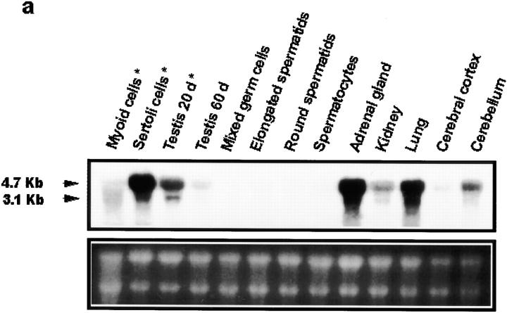
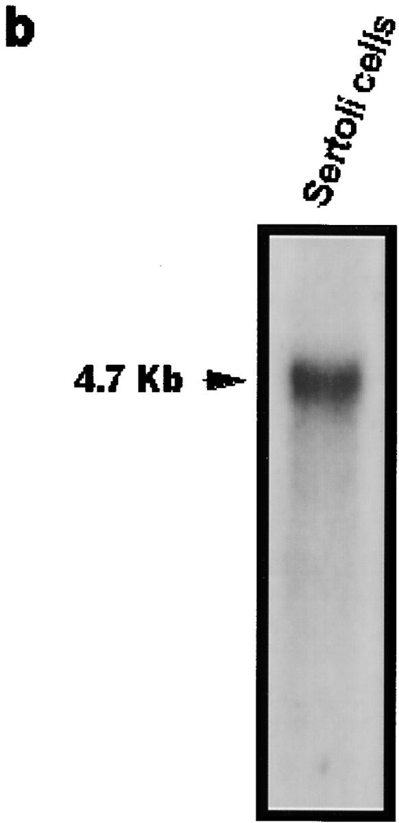
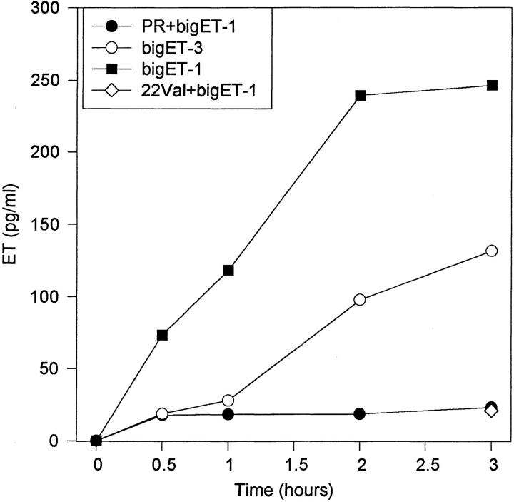
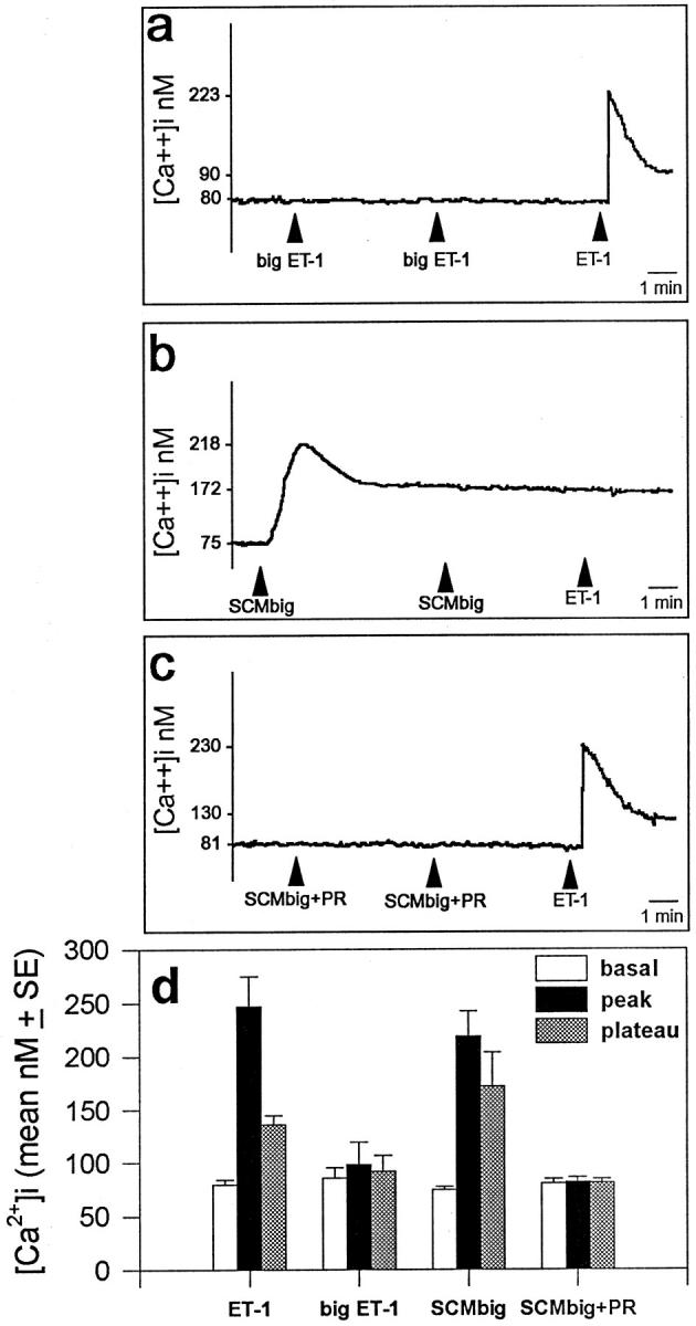
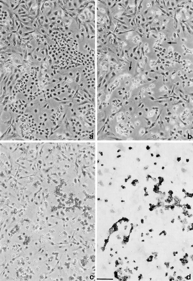
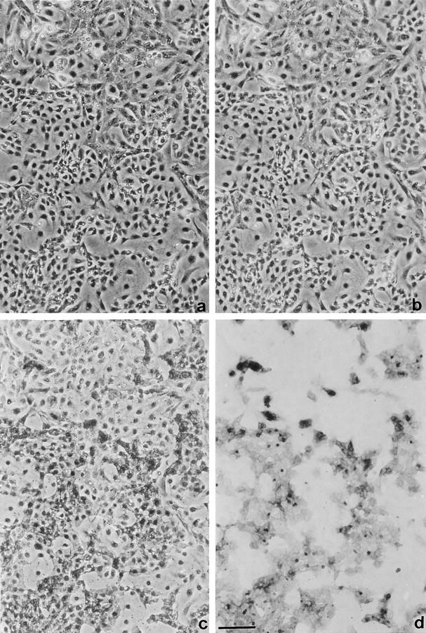
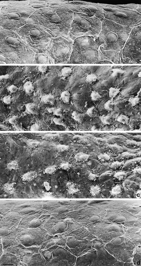
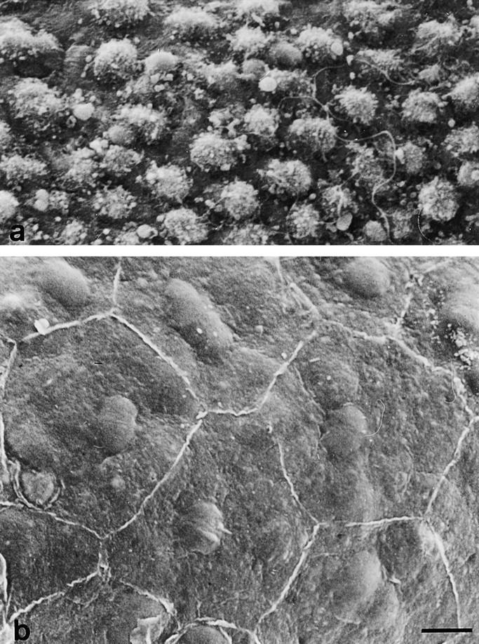
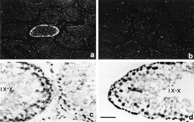
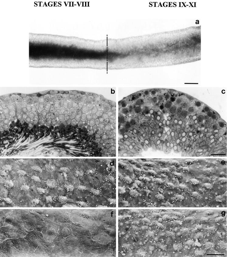
The potent smooth muscle agonist endothelin-1 (ET-1) is involved in the local control of seminiferous tubule contractility, which results in the forward propulsion of tubular fluid and spermatozoa, through its action on peritubular myoid cells. ET-1, known to be produced in the seminiferous epithelium by Sertoli cells, is derived from the inactive intermediate big endothelin-1 (big ET-1) through a specific cleavage operated by the endothelin-converting enzyme (ECE), a membrane-bound metalloprotease with ectoenzymatic activity. The data presented suggest that the timing of seminiferous tubule contractility is controlled locally by the cyclic interplay between different cell types. We have studied the expression of ECE by Sertoli cells and used myoid cell cultures and seminiferous tubule explants to monitor the biological activity of the enzymatic reaction product. Northern blot analysis showed that ECE-1 (and not ECE-2) is specifically expressed in Sertoli cells; competitive enzyme immunoassay of ET production showed that Sertoli cell monolayers are capable of cleaving big ET-1, an activity inhibited by the ECE inhibitor phosphoramidon. Microfluorimetric analysis of intracellular calcium mobilization in single cells showed that myoid cells do not respond to big endothelin, nor to Sertoli cell plain medium, but to the medium conditioned by Sertoli cells in the presence of big ET-1, resulting in cell contraction and desensitization to further ET-1 stimulation; in situ hybridization analysis shows regional differences in ECE expression, suggesting that pulsatile production of endothelin by Sertoli cells (at specific "stages" of the seminiferous epithelium) may regulate the cyclicity of tubular contraction; when viewed in a scanning electron microscope, segments of seminiferous tubules containing the specific stages characterized by high expression of ECE were observed to contract in response to big ET-1, whereas stages with low ECE expression remained virtually unaffected. These data indicate that endothelin-mediated spatiotemporal control of rhythmic tubular contractility might be operated by Sertoli cells through the cyclic expression of ECE-1, which is, in turn, dependent upon the timing of spermatogenesis.


Figures

Figure 1
Figure 1
Northern blot analysis of ECE-1 transcripts in different compartments of three-week-old (a*, b) and adult testes. The blots were prepared with 10 μg total RNA (a) or 4 μg poly(A)+ RNA (b) and hybridized to 32P-labeled ECE cDNA probe; the integrity and equal loading of RNA were ascertained by ethidium bromide staining of the gel before transfer (a, lower panel). Sertoli cells appear to represent the main site of ECE-1 expression in the male gonad.
Figure 1
Figure 1
Northern blot analysis of ECE-1 transcripts in different compartments of three-week-old (a*, b) and adult testes. The blots were prepared with 10 μg total RNA (a) or 4 μg poly(A)+ RNA (b) and hybridized to 32P-labeled ECE cDNA probe; the integrity and equal loading of RNA were ascertained by ethidium bromide staining of the gel before transfer (a, lower panel). Sertoli cells appear to represent the main site of ECE-1 expression in the male gonad.
Figure 2
Figure 2
Immunoenzymatic assay of ET-1 production by intact Sertoli cell monolayers treated with inactive endothelin precursors, as a measure of ECE activity. 0.5 μM big ET-1 is more efficiently converted than 0.5 μM big ET-3, and the conversion is inhibited by 2 mM phosphoramidon (PR) and by the synthetic inhibitory peptide [D-Val22]big ET-1[16-38] (22 val) administered, at a 1 mM concentration, 10 min before precursor addition.
Figure 3
Figure 3
Pattern of [Ca2+]i mobilization in Fura-2–loaded single myoid cells in response to: (a) 100 nM big-ET-1; (b) medium (KHH) preincubated for 30 min with a Sertoli cell monolayer in the presence of 100 nM big ET-1; (c) medium preincubated for 30 min with a Sertoli cell monolayer in the presence of 100 nM big ET-1 plus 2 mM phosphoramidon. Early myoid cell response followed by desensitization, observable in b, demonstrates that Sertoli cells convert big ET-1 into active ET-1; when indicated, 100 nM ET-1 has been added; (d) size of [Ca2+]i increases evaluated in five independent experiments.
Figure 4
Figure 4
Mixed population of tubular (mostly Sertoli cells) and peritubular myoid cells photographed before (a) and 20 min after (b) addition of 100 nM big ET-1. Myoid cells (arrows) appear specifically contracted in response to treatment. To visualize the selective contractile response of myoid cells, the same cells were photographed after cytochemical detection of alkaline phosphatase activity (c and d); (a–c) phase contrast. Bar, 100 μm.
Figure 5
Figure 5
Mixed population of tubular and peritubular cells photographed before (a) and 20 min after (b) addition of 100 nM big ET-1 plus 2 mM phosphoramidon. Owing to inhibition of ECE activity, myoid cells (arrows) fail to respond to big ET-1 (see Fig. 4 for comparison); (c and d) same cells photographed after cytochemical detection of alkaline phosphatase activity in myoid cells. a-c: phase contrast. Bar, 100 μm.
Figure 6
Figure 6
Scanning electron micrograph showing the peritubular surface of adult seminiferous tubules. (a) Control condition. Myoid cell contraction immediately after treatment with 100 nM ET-1 (b) and after 10 min treatment with 100 nM big ET-1 (c). (d) Lack of response after treatment with 100 nM big ET-1 plus 2 mM phosphoramidon. Bar, 10 μm.
Figure 7
Figure 7
Scanning electron micrograph of the peritubular surface of adult seminiferous tubules immediately after treatment with medium preconditioned for 30 min by Sertoli cells in the presence (a) or the absence (b) of 100 nM big ET-1. Bar, 10 μm.
Figure 8
Figure 8
Localization of ECE-1 transcripts in testicular sections hybridized with 32P-labeled antisense (a and c) and sense (b and d) RNA probes. Autoradiographies were exposed for 3 wk. (a and b) Dark field, (c and d) carmalum counterstain. (c) Stage IX-X (left side tubule) shows intense basal labeling, while in the adjacent tubule (at stage IV-V, right side) the labeling is not above background. Bar: (a and b) 200 μm; (c and d) 40 μm.
Figure 9
Figure 9
Different distribution of ECE activity along the seminiferous tubule. a: transilluminated tubular segment in which the transition from stage VIII (left side, dark) to stage IX (right side, light) is apparent. The hatched line indicates the level at which the tubules were dissected to yield segments with expected different ECE activity shown in d–g. (b and c) Toluidine blue–stained Epon sections showing the seminiferous epithelium composition at stage VII-VIII (b) and IX-X (c). Scanning electron micrograph of peritubular surface of seminiferous tubule segments isolated at precise stages of the seminiferous epithelium and treated as follows: (d) stage VII-VIII and (e) stage IX-XI, 10 nM ET-1 followed by immediate fixation; (f) stage VII-VIII and (g) stage IX-XI: 100 nM big ET-1, fixed after 15 min. Only stage IX-XI seminiferous epithelium shows an intrinsic ability to stimulate myoid cell contraction through conversion of big ET-1. Bars: (a) 100 μm; (b and c) 30 μm; (d–g) 25 μm.

Similar articles

Cited by

References

    1. Ackermann A. Substituted naphtol AS phosphate derivatives for the localization of leukocytes alkaline phosphatase activity. Lab Invest. 1962;11:563–566. - PubMed
    1. Arai H, Hori S, Aramori I, Ohkubo H, Nakanishi S. Cloning and expression of a cDNA encoding an endothelin receptor. Nature. 1990;348:730–732. - PubMed
    1. Bardin, C.W., C. Yan Cheng, N.A. Musto, and G.L. Gunsalus. 1988. The Sertoli cell. In The Physiology of Reproduction. E. Knobil and J. Neill, editors. Raven Press, Ltd., New York. 1:933–974.
    1. Barnes K, Brown C, Turner AJ. Endothelin-coverting enzyme. Ultrastructural localization and its recycling from the cell surface. Hypertension. 1998;31:3–9. - PubMed
    1. Boitani C, Palombi F, Stefanini M. Influence of Sertoli cell products upon the in vitro survival of isolated spermatocytes and spermatids. Cell Biol Int Rep. 1983;7:383–393. - PubMed

Publication types