. 1999 May 31;145(5):1077-88.
doi: 10.1083/jcb.145.5.1077.

R-Ras signals through specific integrin alpha cytoplasmic domains to promote migration and invasion of breast epithelial cells

Affiliations

R-Ras signals through specific integrin alpha cytoplasmic domains to promote migration and invasion of breast epithelial cells

P J Keely et al. J Cell Biol. .

Abstract

Specificity and modulation of integrin function have important consequences for cellular responses to the extracellular matrix, including differentiation and transformation. The Ras-related GTPase, R-Ras, modulates integrin affinity, but little is known of the signaling pathways and biological functions downstream of R-Ras. Here we show that stable expression of activated R-Ras or the closely related TC21 (R-Ras 2) induced integrin-mediated migration and invasion of breast epithelial cells through collagen and disrupted differentiation into tubule structures, whereas dominant negative R-Ras had opposite effects. These results imply novel roles for R-Ras and TC21 in promoting a transformed phenotype and in the basal migration and polarization of these cells. Importantly, R-Ras induced an increase in cellular adhesion and migration on collagen but not fibronectin, suggesting that R-Ras signals to specific integrins. This was further supported by experiments in which R-Ras enhanced the migration of cells expressing integrin chimeras containing the alpha2, but not the alpha5, cytoplasmic domain. In addition, a transdominant inhibition previously noted only between integrin beta cytoplasmic domains was observed for the alpha2 cytoplasmic domain; alpha2beta1-mediated migration was inhibited by the expression of excess alpha2 but not alpha5 cytoplasmic domain-containing chimeras, suggesting the existence of limiting factors that bind the integrin alpha subunit. Using pharmacological inhibitors, we found that R-Ras induced migration on collagen through a combination of phosphatidylinositol 3-kinase and protein kinase C, but not MAPK, which is distinct from the other Ras family members, Rac, Cdc42, and N- and K-Ras. Thus, R-Ras communicates with specific integrin alpha cytoplasmic domains through a unique combination of signaling pathways to promote cell migration and invasion.


Figures

Figure 1
Figure 1
Expression of activated R-Ras or TC21 (R-Ras 2) disrupts the organized polarization of breast epithelia. Cells were cultured in three-dimensional collagen gels and the formation of polarized tubule structures was assessed by phase-contrast microscopy. Cells were transfected with: (A) vector only, (B) constitutively active R-Ras(87L), (C) constitutively active TC21(72L), (D) dominant negative R-Ras(43N), or (E) dominant negative TC21(26A). Constitutively active R-Ras and TC21 disrupt tubule formation, whereas dominant negative R-Ras and TC21 enhance formation in a qualitative manner.
Figure 2
Figure 2
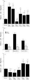
R-Ras and TC21 stimulate integrin-mediated cell migration across collagen but not fibronectin. (a) Migration of cells transfected with Ras family members across collagen I. Expression of constitutively active R-Ras(87L) and R-Ras(38V) stimulate cell migration, whereas dominant negative R-Ras(41A) inhibits migration. Expression of constitutively active isoforms of Ras family members, TC21(72L), N-Ras(12D), and K-Ras(12V) also stimulate cell migration. Haptotactic cell migration was assayed in the absence of serum across filters coated from the underside with collagen I. Values are relative to migration of cells expressing vector alone: *P < 0.05, compared with control cells; (*)P < 0.1. (b) Migration across collagen is mediated by the α2β1 integrin. Cells were preincubated with control IgG or P1E6, an anti–α2 integrin antibody, and allowed to migrate across filters coated with collagen I. P1E6 significantly inhibits migration of control cells as well as cells expressing activated R-Ras(87L) or TC21(72L). *P < 0.05 for P1E6 compared with control IgG. (c) Cell migration across fibronectin. Expression of constitutively active R-Ras(87L) or (38V) decreases migration, whereas constitutively active TC21(72L) does not. In contrast, expression of constitutively active N-Ras(12D) or K-Ras(12V) stimulates migration across fibronectin. *P < 0.05, (*)P < 0.1, compared with control cells transfected with vector alone.
Figure 3
Figure 3
R-Ras and Ras family members stimulate invasion. Cells expressing constitutively active R-Ras(87L) or (38V), TC21(72L), N-Ras(12D), or K-Ras(12V) are significantly more invasive across collagen gels in response to a serum gradient. Values are relative to cells transfected with vector alone (control). *P < 0.05 compared with control invasion.
Figure 4
Figure 4
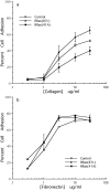
Activated R-Ras enhances adhesion to collagen but not fibronectin. (A) Cells expressing activated R-Ras(87L) (triangles) are more adherent to collagen I than control cells expressing vector alone (open squares). In contrast, cells expressing dominant negative R-Ras(41A) (filled squares) are less adherent to collagen I. (b) Cells expressing activated or dominant negative R-Ras are no different from control cells in their adhesion to fibronectin. Values in a and b are an average of two separate experiments, each of triplet determinations, ± SD.
Figure 5
Figure 5
Flow cytometry of cells expressing α4 integrin chimeras. (A–F) De novo expression of α4 integrin on the surface of control or R-Ras(87L)–expressing cells double-transfected with α4 chimeras. Chimeras consist of the extracellular domain of the α4 subunit and the cytoplasmic domain of α2 (X4C2), α4 (X4C4), or α5 (X4C5). All cell lines expressed similar levels of the X4 chimeras as determined by surface α4 integrin expression. (A) Control cells transfected with X4C2 express α4 on their surface (shaded histogram). In contrast, mock-transfected cells, like untransfected cells, do not express α4 on their surface (open histogram). (B) R-Ras(87L) cells transfected with X4C2 express similar levels of α4 as control X4C2 cells in A. (C) Control cells transfected with X4C4. (D) R-Ras(87L) cells transfected with X4C4. (E) Control cells transfected with X4C5. (F) R-Ras(87L) cells transfected with X4C5. (G and H) Cell surface expression of α2β1 integrin does not change in cells transfected with X4C2. (G) Control cells transfected with X4C2 (shaded histogram) have similar levels of α2 expression on their surface as mock-transfected cells (open histogram). (H) R-Ras(87L) cells transfected with X4C2 also have similar α2 expression levels.
Figure 6
Figure 6
R-Ras specifically enhances migration through effects on the α2 integrin cytoplasmic domain. (a) Migration of cells across ligands specific for the α4β1 integrin: the 40-kD fragment of fibronectin or the CS-1 peptide. Cell lines expressing control vector or activated R-Ras(87L) were double-transfected with α4 chimeras that consist of the extracellular domain of the α4 subunit and the cytoplasmic domain of α2 (X4C2), α4 (X4C4), or α5 (X4C5). R-Ras(87L) enhances migration only for cells expressing the X4C2 or X4C4 chimeras, but not cells expressing the X4C5 chimera. Values are normalized for each chimera set and substratum to control values. *P < 0.05 compared with control cells for that chimera. (b) Migration of cells expressing X4 chimeras across collagen I, which is not a ligand for the α4 extracellular domain. Expression of X4C2 inhibits the increase in cell migration across collagen induced by R-Ras(87L) expression. In contrast, expression of X4C4, X4C5, or X4C0 (α4 extracellular domain with no cytoplasmic domain) did not inhibit R-Ras(87L)–induced cell migration across collagen (i.e., R-Ras still induces a statistically significant increase in the migration of these cells). Values are normalized for each chimera set. *P < 0.05 compared with control values (solid bars) for that chimera. Expression of X4C5 actually increases the effect of R-Ras, but this is not statistically significant, when compared with X4C4, or X4C0-expressing cells.
Figure 6
Figure 6
R-Ras specifically enhances migration through effects on the α2 integrin cytoplasmic domain. (a) Migration of cells across ligands specific for the α4β1 integrin: the 40-kD fragment of fibronectin or the CS-1 peptide. Cell lines expressing control vector or activated R-Ras(87L) were double-transfected with α4 chimeras that consist of the extracellular domain of the α4 subunit and the cytoplasmic domain of α2 (X4C2), α4 (X4C4), or α5 (X4C5). R-Ras(87L) enhances migration only for cells expressing the X4C2 or X4C4 chimeras, but not cells expressing the X4C5 chimera. Values are normalized for each chimera set and substratum to control values. *P < 0.05 compared with control cells for that chimera. (b) Migration of cells expressing X4 chimeras across collagen I, which is not a ligand for the α4 extracellular domain. Expression of X4C2 inhibits the increase in cell migration across collagen induced by R-Ras(87L) expression. In contrast, expression of X4C4, X4C5, or X4C0 (α4 extracellular domain with no cytoplasmic domain) did not inhibit R-Ras(87L)–induced cell migration across collagen (i.e., R-Ras still induces a statistically significant increase in the migration of these cells). Values are normalized for each chimera set. *P < 0.05 compared with control values (solid bars) for that chimera. Expression of X4C5 actually increases the effect of R-Ras, but this is not statistically significant, when compared with X4C4, or X4C0-expressing cells.
Figure 7
Figure 7
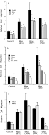
PI3K and PKC contribute to R-Ras induced cell migration. (a) Pretreatment with PI3K inhibitors Wortmannin (Wtm, 30 nM) or LY294002 (LY, 25 μM) partially inhibits the migration of cells expressing activated R-Ras(87L) or (38V), or TC21(72L) across collagen. *P < 0.05 compared with DMSO-treated samples for each cell line shown. (b) Pretreatment of cells with the PI3K inhibitor, Wortmannin (Wtm, 30 nM), or the PKC inhibitor, bisindolylmalemide (Bis, 1 μM), or both. Wortmannin and bisindolylmalemide alone each partially inhibits R-Ras– induced migration of cells across collagen. Together, Wortmannin and bisindolylmalemide have an additive effect and completely inhibit R-Ras–induced migration. *P < 0.05 compared with DMSO-treated samples for each cell line. (c) Pretreatment of cells with the MEK inhibitor, PD98059 (PD98, 25 μM) has no effect on the migration across collagen of cells expressing activated R-Ras(87L) or (38V) or TC21(72L). In contrast, PD98059 completely inhibits migration induced by activated K-Ras(12V). *P < 0.05 compared with DMSO-treated samples for each cell line.

Similar articles

Cited by

References

    1. Balzac F, Retta SF, Albini A, Melchiorri A, Koteliansky VE, Geuna M, Silengo L, Tarone G. Expression of β1B integrin isoform in CHO cells results in a dominant negative effect on cell adhesion and motility. J Cell Biol. 1994;127:557–565. - PMC - PubMed
    1. Batlle E, Verdu J, Dominguez D, del Mont M, Llosad, Diaz V, Loukili N, Paciucci R, Alameda F, de Herreros AG. Protein kinase C-alpha activity inversely modulates invasion and growth of intestinal cells. J Biol Chem. 1998;273:15091–15098. - PubMed
    1. Bazzoni G, Hemler ME. Are changes in integrin affinity and conformation overemphasized? . Trends Biochem Sci. 1998;23:30–34. - PubMed
    1. Burridge K, Chrzanowska-Wodnicka M. Focal adhesions, contractility, and signaling. Annu Rev Cell Dev Biol. 1996;12:463–519. - PubMed
    1. Chan BM, Kassner PD, Schiro JA, Byers HR, Kupper TS, Hemler ME. Distinct cellular functions mediated by different VLA integrin α subunit cytoplasmic domains. Cell. 1992;68:1051–1060. - PubMed

Publication types