. 1999 May 31;145(5):1089-102.
doi: 10.1083/jcb.145.5.1089.

Evidence for apical endocytosis in polarized hepatic cells: phosphoinositide 3-kinase inhibitors lead to the lysosomal accumulation of resident apical plasma membrane proteins

Affiliations

Evidence for apical endocytosis in polarized hepatic cells: phosphoinositide 3-kinase inhibitors lead to the lysosomal accumulation of resident apical plasma membrane proteins

P L Tuma et al. J Cell Biol. .

Abstract

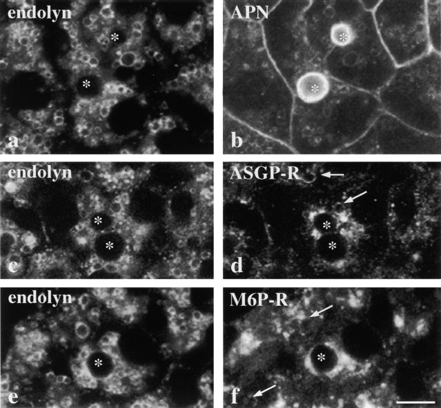
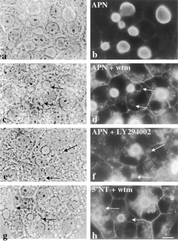
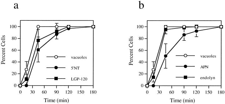
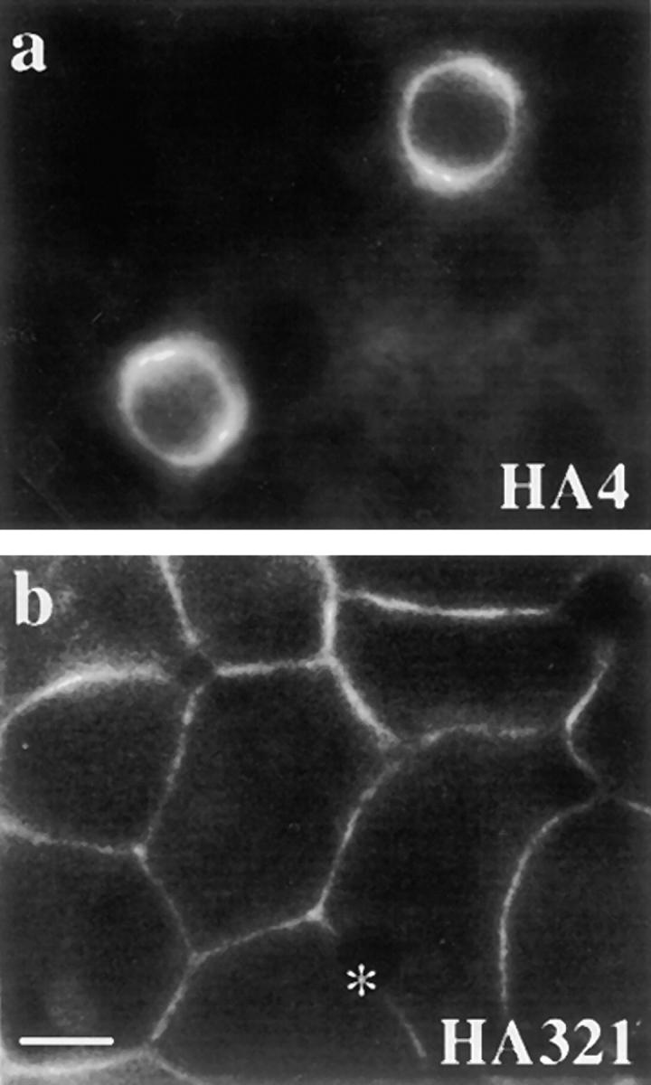
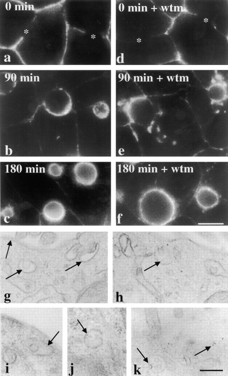
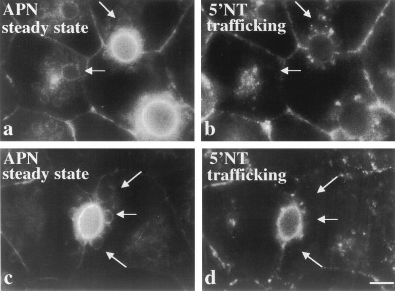
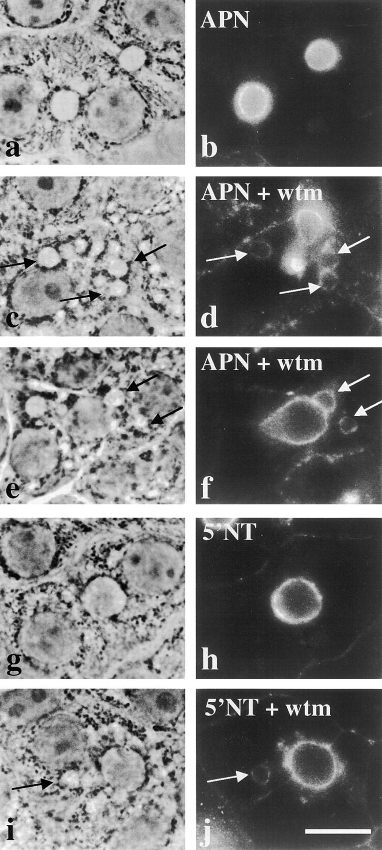
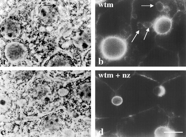
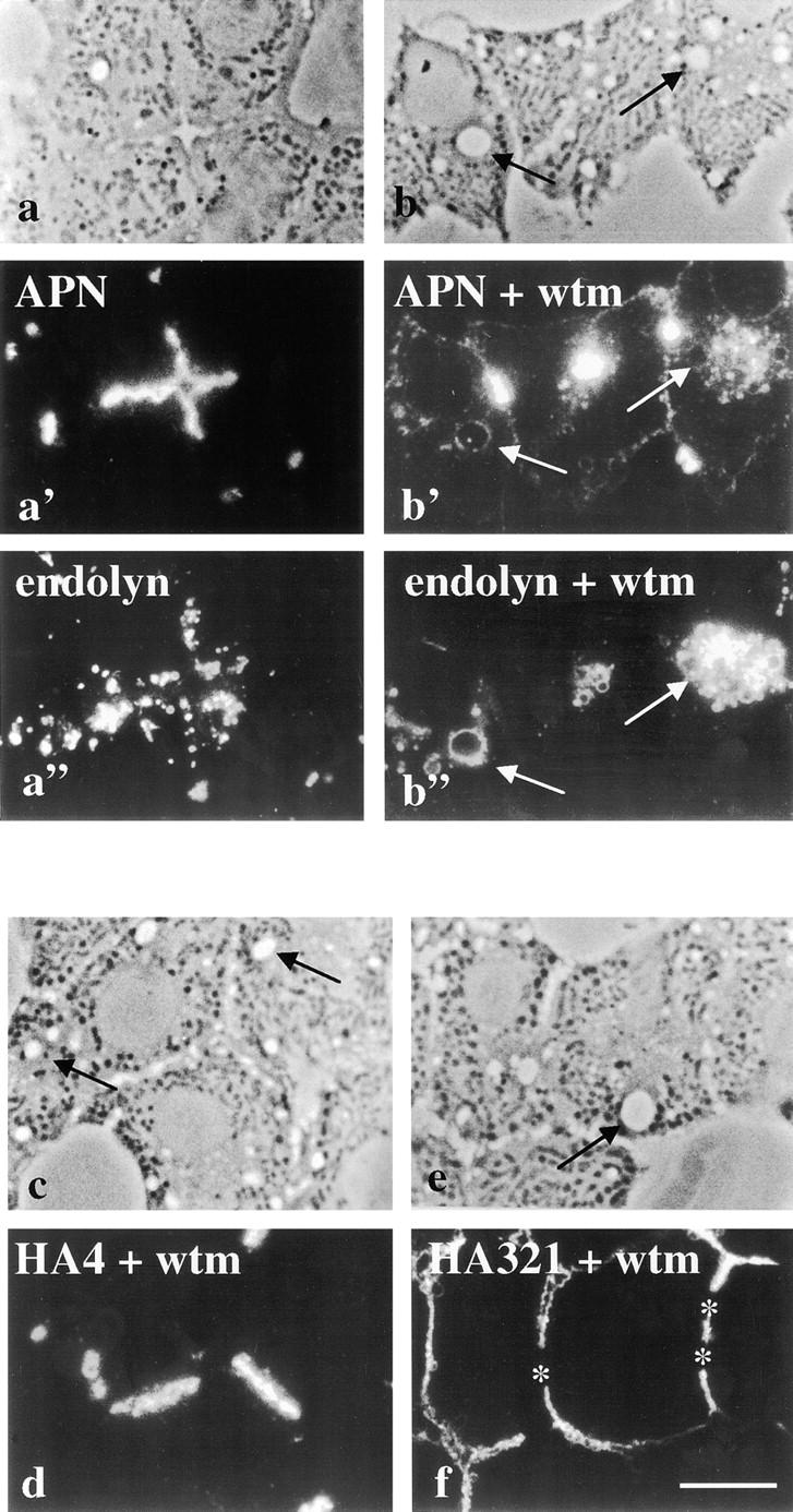
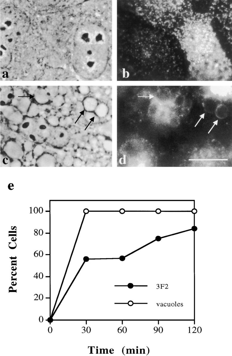
The architectural complexity of the hepatocyte canalicular surface has prevented examination of apical membrane dynamics with methods used for other epithelial cells. By adopting a pharmacological approach, we have documented for the first time the internalization of membrane proteins from the hepatic apical surface. Treatment of hepatocytes or WIF-B cells with phosphoinositide 3-kinase inhibitors, wortmannin or LY294002, led to accumulation of the apical plasma membrane proteins, 5'-nucleotidase and aminopeptidase N in lysosomal vacuoles. By monitoring the trafficking of antibody-labeled molecules, we determined that the apical proteins in vacuoles came from the apical plasma membrane. Neither newly synthesized nor transcytosing apical proteins accumulated in vacuoles. In wortmannin-treated cells, transcytosing apical proteins traversed the subapical compartment (SAC), suggesting that this intermediate in the basolateral-to-apical transcytotic pathway remained functional. Ultrastructural analysis confirmed these results. However, apically internalized proteins did not travel through SAC en route to lysosomal vacuoles, indicating that SAC is not an intermediate in the apical endocytic pathway. Basolateral membrane protein distributions did not change in treated cells, uncovering another difference in endocytosis from the two domains. Similar effects were observed in polarized MDCK cells, suggesting conserved patterns of phosphoinositide 3-kinase regulation among epithelial cells. These results confirm a long-held but unproven assumption that lysosomes are the final destination of apical membrane proteins in hepatocytes. Significantly, they also confirm our hypothesis that SAC is not an apical endosome.


Figures

Figure 4
Figure 4
The apical plasma membrane proteins accumulate in vacuoles that also contain resident lysosomal membrane proteins. Cells were treated for 180 min with 100 nM wortmannin, fixed, permeabilized, and processed for indirect immunofluorescence. Confocal images of cells double labeled for endolyn (a) and APN (b), endolyn (c) and ASGP-R (d), or endolyn (e) and M6P-R (f) are shown. *Bile canaliculi. Arrows are pointing to vacuoles that contain ASGP-R (d) or M6P-R (f). The APN image was overexposed to enhance vacuolar visibility. Bar, 10 μm.
Figure 1
Figure 1
Apical plasma membrane proteins accumulate in vacuoles in the presence of wortmannin (wtm) or LY294002. WIF-B cells were incubated for 180 min in the absence (a and b) or presence (c, d, g, and h) of 100 nM wortmannin or 200 μM LY294002 (e and f). Cells were fixed, permeabilized and stained for APN (b, d, and f) or 5′NT (h). Arrows point to vacuoles containing intracellular apical proteins. Fluorescent images were intentionally overexposed so that vacuolar staining was more visible. The control staining in b was overexposed to the same extent as treated cells. 5′NT staining in control cells is indistinguishable from that of APN shown in a. Bar, 10 μm.
Figure 2
Figure 2
Vacuole formation precedes appearance of apical membrane proteins in vacuoles. WIF-B cells were treated for the indicated times with 100 nM wortmannin, fixed, permeabilized, and double labeled for APN and endolyn (a) or 5′NT and LGP-120 (b). Random phase and fluorescent images were collected for each time point. From phase images, the number of cells containing vacuoles was counted. Vacuoles were defined as the large, spherical intracellular structures induced by wortmannin treatment. From fluorescent images, cells containing vacuoles that stained positive for resident apical membrane (APN or 5′NT) or resident lysosomal membrane proteins (endolyn or LGP-120) were counted. The plotted values are averages from three experiments ± SD.
Figure 3
Figure 3
Plasma membrane polarity is maintained in the presence of wortmannin. WIF-B cells were treated for 180 min with 100 nM wortmannin. The cells were fixed, permeabilized, and processed for indirect immunofluorescence. HA4 staining (a) was detected at the apical membrane with no intracellular vacuolar staining and HA321 staining (b) detected only at the basolateral. Phase images of these cells were indistinguishable from those shown in Fig. 1, d, f, and h. *Bile canalicular space in b. Overexposure of images as in Fig. 1 revealed no vacuolar staining. Bar, 10 μm.
Figure 5
Figure 5
Transcytosing apical plasma membrane proteins bypass vacuoles. (a–f) Cells were pretreated for 15 min in the absence or presence of 100 nM wortmannin (wtm) at 37°C. The cells were chilled to 4°C and labeled with anti–5′NT antibodies for 15 min. After extensive washing, the cells were either fixed and permeabilized directly (0 min of chase; a and d) or placed at 37°C and the antibodies chased for 90 (b and e) or 180 (c and f) min in the continued absence or presence of 100 nM wortmannin. After chase, the cells were fixed, permeabilized, and the trafficked antibodies detected with cy3-conjugated secondary antibodies. The control experiment is shown in a–c and wortmannin-treated cells are shown in d–f. Asterisks in a and d point to unlabeled bile canalicular domains at 0 min of chase in both control and treated cells. Bar, 10 μm. (g–k) Cells were pretreated and labeled as described above, except anti–5′NT antibodies were directly conjugated to 5-nm gold particles. The antibodies were chased for 90 min in the continued absence (g and h) or presence (i–k) of 100 nM wortmannin and processed for electron microscopic visualization (see Methods). Arrows are pointing to gold particles located at the apical cell surface or in the SAC in both control and treated cells. Bar, 250 nm.
Figure 6
Figure 6
Longer pretreatment of cells with wortmannin does not lead to the vacuolar accumulation of transcytosing apical plasma membrane proteins. WIF-B cells were pretreated with 100 nM wortmannin for 120 min at 37°C, subsequently chilled to 4°C, and labeled with anti–5′NT antibodies. After extensive washing the cells were returned to 37°C and the antibodies chased for 60 (a and b) or 120 (c and d) min. Anti–5′NT antibodies were detected with cy3-conjugated secondary antibodies (b and d). The corresponding steady state APN distributions were determined in the same cells (a and c). Arrows point to vacuoles that contain APN in a and c and the corresponding areas in b and d. Note the lack of colocalization between the steady state APN labeling and the trafficking 5′NT molecules. Bar, 10 μm.
Figure 7
Figure 7
The apical proteins accumulated in vacuoles originate from the apical cell surface. WIF-B cells were chilled to 4°C and labeled with anti–APN (a–f) or anti–5′NT antibodies (g–h). After extensive washing, the cells were returned to 37°C and the antibodies allowed to chase for 5 h (in the case of APN antibodies) or 3 h (for 5′NT antibodies). Cells were then incubated in the absence (a, b, g, and h) or presence (c, d, i, and j) of 100 nM wortmannin (wtm) for an additional 2 h. Cells were fixed, permeabilized, and labeled with secondary antibodies. The corresponding phase images are shown in the left-hand column. In b and h, no vacuolar staining is observed in untreated cells. Arrows are pointing to vacuoles that stained positive for apical plasma membrane proteins after the additional incubation with wortmannin in c–f and i–j. Bar, 10 μm.
Figure 8
Figure 8
Vacuolar formation and apical redistribution are microtubule-dependent processes. WIF-B cells were treated for 60 min in the absence (a and b) or presence (c and d) of 33 μM nocodazole (nz) to depolymerize microtubules. Cells were then incubated with 100 nM wortmannin (wtm) in the continued absence (a and b) or presence (c and d) of nocodazole. Cells were fixed, permeabilized, and processed for indirect immunofluorescent detection of APN steady state distributions. The corresponding phase images are shown. Arrows in a and b point to vacuoles that are positive for APN staining. In c and d, vacuole formation and redistribution of APN are inhibited by nocodazole treatment. Bar, 10 μm.
Figure 9
Figure 9
Apical plasma membrane proteins redistribute to lysosomally derived vacuoles in intact liver hepatocytes. Intact livers were perfused in the absence or presence of 2 μM wortmannin (wtm) and 50 μM leupeptin for 180 min at 37°C. Biopsies were removed and fixed by immersion. Semi-thin sections (0.5 μm) were cut and processed for indirect immunofluorescence. Control perfused liver sections (a–a”) and wortmannin-perfused sections (b–b”) are shown. APN staining is shown (a' and b') with corresponding double-labeled images for endolyn (a” and b”). Phase images for both conditions are shown at the top of each column. HA4 (c) and HA321 (f) staining patterns are shown as single labels with corresponding phase images. Arrows in a–a” and b–b” point to vacuoles that are visible by phase and are positive for both APN and endolyn staining. Arrows in c and e point to vacuoles with no detectable HA4 or HA321 staining. The image showing APN staining in treated hepatocytes (b') was overexposed to enhance vacuolar staining visualization. Bar, 10 μm.
Figure 10
Figure 10
Apical plasma membrane proteins in MDCK cells accumulate in vacuoles in the presence of wortmannin. (a–d) MDCK cells were treated in the absence (a and b) or presence (c and d) of 2 μM wortmannin and 50 μM leupeptin for 120 min at 37°C. Cells were fixed, permeabilized, and processed for indirect immunofluorescent detection of the apical plasma membrane protein, 3F2 (b and d). The corresponding phase images are shown (a and c). The nuclear focal plane is shown in a and the cell surface focal plane is shown in b. Arrows point to vacuoles that contain 3F2 in wortmannin-treated cells. Bar, 10 μm. (e) Vacuole formation and appearance of positive 3F2 vacuole staining was measured as described in Fig. 2. The average of two experiments is plotted for each time point.

Similar articles

Cited by

References

    1. Apodaca G, Katz LA, Mostov KE. Receptor-mediated transcytosis of IgA in MDCK cells is via apical recycling endosomes. J Cell Biol. 1994;125:67–86. - PMC - PubMed
    1. Barr VA, Hubbard AL. Newly synthesized hepatocyte plasma membrane proteins are transported in transcytotic vesicles in the bile duct-ligated rat. Gastroenterology. 1993;105:554–571. - PubMed
    1. Barr VA, Scott LJ, Hubbard AL. Immunoadsorption of hepatic vesicles carrying newly synthesized dipeptidyl peptidase IV and polymeric IgA receptor. J Biol Chem. 1995;270:27834–27844. - PubMed
    1. Barroso M, Sztul E. Basolateral to apical transcytosis in polarized cells is indirect and involves BFA and trimeric G-sensitive passage through the apical endosome. J Cell Biol. 1994;124:83–100. - PMC - PubMed
    1. Bartles JR, Braiterman LT, Hubbard AL. Biochemical characterization of domain-specific glycoproteins of the rat hepatocyte plasma membrane. J Biol Chem. 1985;260:12792–12802. - PubMed

Publication types