Review
. 1999 Jun;63(2):293-307.
doi: 10.1128/MMBR.63.2.293-307.1999.

Structural features of the glutamate transporter family

Affiliations
Review

Structural features of the glutamate transporter family

D J Slotboom et al. Microbiol Mol Biol Rev. 1999 Jun.

Abstract

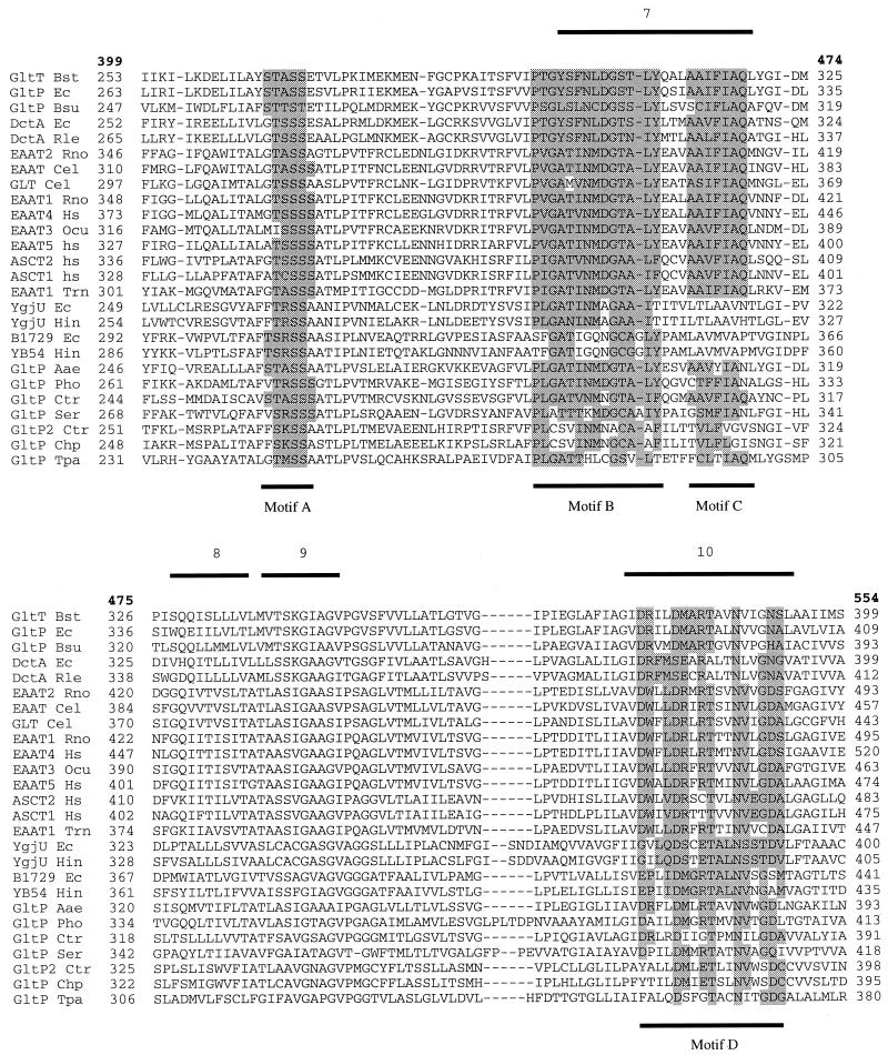
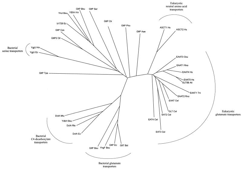
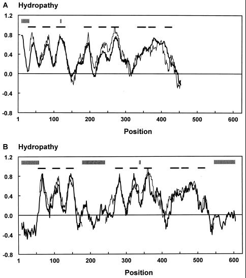
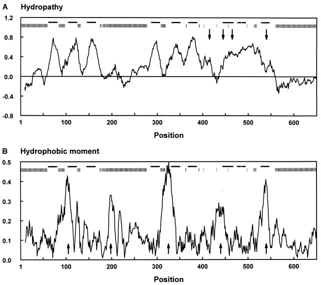
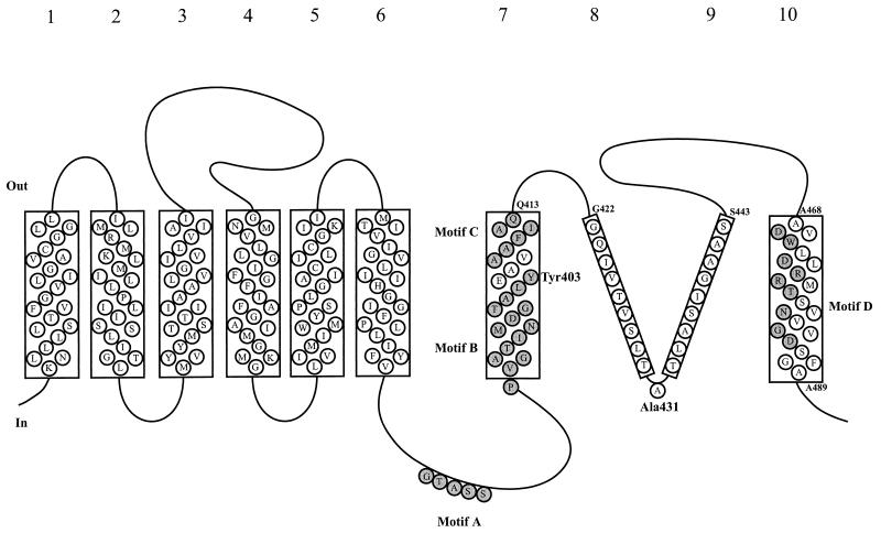
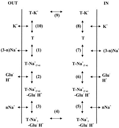
Neuronal and glial glutamate transporters remove the excitatory neurotransmitter glutamate from the synaptic cleft and thus prevent neurotoxicity. The proteins belong to a large and widespread family of secondary transporters, including bacterial glutamate, serine, and C4-dicarboxylate transporters; mammalian neutral-amino-acid transporters; and an increasing number of bacterial, archaeal, and eukaryotic proteins that have not yet been functionally characterized. Sixty members of the glutamate transporter family were found in the databases on the basis of sequence homology. The amino acid sequences of the carriers have diverged enormously. Homology between the members of the family is most apparent in a stretch of approximately 150 residues in the C-terminal part of the proteins. This region contains four reasonably well-conserved sequence motifs, all of which have been suggested to be part of the translocation pore or substrate binding site. Phylogenetic analysis of the C-terminal stretch revealed the presence of five subfamilies with characterized members: (i) the eukaryotic glutamate transporters, (ii) the bacterial glutamate transporters, (iii) the eukaryotic neutral-amino-acid transporters, (iv) the bacterial C4-dicarboxylate transporters, and (v) the bacterial serine transporters. A number of other subfamilies that do not contain characterized members have been defined. In contrast to their amino acid sequences, the hydropathy profiles of the members of the family are extremely well conserved. Analysis of the hydropathy profiles has suggested that the glutamate transporters have a global structure that is unique among secondary transporters. Experimentally, the unique structure of the transporters was recently confirmed by membrane topology studies. Although there is still controversy about part of the topology, the most likely model predicts the presence of eight membrane-spanning alpha-helices and a loop-pore structure which is unique among secondary transporters but may resemble loop-pores found in ion channels. A second distinctive structural feature is the presence of a highly amphipathic membrane-spanning helix that provides a hydrophilic path through the membrane. Recent data from analysis of site-directed mutants and studies on the mechanism and pharmacology of the transporters are discussed in relation to the structural model.


Figures

FIG. 1
FIG. 1
Multiple-sequence alignment of a stretch of approximately 150 residues near the C terminus of the transporters. The alignment was made with the program CLUSTALX (84). A representative set of 26 members of the glutamate transporter family is shown. Bold numbers refer to the positions in the multiple-sequence alignment and correspond to the numbers in Fig. 4. Other numbers refer to the residue numbers of the individual sequences. Bars below and above the sequences indicate the positions of the conserved motifs (motifs A to D, highlighted) and the positions of the transmembrane segments (as published by Grunewald et al. [32]), respectively. Abbreviated transporter names are taken from Table 2.
FIG. 2
FIG. 2
Phylogenetic tree of 35 members of the glutamate transporter family. The set of 35 members does not contain pairs of sequences with more than 70% identical residues. The tree is based on the part of the multiple-sequence alignment shown in Fig. 1. It was drawn with the DRAWTREE program from the PHYLIP package (27). Abbreviated transporter names are taken from Table 2.
FIG. 3
FIG. 3
Alignment of the average hydropathy profiles of the subfamily of bacterial glutamate transporters (thin line) and the subfamily consisting of b1729 of E. coli, YB54 of H. influenzae, GltP of B. burgdorferi, and YhcL of B. subtilis (thick line) (A) and the subfamilies of bacterial and eukaryotic glutamate transporters (thin and thick lines, respectively) (B). The profiles were aligned as specified by Lolkema and Slotboom (53) with a window of 19 amino acids. Vertical and horizontal bars above the profiles indicate the positions of gaps in the sequences and the positions of the transmembrane segments (32), respectively. The profiles are almost superimposable even though the sequences have considerably diverged. The bacterial glutamate transporters and the subfamily containing YB54 of H. influenzae have 18 to 24% identical residues (A), whereas the subfamilies of bacterial and eukaryotic glutamate transporters have 22 to 29% identical residues (B). The subfamily containing YB54 of H. influenzae has an extra hydrophobic segment at the N terminus.
FIG. 4
FIG. 4
Average profiles of hydropathy (A) and hydrophobic moment (B) of the glutamate transporter family. The set of 35 members (Fig. 2) which does not contain pairs of sequences with more than 70% identical residues was used. Vertical and horizontal bars above the profiles indicate the positions of gaps in the sequences and the positions of the transmembrane segments (32), respectively. Position numbers refer to positions in the multiple-sequence alignment and correspond to the bold numbers in Fig. 1. In panel A, a window of 19 residues was used and the arrows point to the positions of the conserved motifs A to D. In panel B, a window of 21 residues and a period of 3.6 residues, appropriate for α-helical structures, was used and the arrows point to the five conserved putative amphipatic helices (AH1 to AH5 from left to right).
FIG. 5
FIG. 5
Model for the membrane topology of the C-terminal part of the glutamate transporters comprising membrane-spanning segments 7 to 10 (based on the model of Grunewald et al. [32]). The sequence of rat EAAT2 (GLT-1) is shown. Membrane segments 7 and 10 are likely to be helices and were also proposed in the model of Slotboom et al. (77). Membrane segments 8 and 9 form a reentrant loop or loop-pore structure according to Grunewald et al. (32). Conserved motifs B and C and the conserved hydrophilic face of helix 10 (motif D) are shaded. Tyrosine 403, which is involved in potassium binding and is accessible from either side of the membrane depending on the presence of substrates, is located in the middle of helix 7.
FIG. 6
FIG. 6
Schematic representation of the transport cycle of the eukaryotic glutamate transporters (44). T, transporter; glu, glutamate; n, the number of sodium ions that bind after glutamate binding. A description is given in the text.
FIG. 7
FIG. 7
Structures of some transported substrates (A, B, D, E, and F) and competitive inhibitors (G and H) of the glutamate transporters. 4-Methylglutamate (C) is a substrate of some transporters but a competitive inhibitor of others. (A) Glutamate; (B) β-hydroxyaspartate; (C) 4-methylglutamate; (D) serine-O-sulfate; (E) cysteate; (F) pyrrodiline-2,4-dicarboxylate; (G) kainate; (H) β-benzyloxyaspartate.
FIG. 8
FIG. 8
Model for hydrogen bridge formation between a residue in the substrate binding site of the glutamate transporters (histidine is used as an example) and the substrates glutamate (A) and cysteine (B). Hydrogen bridge formation can explain the observed substrate-dependent proton stoichiometry (112) (see the text).

References

    1. Altschul S F, Madden T L, Schaffer A A, Zhang J, Zhang Z, Miller W, Lipman D J. Gapped BLAST and PSI-BLAST: a new generation of protein database search programs. Nucleic Acids Res. 1997;25:3389–3402. - PMC - PubMed
    1. Amato A, Barbour B, Szatkowski M, Attwell D. Counter-transport of potassium by the glutamate uptake carrier in glial cells isolated from the tiger salamander retina. J Physiol (London) 1994;479:371–380. - PMC - PubMed
    1. Arriza J L, Eliasof S, Kavanaugh M P, Amara S G. Excitatory amino acid transporter 5, a retinal glutamate transporter coupled to a chloride conductance. Proc Natl Acad Sci USA. 1997;94:4155–4160. - PMC - PubMed
    1. Arriza J L, Fairman W A, Wadiche J I, Murdoch G H, Kavanaugh M P, Amara S G. Functional comparisons of three glutamate transporter subtypes cloned from human motor cortex. J Neurosci. 1994;14:5559–5569. - PMC - PubMed
    1. Arriza J L, Kavanaugh M P, Fairman W A, Wu Y N, Murdoch G H, North R A, Amara S G. Cloning and expression of a human neutral amino acid transporter with structural similarity to the glutamate transporter gene family. J Biol Chem. 1993;268:15329–15332. - PubMed

Publication types

Substances

LinkOut - more resources