Induction of MHC class I expression on immature thymocytes in HIV-1-infected SCID-hu Thy/Liv mice: evidence of indirect mechanisms
- PMID: 10358212
- PMCID: PMC4435947
Induction of MHC class I expression on immature thymocytes in HIV-1-infected SCID-hu Thy/Liv mice: evidence of indirect mechanisms
Abstract
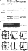
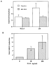
The SCID-hu Thy/Liv mouse and human fetal thymic organ culture (HF-TOC) models have been used to explore the pathophysiologic mechanisms of HIV-1 infection in the thymus. We report here that HIV-1 infection of the SCID-hu Thy/Liv mouse leads to the induction of MHC class I (MHCI) expression on CD4+CD8+ (DP) thymocytes, which normally express low levels of MHCI. Induction of MHCI on DP thymocytes in HIV-1-infected Thy/Liv organs precedes their depletion and correlates with the pathogenic activity of the HIV-1 isolates. Both MHCI protein and mRNA are induced in thymocytes from HIV-1-infected Thy/Liv organs, indicating induction of MHCI gene expression. Indirect mechanisms are involved, because only a fraction (<10%) of the DP thymocytes were directly infected by HIV-1, although the majority of DP thymocytes are induced to express high levels of MHCI. We further demonstrate that IL-10 is induced in HIV-1-infected thymus organs. Similar HIV-1-mediated induction of MHCI expression was observed in HF-TOC assays. Exogenous IL-10 in HF-TOC induces MHCI expression on DP thymocytes. Therefore, HIV-1 infection of the thymus organ leads to induction of MHCI expression on immature thymocytes via indirect mechanisms involving IL-10. Overexpression of MHCI on DP thymocytes can interfere with thymocyte maturation and may contribute to HIV-1-induced thymocyte depletion.
Figures


Similar articles
-
Activation of the signal transducer and activator of transcription 1 signaling pathway in thymocytes from HIV-1-infected human thymus.AIDS. 2003 Jun 13;17(9):1269-77. doi: 10.1097/00002030-200306130-00001. AIDS. 2003. PMID: 12799548 Free PMC article.
-
Divergent effects of chronic HIV-1 infection on human thymocyte maturation in SCID-hu mice.J Immunol. 1995 Jan 15;154(2):907-21. J Immunol. 1995. PMID: 7814892
-
Effects of IL-7 on early human thymocyte progenitor cells in vitro and in SCID-hu Thy/Liv mice.J Immunol. 2003 Jul 15;171(2):645-54. doi: 10.4049/jimmunol.171.2.645. J Immunol. 2003. PMID: 12847229
-
HIV-1 replication and pathogenesis in the human thymus.Curr HIV Res. 2003 Jul;1(3):275-85. doi: 10.2174/1570162033485258. Curr HIV Res. 2003. PMID: 15046252 Free PMC article. Review.
-
SCID-hu mice: a model for studying disseminated HIV infection.Semin Immunol. 1996 Aug;8(4):223-31. doi: 10.1006/smim.1996.0028. Semin Immunol. 1996. PMID: 8883145 Review.
Cited by
-
Type I interferon contributes to CD4+ T cell depletion induced by infection with HIV-1 in the human thymus.J Virol. 2011 Sep;85(17):9243-6. doi: 10.1128/JVI.00457-11. Epub 2011 Jun 22. J Virol. 2011. PMID: 21697497 Free PMC article.
-
Validation of the SCID-hu Thy/Liv mouse model with four classes of licensed antiretrovirals.PLoS One. 2007 Aug 1;2(7):e655. doi: 10.1371/journal.pone.0000655. PLoS One. 2007. PMID: 17668043 Free PMC article.
-
Human immunodeficiency virus type 1 IIIB selected for replication in vivo exhibits increased envelope glycoproteins in virions without alteration in coreceptor usage: separation of in vivo replication from macrophage tropism.J Virol. 2001 Sep;75(18):8498-506. doi: 10.1128/jvi.75.18.8498-8506.2001. J Virol. 2001. PMID: 11507195 Free PMC article.
-
Effect of latent human immunodeficiency virus infection on cell surface phenotype.J Virol. 2002 Feb;76(4):1673-81. doi: 10.1128/jvi.76.4.1673-1681.2002. J Virol. 2002. PMID: 11799162 Free PMC article.
-
Separation of human immunodeficiency virus type 1 replication from nef-mediated pathogenesis in the human thymus.J Virol. 2001 Apr;75(8):3916-24. doi: 10.1128/JVI.75.8.3916-3924.2001. J Virol. 2001. PMID: 11264380 Free PMC article.
References
-
- Courgnaud V, Laure F, Brossard A, Bignozzi C, Goudeau A, Barin F, Brechot C. Frequent and early in utero HIV-1 infection. AIDS Res Hum Retroviruses. 1991;7:337. - PubMed
-
- Harris PJ, Candeloro PD, Bunn JE. HIV infection of the adult thymus: an even more conventional theory explaining CD cell decrease and CD8 cell increase in AIDS. Med Hypotheses. 1991;36:379. - PubMed
-
- Tremblay M, Numazaki K, Goldman H, Wainberg MA. Infection of human thymic lymphocytes by HIV-1. J Acquired Immune Defic Syndr. 1990;3:356. - PubMed
-
- Joshi V, Oleske JM. Pathologic appraisal of the thymus gland in acquired immunodeficiency syndrome in children. Arch Pathol Lab Med. 1985;109: 142. - PubMed
-
- Rosenzweig M, Clark DP, Gaulton GN. Selective thymocyte depletion in neonatal HIV-1 thymic infection. AIDS. 1993;7:1601. - PubMed
Publication types
MeSH terms
Substances
Grants and funding
LinkOut - more resources
Full Text Sources
Other Literature Sources
Research Materials
Miscellaneous
