. 1999 Jun;103(11):1561-70.
doi: 10.1172/JCI5754.

The IL-1 receptor and Rho directly associate to drive cell activation in inflammation

Affiliations

The IL-1 receptor and Rho directly associate to drive cell activation in inflammation

R Singh et al. J Clin Invest. 1999 Jun.

Abstract

IL-1-stimulated mesenchymal cells model molecular mechanisms of inflammation. Binding of IL-1 to the type I IL-1 receptor (IL-1R) clusters a multi-subunit signaling complex at focal adhesion complexes. Since Rho family GTPases coordinately organize actin cytoskeleton and signaling to regulate cell phenotype, we hypothesized that the IL-1R signaling complex contained these G proteins. IL-1 stimulated actin stress fiber formation in serum-starved HeLa cells in a Rho-dependent manner and rapidly activated nucleotide exchange on RhoA. Glutathione S-transferase (GST) fusion proteins, containing either the full-length IL-1R cytosolic domain (GST-IL-1Rcd) or the terminal 68 amino acids of IL-1R required for IL-1-dependent signal transduction, specifically coprecipitated both RhoA and Rac-1, but not p21(ras), from Triton-soluble HeLa cell extracts. In whole cells, a small-molecular-weight G protein coimmunoprecipitated by anti-IL-1R antibody was a substrate for C3 transferase, which specifically ADP-ribosylates Rho GTPases. Constitutively activated RhoA, loaded with [gamma-32P]GTP, directly interacted with GST-IL-1Rcd in a filter-binding assay. The IL-1Rcd-RhoA interaction was functionally important, since a dominant inhibitory mutant of RhoA prevented IL-1Rcd-directed transcriptional activation of the IL-6 gene. Consistent with our previous data demonstrating that IL-1R-associated myelin basic protein (MBP) kinases are necessary for IL-1-directed gene expression, cellular incorporation of C3 transferase inhibited IL-1R-associated MBP kinase activity both in solution and in gel kinase assays. In summary, IL-1 activated RhoA, which was physically associated with IL-1Rcd and necessary for activation of cytosolic nuclear signaling pathways. These findings suggest that IL-1-stimulated, Rho-dependent cytoskeletal reorganization may cluster signaling molecules in specific architectures that are necessary for persistent cell activation in chronic inflammatory disease.


Figures

Figure 1
Figure 1
GST-IL-1Rcd fusion proteins. A series of GST-IL-1R fusion proteins were purified from E. coli as described in Methods. (a) The full-length, 569–amino acid human IL-1R is shown for reference. The mature single-transmembrane protein is 552 amino acids. The extracellular domain (open region, ED), including a 17–amino acid signal peptide, spans the amino acids from 1 to 336; the transmembrane region (filled region, TM) from 337 to 357; and the cytosolic domain (hatched region, CD) from 358 to 569. (b) GST and GST-IL-1R fusion proteins are shown. GST-IL-1Rcd contains amino acids 369–569 of IL-1Rcd, fused to GST. The ClaI site in the IL-1Rcd cDNA sequence, used to remove the sequences encoding the COOH-terminal amino acid residues required for downstream signaling, is indicated. The resulting fusion protein, GST-IL-1RcdΔ, contains membrane-proximal cytosolic domain region amino acids 369–501 fused to GST. GST-IL-1Rkb contains the COOH-terminus signaling domain (amino acids 501–569), and its construction is described in Methods.
Figure 2
Figure 2
RhoA is necessary for IL-1–mediated actin stress fiber formation. Control-scraped HeLa cells and cells scrape-loaded in the presence of C3 transferase were plated on coverslips and allowed to recover for 12 hours, as described in Methods. Serum-starved cells were stimulated with or without IL-1α for 10 minutes, and actin stress fibers were viewed by rhodamine-conjugated phalloidin. (a) Few actin stress fibers were seen in vehicle-stimulated HeLa cells. (b) Cells stimulated with IL-1 organize actin stress fibers. (c) In contrast, IL-1 failed to stimulate stress fiber formation in HeLa cells previously scrape-loaded with C3 transferase. Each photomicrograph is representative of a blinded evaluation of photographs taken from 10 fields for each condition from 4 separate experiments.
Figure 3
Figure 3
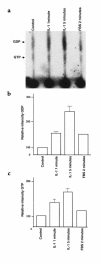
IL-1 activates RhoA nucleotide exchange. HeLa cells were rendered quiescent for 18 hours and then labeled with [32P]orthophosphate (700 μCi/mL) for 4 hours. Cells were stimulated with vehicle, with IL-1α for 1 minute or 5 minutes, or with 10% FBS for 2 minutes. The cells were lysed and RhoA was immunoprecipitated. The [32P]GTP and [32P]GDP associated with RhoA were eluted, separated by TLC, and viewed by autoradiography (a) as described in Methods. Positions of unlabeled GTP and GDP standards, viewed under 254-nm ultraviolet light, are indicated at left. After autoradiography, changes in GDP (b) and GTP (c) bound to immunoprecipitated RhoA were quantified by scanning densitometry. The results are representative of 3 independent experiments.
Figure 4
Figure 4
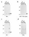
GST-IL-1R fusion proteins precipitate RhoA and Rac-1 GTPases from HeLa cell extracts. Proteins coprecipitated by GST-IL-1R fusion proteins from HeLa cell lysates were analyzed by immunoblotting for Rho GTPases, as described in Methods. Proteins were resolved by SDS-PAGE. Molecular mass markers (in kDa) are indicated at left, and the position of GST-IL-1R–associated Rho GTPase is indicated by an arrow at the bottom right of each panel. GST-IL-1Rcd fusion proteins specifically associate with Rho family GTPases RhoA (a) and Rac-1 (c), but not H-Ras (d). RhoA associated with GST-IL-1Rcd fusion proteins is blocked by preincubation of the RhoA antibody with the RhoA peptide immunogen (b). Results are representative of 4 independent experiments.
Figure 5
Figure 5
A substrate for C3 transferase coprecipitates with native IL-1R. Quiescent HeLa cells were stimulated with IL-1α (20 ng/mL) for 10 minutes, and HeLa membranes were prepared as described in Methods. Membrane-associated Rho proteins in HeLa membranes were ADP-ribosylated in the presence (+) or absence (–) of C3 transferase and [32P]NAD. Ribosylated membrane extracts were used directly (Total lysate), or were subsequently analyzed by immunoprecipitation using an anti–IL-1R antibody (IL-1RAb) or specific small GTPase antibodies directed against H-Ras (H-Ras-Ab), Rac-1 (Rac-1-Ab), or RhoA (RhoA-Ab). Immunoprecipitated proteins were separated by SDS-PAGE, and [32P]NAD-ribosylated RhoA GTPases were viewed by autoradiography. The [32P]NAD-ribosylated Rho proteins are indicated by the arrow at right, and molecular mass markers (in kilodaltons) are shown at left. These autoradiographs are representative of 3 independent experiments.
Figure 6
Figure 6
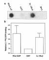
GST-IL-1Rcd interacts directly with GTP-loaded V14RhoA in a filter-binding assay. GST, GST fusion proteins (GST-RhoGAP and GST-IL-1Rcd), and BSA were spotted on nitrocellulose and probed with [γ-32P]GTP–loaded V14RhoA as described in Methods. Interacting proteins are viewed by autoradiography. The results shown are representative of 7 independent experiments. (a) [γ-32P]GTP-V14RhoA specifically binds RhoGAP (positive control) and GST-IL-1Rcd, but not the negative controls GST and BSA. (b) [γ-32P]GTP-RhoV14 bound to GST, GST-RhoGAP, and GST-IL-1Rcd was quantified by scintillation counting, and cpm bound to GST fusion proteins are normalized to those bound to GST in each of 7 independent experiments. Binding of [γ-32P]GTP-RhoV14 to BSA was not observed.
Figure 7
Figure 7
RhoA is required for assembly and activation of MBP kinases by IL-1Rcd. (a) Triton X-100 extracts were prepared from serum-deprived HeLa cells that had been treated with or without C3 transferase, and were incubated overnight with GST-IL-1Rcd fusion proteins. Fusion protein complexes were then assayed in solution for MBP kinase activity as described in Methods. Molecular mass markers (in kDa) are indicated at left, and the migration of GST and the GST-IL-1R fusion proteins, confirmed by Coomassie blue staining, is shown on the right. The autoradiogram is representative of 2 independent experiments. (b) HeLa cells were scrape-loaded in the presence (+) or absence (–) of C3 transferase. Extracts were prepared, and residual RhoA proteins available for ADP-ribosylation were determined using an in vitro reaction in the presence of C3 transferase, as described in Methods. Proteins were separated by SDS-PAGE, and [32P]NAD-labeled RhoA was viewed by autoradiography. Molecular mass markers are indicated at left, and migration of [32P]NAD-labeled RhoA proteins is indicated by the arrow on the right. (c) HeLa cell extracts were prepared from IL-1–stimulated cells that had been scrape-loaded in the absence (–) or presence (+) of C3 exoenzyme. MBP kinases associating with IL-1R immune complexes were identified by an in-gel kinase assay as described in Methods. Proteins were separated by SDS-PAGE and viewed by autoradiography. Molecular weight markers are indicated at left, and positions of the kinases p100, p83, and p63 are indicated on the right. IL-1R immunocomplexes coprecipitated 63-, 83-, and 100-kDa MBP kinases, as we have reported previously (8). In contrast, no MBP kinase activity coprecipitated with IL-1R immunocomplexes from HeLa cells scrape-loaded with C3 transferase. The autoradiogram is representative of 2 independent experiments.

References

    1. Dinarello CA. Biologic basis for interleukin-1 in disease. Blood. 1996;87:2095–2147. - PubMed
    1. Heguy A, Baldari CT, Macchia G, Telford JL, Melli M. Amino acids conserved in interleukin-1 receptors (IL-1Rs) and the Drosophila toll protein are essential for IL-1R signal transduction. J Biol Chem. 1992;267:2605–2609. - PubMed
    1. Kuno K, Okamoto S, Hirose K, Murakami S, Matsushima K. Structure and function of the intracellular portion of the mouse interleukin 1 receptor (type I). Determining the essential region for transducing signals to activate the interleukin 8 gene. J Biol Chem. 1993;268:13510–13518. - PubMed
    1. Sims JE, et al. Interleukin 1 signaling occurs exclusively via the type I receptor. Proc Natl Acad Sci USA. 1993;90:6155–6159. - PMC - PubMed
    1. Greenfeder SA, et al. Molecular cloning and characterization of a second subunit of the interleukin 1 receptor complex. J Biol Chem. 1995;270:13757–13765. - PubMed

Publication types

MeSH terms