. 1999 Jun;103(11):1587-95.
doi: 10.1172/JCI5343.

Exocrine pancreatic disorders in transsgenic mice expressing human keratin 8

Affiliations

Exocrine pancreatic disorders in transsgenic mice expressing human keratin 8

M L Casanova et al. J Clin Invest. 1999 Jun.

Abstract

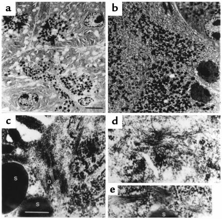
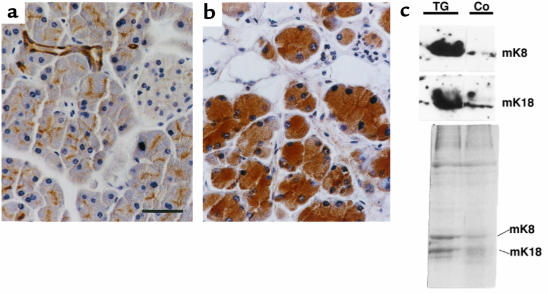
Keratins K8 and K18 are the major components of the intermediate-filament cytoskeleton of simple epithelia. Increased levels of these keratins have been correlated with various tumor cell characteristics, including progression to malignancy, invasive behavior, and drug sensitivity, although a role for K8/K18 in tumorigenesis has not yet been demonstrated. To examine the function of these keratins, we generated mice expressing the human K8 (hk8) gene, which leads to a moderate keratin-content increase in their simple epithelia. These mice displayed progressive exocrine pancreas alterations, including dysplasia and loss of acinar architecture, redifferentiation of acinar to ductal cells, inflammation, fibrosis, and substitution of exocrine by adipose tissue, as well as increased cell proliferation and apoptosis. Histological changes were not observed in other simple epithelia, such as the liver. Electron microscopy showed that transgenic acinar cells have keratins organized in abundant filament bundles dispersed throughout the cytoplasm, in contrast to control acinar cells, which have scarce and apically concentrated filaments. The phenotype found was very similar to that reported for transgenic mice expressing a dominant-negative mutant TGF-beta type II receptor (TGFbetaRII mice). We show that these TGFbetaRII mutant mice also have elevated K8/K18 levels. These results indicate that simple epithelial keratins play a relevant role in the regulation of exocrine pancreas homeostasis and support the idea that disruption of mechanisms that normally regulate keratin expression in vivo could be related to inflammatory and neoplastic pancreatic disorders.


Figures

Figure 1
Figure 1
Phenotype of transgenic mice carrying HK8 constructs. (a) DNA constructs injected. Arrows indicate the position of the transcriptional start site; exons are denoted by filled boxes. RI represents EcoRI restriction site and is included for orientation. (b) Dwarfism of transgenic HK8 mice. Eight-week-old transgenic animal (right) and nontransgenic littermate (left). (c) Reduction of pancreas size. Pancreata from 8-week-old control (left) and transgenic mice (TGK8-4; right) were photographed. The transgenic pancreas is about 30% the size of that in nontransgenic littermates. D, duodenum; S, stomach; P, pancreas. Scale bar: 3 mm.
Figure 2
Figure 2
Pancreatic histopathology of HK8 transgenic mice. Pancreas sections from 2-month-old (ac) and 1-year-old (d) animals stained with hematoxylin/eosin. (a) Control pancreas; (b) TGK8-8; (c and d) TGK8-4. (a and b) Loss of normal exocrine pancreas architecture and dysplastic changes in transgenic acinar cells. Arrowheads denote apoptotic figures; the arrow depicts diffuse mononuclear inflammatory infiltrate cells; asterisks show intralobular sclerosis; open arrows denote dysplastic cells with enlarged cellular and nuclear size. (c and d) Pancreata from TGK8-4 mice. (c) Extensive metaplasia of acinar cells, giving rise to aberrant ductules (arrowheads). A diffuse mononuclear inflammatory infiltrate (arrows), interlobular sclerosis (asterisk), and apoptotic cells (open triangles) were also frequent. (d) Replacement of acinar tissue by adipose cells, presence of a perivascular mononuclear infiltrate (arrow), and eosinophilic foci of acinar cells (oval structures) observed in aging mice. L, islets of Langerhans; B, blood vessels. Scale bar: (a and b) 100 μm; (c) 125 μm; (d) 200 μm.
Figure 3
Figure 3
Analysis of HK8 protein and mRNA in transgenic mouse pancreas. (a) Western blot analysis of total protein extracts (10 μg) from pancreata of 6-week-old nontransgenic mice (Co), nonphenotypic transgenic mice (line TGK8-6), and phenotypic transgenic (line TGK8-4) mice. Triplicate gels were immunoblotted using anti-HK8 mAb M20 (top); rat mAb TROMA-1, which, under these conditions, recognizes mouse K8 strongly (double asterisk) and human K8 weakly (single asterisk) (middle); or the anti-K18 polyclonal antibody 1589 (bottom). (b) Northern blot analysis using 15 μg of total RNA from pancreata of 6-week-old animals bearing different hk8 transgene copy numbers. The filters were sequentially hybridized for HK8 (top), mK18 (middle), and 7S (bottom). (c) Coomassie blue–stained gel. Equal amounts of cytoskeletal extracts from the same amount (in grams) of pancreata and livers from 6-week-old control and transgenic mice were loaded. (d and e) Immunodetection of K8 in pancreas sections. Paraffin-embedded sections from 2-month-old control (d) and transgenic (e) pancreata. Control pancreas was stained with the TROMA-1 mAb to view endogenous mK8. The signal is restricted to the apical region of acinar cells and to ductal cells. Transgenic pancreas sections were labeled with the HK8-specific CAM 5.2 antibody. The staining is stronger and appears throughout the cytoplasm of acinar cells. The inset shows an area of ductules reacting with antibody CAM 5.2; the arrow depicts an acinus undergoing dedifferentiation into a tubular complex. L, islet of Langerhans. Scale bar: 60 μm.
Figure 4
Figure 4
Increased apoptosis in the transgenic pancreas. (a and b) Toluidine blue staining of semithin sections (1 μm). (c and d) Confocal microscopy of PI-stained nuclei. (e and f) DNA content determination by flow cytometry. Integrated and peak DNA signals were used for aggregated discrimination. To avoid cellular debris, events were gated out 1 log below the 2-N DNA peak. (a, c, and e) Control pancreas; (b, d, and f) transgenic pancreas. Arrowheads and asterisks denote apoptotic and normal nuclei, respectively. Scale bar: (a and b) 10 μm; (c and d) 30μm.
Figure 5
Figure 5
Ultrastructural analysis of pancreatic acinar transgenic cells. (a and b) Low-magnification electron microscopy survey of control (a) and TGK8-4 (b) acinar cells from 2-month-old littermates. Arrowheads mark the lumen in the acini of control cells. Note the accumulation and loss of the apical localization of zymogen granules in transgenic cells. Apoptotic transgenic cells with condensed chromatin, indented nuclei, and swollen endoplasmic reticulum are also frequent. (c) Electron micrograph showing the apical region of 2 cells around the acinar lumen in the control pancreas. IFs, frequently anchored to desmosomes, are scarce and are restricted to this region in these cells. (d and e) Bundles of IFs are abundant and found dispersed throughout the cytoplasm in transgenic acinar cells. S, secretory granules; D, desmosome. Scale bar: (a and b) 5 μm; (ce) 250 nm.
Figure 6
Figure 6
Analysis of K8 and K18 protein expression in pancreata from transgenic TGFβRII mice. (a and b) Immunodetection of K8. Paraffin-embedded sections from control (a) and transgenic (b) pancreas sections from 6-month-old littermates were stained with TROMA-1 antibody. Scale bar: 50 μm. (c) Western blot analysis of K8 and K18 in TGFβRII mice (TG) and control nontransgenic mice (Co) (top). Total protein extracts (10 μg) from pancreata of 4-month-old control and transgenic mice (which presented a moderate phenotype) were blotted with antibodies TROMA-1 (recognizing K8) and 1589 (recognizing K18). Note the increased level of these keratins in TGFβRII mice, also detected in Coomassie blue–stained gels of cytoskeletal fractions (bottom).

Similar articles

Cited by

References

    1. Moll R, Franke WW, Schiller D. The catalog of human cytokeratins: patterns of expression in normal epithelia, tumors and cultured cells[review] Cell. 1982;31:11–24. - PubMed
    1. Fuchs E, Weber K. Intermediate filaments: structure, dynamics, function and disease. Annu Rev Biochem. 1994;63:345–382. - PubMed
    1. McLean WH, Lane EB. Intermediate filaments in disease[review] Curr Opin Cell Biol. 1995;7:118–125. - PubMed
    1. Fuchs E. The cytoskeleton and disease: genetic disorders of IFs. Annu Rev Genet. 1996;30:197–231. - PubMed
    1. Brûlet P, Babinet C, Kemler R, Jacob F. Monoclonal antibodies against trophectoderm-specific markers during mouse blastocyst formation. Proc Natl Acad Sci USA. 1980;77:4113–4117. - PMC - PubMed

Publication types

MeSH terms

Substances