. 1999 Jun 7;189(11):1707-14.
doi: 10.1084/jem.189.11.1707.

Identification of Grb2 as a novel binding partner of tumor necrosis factor (TNF) receptor I

Affiliations

Identification of Grb2 as a novel binding partner of tumor necrosis factor (TNF) receptor I

E Hildt et al. J Exp Med. .

Abstract

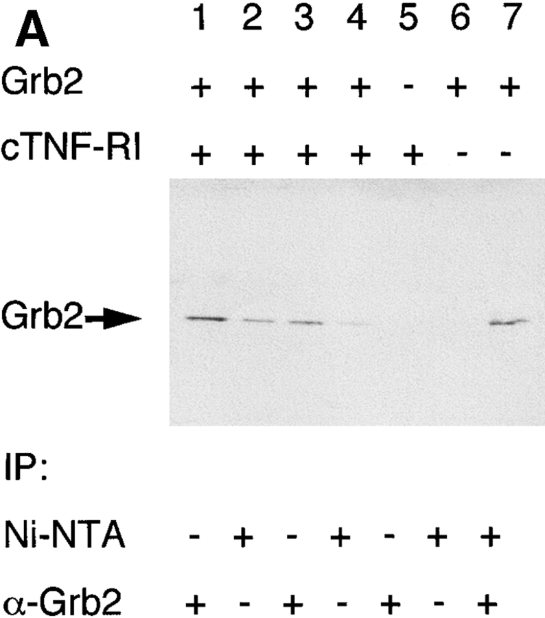
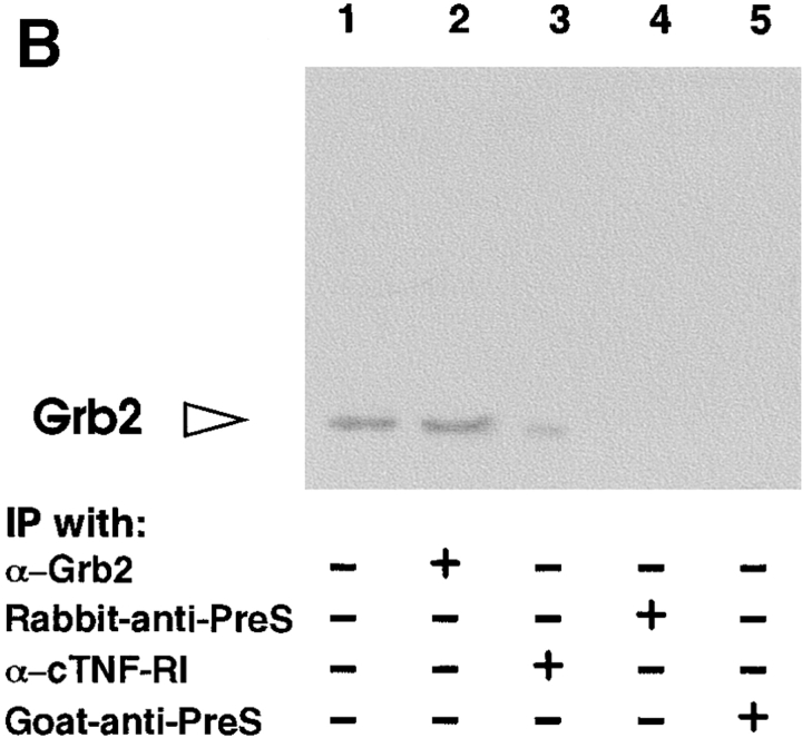
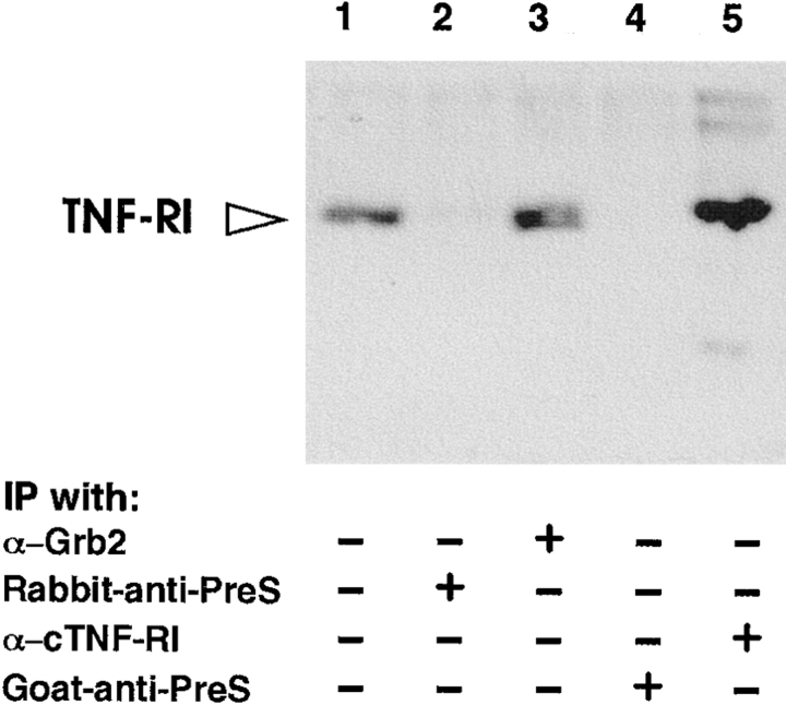
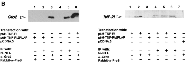
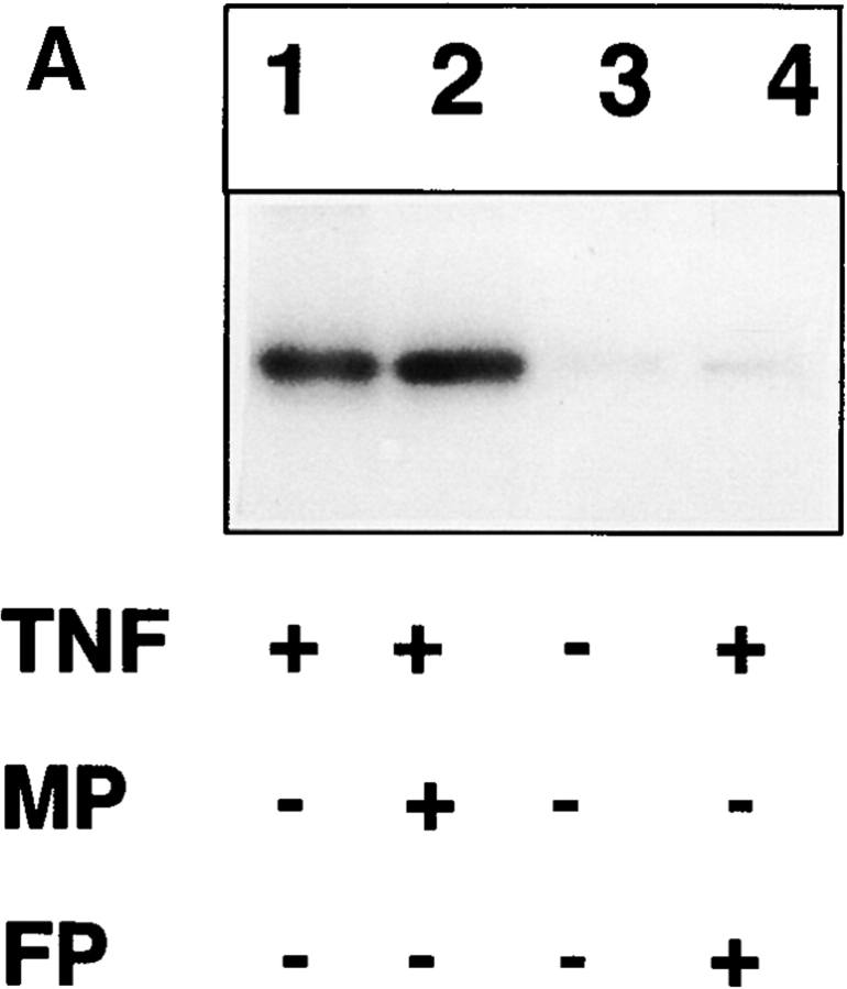
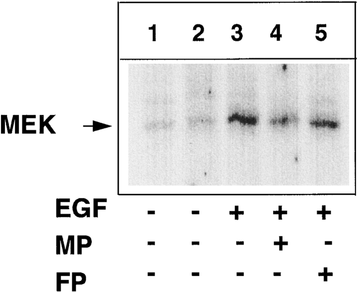
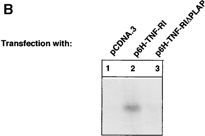
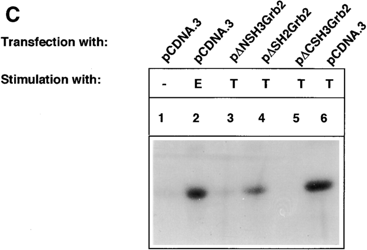
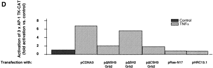
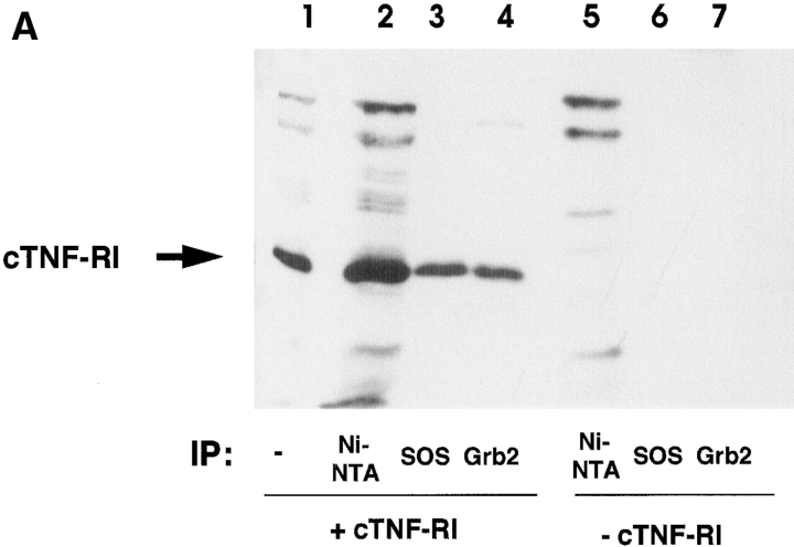
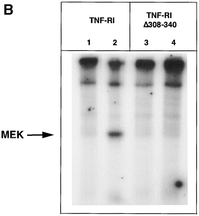
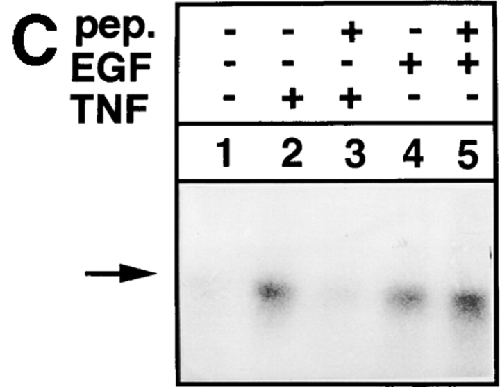
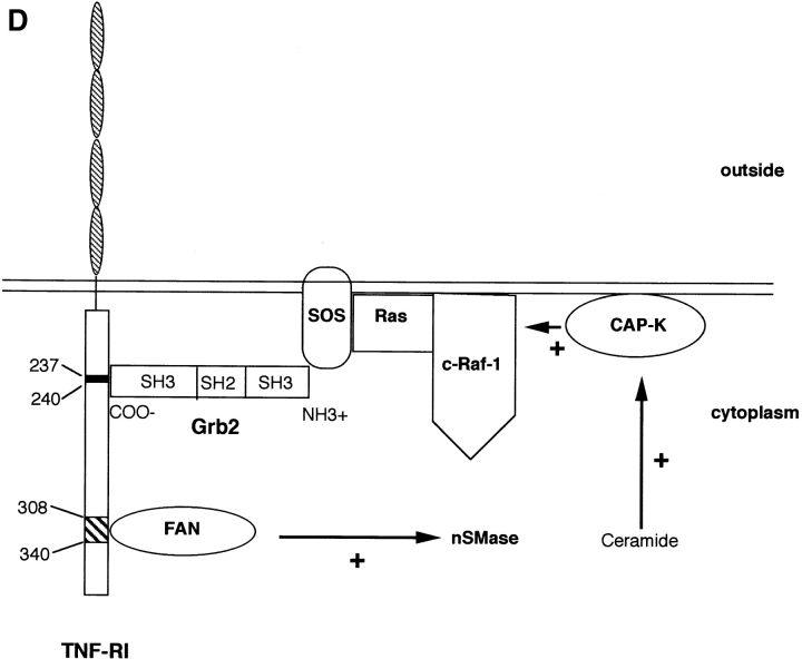
Tumor necrosis factor alpha (TNF-alpha) is a proinflammatory cytokine. Its pleiotropic biological properties are signaled through two distinct cell surface receptors: the TNF receptor type I (TNFR-I) and the TNF receptor type II. Neither of the two receptors possesses tyrosine kinase activity. A large majority of TNF-alpha-dependent activities can be mediated by TNFR-I. Recently, c-Raf-1 kinase was identified as an intracellular target of a signal transduction cascade initiated by binding of TNF-alpha to TNFR-I. However, the mechanism engaged in TNF-alpha-dependent activation of c-Raf-1 kinase is still enigmatic. Here we report that the cytosolic adapter protein Grb2 is a novel binding partner of TNFR-I. Grb2 binds with its COOH-terminal SH3 domain to a PLAP motif within TNFR-I and with its NH2-terminal SH3 domain to SOS (son of sevenless). A PLAP deletion mutant of TNFR-I fails to bind Grb2. The TNFR-I/Grb2 interaction is essential for the TNF-alpha-dependent activation of c-Raf-1 kinase; activation of c-Raf-1 kinase by TNF-alpha can be blocked by coexpression of Grb2 mutants harboring inactivating point mutations in the NH2- or COOH-terminal SH3 domain, cell-permeable peptides that disrupt the Grb2/TNFR-I interaction or transdominant negative Ras. Functionality of the TNFR-I/Grb2/SOS/Ras interaction is a prerequisite but not sufficient for TNF-alpha-dependent activation of c-Raf-1 kinase. Inhibition of the TNFR-I/FAN (factor associated with neutral sphingomyelinase) interaction, which is essential for TNF-alpha-dependent activation of the neutral sphingomyelinase, either by cell-permeable peptides or by deletion of the FAN binding domain, prevents activation of c-Raf-1 kinase. In conclusion, binding of the Grb2 adapter protein via its COOH-terminal SH3 domain to the nontyrosine kinase receptor TNFR-I results in activation of a signaling cascade known so far to be initiated, in the case of the tyrosine kinase receptors, by binding of the SH2 domain of Grb2 to phosphotyrosine.


Figures

Figure 1
Figure 1
Grb2 and TNFR-I are direct binding partners. (A) Purified hexa-His–tagged cytoplasmic domain of TNFR-I (cTNFR-I; aa 205–426) (lanes 1–3 and 5, 1 μg; lane 4, 0.1 μg) was added to purified Grb2 (lanes 1–4, 6, and 7, 0.8 μg) in a volume of 300 μl. The mixtures were precipitated by addition of Ni-NTA–agarose (lanes 2, 4, 6, and 7) (specific for hexa-His–tagged c-TNFR-I), by a polyclonal sera specific for Grb2. Analysis of the precipitates for the presence of Grb2 was performed by Western blot using a monoclonal Grb2-specific antiserum. In lane 7, one-fifth of the precipitate was loaded to avoid overloading. Lanes 5 and 6 served as negative controls. (B) Coimmunoprecipitation of Grb2 and TNFR-I from the lysate of 106 CCL13 cells. The lysates were precipitated by polyclonal rabbit-derived sera against Grb2 or goat-derived serum against TNFR-I and analyzed for the presence of Grb2 (left) or TNFR-I (right) by Western blot, using a mouse mAb specific for Grb2 or a polyclonal rabbit-derived serum specific for TNFR-I. Precipitations with an unrelated rabbit serum (rabbit anti-PreS2; left, lane 4; right, lane 2) or an unrelated goat serum (goat anti-PreS2; left, lane 5; right, lane 4) served as negative control. Total cellular lysates (left and right, lane 1) were used as positive controls.
Figure 1
Figure 1
Grb2 and TNFR-I are direct binding partners. (A) Purified hexa-His–tagged cytoplasmic domain of TNFR-I (cTNFR-I; aa 205–426) (lanes 1–3 and 5, 1 μg; lane 4, 0.1 μg) was added to purified Grb2 (lanes 1–4, 6, and 7, 0.8 μg) in a volume of 300 μl. The mixtures were precipitated by addition of Ni-NTA–agarose (lanes 2, 4, 6, and 7) (specific for hexa-His–tagged c-TNFR-I), by a polyclonal sera specific for Grb2. Analysis of the precipitates for the presence of Grb2 was performed by Western blot using a monoclonal Grb2-specific antiserum. In lane 7, one-fifth of the precipitate was loaded to avoid overloading. Lanes 5 and 6 served as negative controls. (B) Coimmunoprecipitation of Grb2 and TNFR-I from the lysate of 106 CCL13 cells. The lysates were precipitated by polyclonal rabbit-derived sera against Grb2 or goat-derived serum against TNFR-I and analyzed for the presence of Grb2 (left) or TNFR-I (right) by Western blot, using a mouse mAb specific for Grb2 or a polyclonal rabbit-derived serum specific for TNFR-I. Precipitations with an unrelated rabbit serum (rabbit anti-PreS2; left, lane 4; right, lane 2) or an unrelated goat serum (goat anti-PreS2; left, lane 5; right, lane 4) served as negative control. Total cellular lysates (left and right, lane 1) were used as positive controls.
Figure 2
Figure 2
A PLAP motif in the cytoplasmic domain of TNFR-I mediates the interaction with Grb2. (A) Purified hexa-His–tagged cytTNFR-I (1 μg) was added to cellular lysate (500 μg) derived from TNF-α–stimulated 293 cells. Coprecipitation of Grb2 by addition of Ni-NTA–agarose, which precipitates the 6H-cytTNFR-I, was inhibited by increasing concentrations of a peptide (functional peptide [FP], lanes 4–6) harboring the recognition sequence TTKPLAP, whereas a mutated peptide (MP) (TTKKLAP, lanes 7–9) did not significantly affect the interaction of Grb2 and TNFR-I. An unrelated peptide served as additional control (CP) (DRQIKIWFQNRRMKWKK; lanes 1–3). The peptides were used in concentrations of  0.1 (lanes 2, 5, and 8) and 4 μg/ml (lanes 3, 6, and 9). The precipitates were analyzed by Western blot using a Grb2-specific antiserum. (B) Left: after transient transfection of 293 cells with p6H-TNFR-I (encoding for a fusion protein of an NH2-terminal hexa-His tag and full length TNFR-I) (lanes 5 and 6), with p6H-TNFR-IΔ-PLAP (encoding for the hexa-His–tagged PLAP deletion mutant (lanes 1–3), or with the vector pCDNA.3 (lane 4), cellular lysates were precipitated by addition of Ni-NTA–agarose (lanes 2, 4, and 5) by a Grb2-specific polyclonal serum (lanes 3 and 6) or by an unrelated rabbit-derived serum (anti-PreS2; lane 1). The subsequent Western blot analysis was performed using a Grb2-specific, mouse-derived mAb. Right: lysates derived from p6H-TNFR-I– (lanes 1, 2, 4, and 5), p6H-TNFR-IΔPLAP– (lanes 3 and 6), or pCDNA.3- (lane 7) transfected cells were precipitated with Ni-NTA–agarose (lanes 4–6), a Grb2-specific polyclonal rabbit derived serum (lanes 2, 3, and 7), or an unrelated rabbit serum (anti-PreS2; lane 1). The precipitates were analyzed using a hexa-His tag–specific antibody.
Figure 2
Figure 2
A PLAP motif in the cytoplasmic domain of TNFR-I mediates the interaction with Grb2. (A) Purified hexa-His–tagged cytTNFR-I (1 μg) was added to cellular lysate (500 μg) derived from TNF-α–stimulated 293 cells. Coprecipitation of Grb2 by addition of Ni-NTA–agarose, which precipitates the 6H-cytTNFR-I, was inhibited by increasing concentrations of a peptide (functional peptide [FP], lanes 4–6) harboring the recognition sequence TTKPLAP, whereas a mutated peptide (MP) (TTKKLAP, lanes 7–9) did not significantly affect the interaction of Grb2 and TNFR-I. An unrelated peptide served as additional control (CP) (DRQIKIWFQNRRMKWKK; lanes 1–3). The peptides were used in concentrations of  0.1 (lanes 2, 5, and 8) and 4 μg/ml (lanes 3, 6, and 9). The precipitates were analyzed by Western blot using a Grb2-specific antiserum. (B) Left: after transient transfection of 293 cells with p6H-TNFR-I (encoding for a fusion protein of an NH2-terminal hexa-His tag and full length TNFR-I) (lanes 5 and 6), with p6H-TNFR-IΔ-PLAP (encoding for the hexa-His–tagged PLAP deletion mutant (lanes 1–3), or with the vector pCDNA.3 (lane 4), cellular lysates were precipitated by addition of Ni-NTA–agarose (lanes 2, 4, and 5) by a Grb2-specific polyclonal serum (lanes 3 and 6) or by an unrelated rabbit-derived serum (anti-PreS2; lane 1). The subsequent Western blot analysis was performed using a Grb2-specific, mouse-derived mAb. Right: lysates derived from p6H-TNFR-I– (lanes 1, 2, 4, and 5), p6H-TNFR-IΔPLAP– (lanes 3 and 6), or pCDNA.3- (lane 7) transfected cells were precipitated with Ni-NTA–agarose (lanes 4–6), a Grb2-specific polyclonal rabbit derived serum (lanes 2, 3, and 7), or an unrelated rabbit serum (anti-PreS2; lane 1). The precipitates were analyzed using a hexa-His tag–specific antibody.
Figure 3
Figure 3
The TNFR-I/Grb2 interaction is essential for TNF-dependent activation of c-Raf-1 kinase and subsequent activation of AP-1. (A) 12-h serum-deprived 293 cells were grown for 6 h in the presence of cell-permeable peptides (15 μg/ml) covering the PLAP sequence (functional peptide [FP]: DRQIKIWFQNRRMKWKKTTKPLAP; left, lane 4; right, lane 5) or a mutated peptide ([MP]: DRQIKIWFQNRRMKWKKTTKKLAP; left, lane 2; right, lane 4). Thereafter, cells were exposed for 15 min to TNF-α (100 U/ml) (left, lanes 1, 2, and 4) or to EGF (5 ng/ml) (right, lanes 3–5) in addition to the cell-permeable peptides. Activity of c-Raf-1 kinase was determined by immunocomplex assay using 6H-MEK as substrate and compared with unstimulated cells (left, lane 3; right, lanes 1 and 2). (B) 70Z3 cells were transfected with pCDNA.3 (lane 1), p6H-TNFR-I (lane 2), and p6H-TNFR-IΔPLAP (lane 3). After stimulation with TNF-α (200 U/ml), the activity of c-Raf-1 kinase was determined by immunocomplex assay. Activity of c-Raf-1 kinase in pCDNA.3-transfected cells was set arbitrarily as 1. (C) Immunocomplex assay of c-Raf-1 kinase activity in 293 cells transiently transfected with pCDNA.3 (lanes 1, 2, and 6), pΔNSH3-Grb2 (lane 3), pΔSH2Grb2 (lane 4), and pΔCSH3Grb2 (lane 5) after TNF-α (lanes 3–6) or EGF (lane 2) stimulation. Unstimulated cells (lane 1) served as negative control. (D) Reporter gene assay in 293 cells transiently cotransfected with pCDNA.3, pΔNSH3-Grb2, pΔSH2Grb2, pΔCSH3Grb2, and pHCR13.1 encoding a transdominant negative mutant of c-Raf-1 kinase 20 or pRasN17 and the reporter plasmid p3xAP-1–CAT after TNF-α stimulation (100 U/ml) for 24 h. The determination of the induced CAT amount was performed using a commercially available ELISA system. Activities given as fold induction are mean values of two independent experiments.
Figure 3
Figure 3
The TNFR-I/Grb2 interaction is essential for TNF-dependent activation of c-Raf-1 kinase and subsequent activation of AP-1. (A) 12-h serum-deprived 293 cells were grown for 6 h in the presence of cell-permeable peptides (15 μg/ml) covering the PLAP sequence (functional peptide [FP]: DRQIKIWFQNRRMKWKKTTKPLAP; left, lane 4; right, lane 5) or a mutated peptide ([MP]: DRQIKIWFQNRRMKWKKTTKKLAP; left, lane 2; right, lane 4). Thereafter, cells were exposed for 15 min to TNF-α (100 U/ml) (left, lanes 1, 2, and 4) or to EGF (5 ng/ml) (right, lanes 3–5) in addition to the cell-permeable peptides. Activity of c-Raf-1 kinase was determined by immunocomplex assay using 6H-MEK as substrate and compared with unstimulated cells (left, lane 3; right, lanes 1 and 2). (B) 70Z3 cells were transfected with pCDNA.3 (lane 1), p6H-TNFR-I (lane 2), and p6H-TNFR-IΔPLAP (lane 3). After stimulation with TNF-α (200 U/ml), the activity of c-Raf-1 kinase was determined by immunocomplex assay. Activity of c-Raf-1 kinase in pCDNA.3-transfected cells was set arbitrarily as 1. (C) Immunocomplex assay of c-Raf-1 kinase activity in 293 cells transiently transfected with pCDNA.3 (lanes 1, 2, and 6), pΔNSH3-Grb2 (lane 3), pΔSH2Grb2 (lane 4), and pΔCSH3Grb2 (lane 5) after TNF-α (lanes 3–6) or EGF (lane 2) stimulation. Unstimulated cells (lane 1) served as negative control. (D) Reporter gene assay in 293 cells transiently cotransfected with pCDNA.3, pΔNSH3-Grb2, pΔSH2Grb2, pΔCSH3Grb2, and pHCR13.1 encoding a transdominant negative mutant of c-Raf-1 kinase 20 or pRasN17 and the reporter plasmid p3xAP-1–CAT after TNF-α stimulation (100 U/ml) for 24 h. The determination of the induced CAT amount was performed using a commercially available ELISA system. Activities given as fold induction are mean values of two independent experiments.
Figure 4
Figure 4
SOS coprecipitates with Grb2 and TNFR-I. (A) In the presence (+; lanes 1–4) or absence (−; lanes 5–7) of purified hexa-His–tagged cyt(c)TNFR-I (1 μg), cellular lysates derived from 293 cells were precipitated with Ni-NTA–agarose (lanes 2 and 5), SOS1/2-specific antiserum (lanes 3 and 6), and Grb2-specific antiserum (lanes 4 and 7). The precipitates were analyzed for the presence of cytTNFR-I by Western blot using a hexa-His tag–specific antibody. In lane 1, the mixture of cytTNFR-I combined with cellular lysate was loaded directly as the positive control. (B) 70Z3 cells stably producing wild-type TNFR-I or a deletion mutant (TNFRID308–340) lacking the nSMase-activating domain were stimulated with TNF-α (200 U/ml) for 15 min, and c-Raf-1 kinase activity was determined by immunocomplex assay. (C) 12-h serum-deprived 293 cells were grown for 6 h in the presence of cell-permeable peptides (lanes 2 and 4) covering the FAN binding sequence (functional peptide [FP], 5 μg/ml). Thereafter, cells were exposed for 15 min to TNF-α (100 U/ml; lanes 2 and 3) or EGF (5 ng/ml; lanes 1 and 5) in addition to the cell-permeable peptides. Activity of c-Raf-1 kinase was determined by immunocomplex assay using 6H-MEK as substrate. (D) Schematic representation of the proposed model of TNF-α–dependent activation of c-Raf-1 kinase. A PLAP motif recruits the Grb2 adapter protein through its COOH-terminal SH3 domain to TNFR-I. The NH2-terminal SH3 domain of Grb2 interacts with SOS, which recruits via Ras c-Raf-1 kinase to the membrane. TNF-α–dependent activation of nSMase is mediated by binding of FAN to a distinct domain of TNFR-I (12, 13). Activation of nSMase results in the production of ceramide and, consequently, in an activation of CAP kinase. Both TNF-α–responsive pathways, the activation of Ras via Grb2 and the activation of nSMase, cooperatively stimulate c-Raf-1 kinase.
Figure 4
Figure 4
SOS coprecipitates with Grb2 and TNFR-I. (A) In the presence (+; lanes 1–4) or absence (−; lanes 5–7) of purified hexa-His–tagged cyt(c)TNFR-I (1 μg), cellular lysates derived from 293 cells were precipitated with Ni-NTA–agarose (lanes 2 and 5), SOS1/2-specific antiserum (lanes 3 and 6), and Grb2-specific antiserum (lanes 4 and 7). The precipitates were analyzed for the presence of cytTNFR-I by Western blot using a hexa-His tag–specific antibody. In lane 1, the mixture of cytTNFR-I combined with cellular lysate was loaded directly as the positive control. (B) 70Z3 cells stably producing wild-type TNFR-I or a deletion mutant (TNFRID308–340) lacking the nSMase-activating domain were stimulated with TNF-α (200 U/ml) for 15 min, and c-Raf-1 kinase activity was determined by immunocomplex assay. (C) 12-h serum-deprived 293 cells were grown for 6 h in the presence of cell-permeable peptides (lanes 2 and 4) covering the FAN binding sequence (functional peptide [FP], 5 μg/ml). Thereafter, cells were exposed for 15 min to TNF-α (100 U/ml; lanes 2 and 3) or EGF (5 ng/ml; lanes 1 and 5) in addition to the cell-permeable peptides. Activity of c-Raf-1 kinase was determined by immunocomplex assay using 6H-MEK as substrate. (D) Schematic representation of the proposed model of TNF-α–dependent activation of c-Raf-1 kinase. A PLAP motif recruits the Grb2 adapter protein through its COOH-terminal SH3 domain to TNFR-I. The NH2-terminal SH3 domain of Grb2 interacts with SOS, which recruits via Ras c-Raf-1 kinase to the membrane. TNF-α–dependent activation of nSMase is mediated by binding of FAN to a distinct domain of TNFR-I (12, 13). Activation of nSMase results in the production of ceramide and, consequently, in an activation of CAP kinase. Both TNF-α–responsive pathways, the activation of Ras via Grb2 and the activation of nSMase, cooperatively stimulate c-Raf-1 kinase.

Similar articles

Cited by

References

    1. Fiers W. Tumor necrosis factor; characterization at the molecular, cellular and in vivo level. FEBS (Fed Eur Biochem Soc) Lett. 1991;285:199–212. - PubMed
    1. Schoenfeldt HJ, Poeschl B, Frey JR, Loetscher J, Hunziker W, Lustig A, Zulauf M. Efficient purification of recombinant human tumor necrosis factor beta from E. coliyields biological active protein with a trimeteric structure that binds to both tumor necrosis factor receptors. J Biol Chem. 1991;266:3863–3869. - PubMed
    1. Bazzoni, R., and T. Beuteler. 1996. The tumor necrosis factor ligand and receptor families. In Cytokines. J.S. Flier, editor. Massachusetts Medical Society, Boston, MA. 334:1717– 1725. - PubMed
    1. Lewis M, Tartaglia LA, Lee A, Bennett GL, Rice GC, Wong GH, Chen EY, Goeddel DV. Cloning and expression of cDNAs for two distinct murine tumor necrosis factor receptors demonstrate one receptor is species specific. Proc Natl Acad Sci USA. 1991;88:2830–2834. - PMC - PubMed
    1. Pfeffer K, Matsuyama T, Kundig TM, Wakeham A, Kishihara K, Shahinian A, Wiegmann K, Ohashi PS, Kroenke M, Mak TW. Mice deficient for the 55 kd tumor necrosis factor receptor are resistant to endotoxic shock, yet succumb to L. monocytogenesinfection. Cell. 1993;73:457–467. - PubMed

Publication types

MeSH terms