. 1999 Jun 7;189(11):1747-56.
doi: 10.1084/jem.189.11.1747.

BAFF, a novel ligand of the tumor necrosis factor family, stimulates B cell growth

Affiliations

BAFF, a novel ligand of the tumor necrosis factor family, stimulates B cell growth

P Schneider et al. J Exp Med. .

Abstract

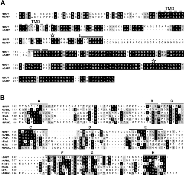
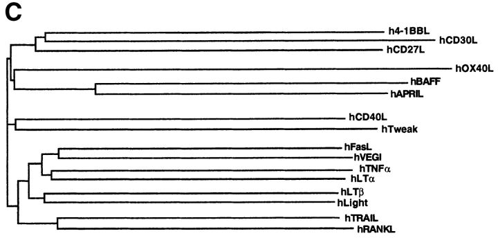
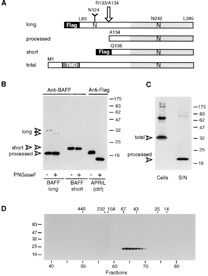
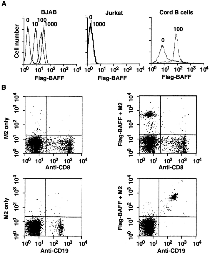
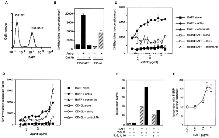
Members of the tumor necrosis factor (TNF) family induce pleiotropic biological responses, including cell growth, differentiation, and even death. Here we describe a novel member of the TNF family, designated BAFF (for B cell activating factor belonging to the TNF family), which is expressed by T cells and dendritic cells. Human BAFF was mapped to chromosome 13q32-34. Membrane-bound BAFF was processed and secreted through the action of a protease whose specificity matches that of the furin family of proprotein convertases. The expression of BAFF receptor appeared to be restricted to B cells. Both membrane-bound and soluble BAFF induced proliferation of anti-immunoglobulin M-stimulated peripheral blood B lymphocytes. Moreover, increased amounts of immunoglobulins were found in supernatants of germinal center-like B cells costimulated with BAFF. These results suggest that BAFF plays an important role as costimulator of B cell proliferation and function.


Figures

Figure 1
Figure 1
(A) Predicted aa sequence of human and mouse BAFF. The predicted transmembrane domain (TMD, dashed line), the potential N-linked glycosylation sites (stars), and the natural processing site of hBAFF (arrow) are indicated. The double line above hBAFF indicates the sequence obtained by Edman degradation of the processed form of hBAFF. (B) Comparison of the extracellular protein sequence of BAFF and some members of the TNF ligand family. Identical and homologous residues are represented in black and shaded boxes, respectively. (C) Dendrogram of TNF family ligands.
Figure 1
Figure 1
(A) Predicted aa sequence of human and mouse BAFF. The predicted transmembrane domain (TMD, dashed line), the potential N-linked glycosylation sites (stars), and the natural processing site of hBAFF (arrow) are indicated. The double line above hBAFF indicates the sequence obtained by Edman degradation of the processed form of hBAFF. (B) Comparison of the extracellular protein sequence of BAFF and some members of the TNF ligand family. Identical and homologous residues are represented in black and shaded boxes, respectively. (C) Dendrogram of TNF family ligands.
Figure 2
Figure 2
Characterization of recombinant BAFF. (A) Schematic representation of recombinant BAFF constructs. Soluble recombinant BAFFs starting at Leu83 and Gln136 are expressed fused to an NH2-terminal Flag tag and a 6–amino acid linker. The long form is cleaved between Arg133 and Ala134 (arrow) in 293 T cells, to yield a processed form of BAFF. Asn124 and Asn242 belong to N-glycosylation consensus sites. N-linked glycan present on Asn124 is shown as a Y. TMD, transmembrane domain. (B) PNGase F treatment of recombinant BAFF. Concentrated supernatants containing Flag-tagged BAFFs and APRIL were deglycosylated and analyzed by Western blotting using polyclonal anti-BAFF antibodies or anti-Flag M2, as indicated. All bands except processed BAFF also reacted with anti-Flag M2 (data not shown). (C) Full-length BAFF is processed to a soluble form. 293 T cells were transiently transfected with full-length BAFF. Transfected cells and their concentrated supernatants (S/N) were analyzed by Western blotting using polyclonal anti-BAFF antibodies. Supernatants corresponding to 10 times the amount of cells were loaded onto the gel. (D) Size exclusion chromatography of sBAFF on Superdex-200. Concentrated supernatants containing sBAFF/short were fractionated on a Superdex-200 column, and the eluted fractions were analyzed by Western blotting using anti-Flag M2 antibody. The migration positions of the molecular mass markers (in kD) are indicated on the left-hand side for SDS-PAGE and at the top of the figure for size exclusion chromatography.
Figure 3
Figure 3
Expression of BAFF. (A) Northern blots (2 μg poly A+ RNA per lane) of various human tissues were probed with BAFF antisense mRNA. (B) Reverse transcriptase amplification of BAFF, IL-2 receptor α chain (IL2-Rα), and actin from RNA of purified blood T cells at various time points of PHA activation, E-rosetting–negative blood cells (mostly B cells), in vitro–derived immature dendritic cells, 293 cells, and 293 cells stably transfected with full-length BAFF (293-BAFF). Control amplifications were performed in the absence of added cDNA. IL-2 receptor α chain was amplified as a marker of T cell activation.
Figure 4
Figure 4
BAFF binds to mature B cells. (A) Binding of sBAFF to BJAB and Jurkat cell lines, and to purified CD19+ cells of cord blood. Cells were stained with the indicated amount (in ng/50 μl) of Flag-BAFF and analyzed by flow cytometry. (B) Binding of sBAFF to PBLs. PBLs were stained with anti-CD8–FITC or with anti-CD19–FITC (x axis) and with Flag-BAFF plus M2-biotin and avidin-PE (y axis). Flag-BAFF was omitted in controls.
Figure 5
Figure 5
BAFF costimulates B cell proliferation. (A) Surface expression of BAFF in stably transfected 293 cells. 293-BAFF and 293 wt cells were stained with anti-BAFF mAb 43.9 and analyzed by flow cytometry. (B) Costimulation of PBLs by 293-BAFF cells. PBLs (105/well) were incubated with 15,000 paraformaldehyde-fixed 293 cells (293 wt or 293-BAFF) in the presence or absence of anti-B cell receptor antibody (anti-μ). Fixed 293 cells alone incorporated 100 cpm. (C) Dose-dependent costimulation of PBL proliferation by sBAFF in the presence of anti-μ. Proliferation was determined after 72 h incubation by [3H]thymidine incorporation. Controls include cells treated with BAFF alone, with heat-denatured BAFF, or with an irrelevant isotype-matched antibody in place of anti-μ. (D) Comparison of (co)stimulatory effects of sCD40L and sBAFF on PBL proliferation. Experiment was performed as described in panel C. (E) BAFF costimulates Ig secretion of preactivated human B cells. Purified CD19+ B cells were activated by coculture with EL-4 T cells and activated T cell supernatants for 5–6 d, then reisolated and cultured for another 7 d in the presence of medium only (−) or containing 5% activated T cell supernatants (T-SUP) or a blend of cytokines (IL-2, IL-4, IL-10). The columns represent means of Ig concentrations for cultures with or without 1 μg/ml BAFF. Means of fold increase ± SD were 1.23 ± 0.11 for medium only, 2.06 ± 0.18 with T cell supernatants (four experiments), and 1.45 ± 0.06 with IL-2, IL-4, and IL-10 (two experiments). These were performed with peripheral blood (three experiments) or cord blood B cells (one experiment; 2.3-fold increase with T cell supernatants, 1.5-fold increase with IL-2, IL-4, and IL-10). (F) Dose–response curve for the effect of BAFF in cultures with T cell supernatants, as shown in panel D. Mean ± SD of three experiments.

References

    1. Smith CA, Farrah T, Goodwin RG. The TNF-receptor superfamily of cellular and viral proteins: activation, costimulation, and death. Cell. 1994;76:959–962. - PubMed
    1. Vassalli P. The pathophysiology of tumor necrosis factors. Annu Rev Immunol. 1992;10:411–452. - PubMed
    1. De Togni P, Goellner J, Ruddle NH, Streeter PR, Fick A, Mariathasan S, Smith SC, Carlson R, Shornick LP, Strauss-Schoenberger J, et al. Abnormal development of peripheral lymphoid organs in mice deficient in lymphotoxin. Science. 1994;264:703–707. - PubMed
    1. Koni PA, Sacca R, Lawton P, Browning JL, Ruddle NH, Flavell RA. Distinct roles in lymphoid organogenesis for lymphotoxins alpha and beta revealed in lymphotoxin beta-deficient mice. Immunity. 1997;6:491–500. - PubMed
    1. Amakawa R, Hakem A, Kundig TM, Matsuyama T, Simard JJ, Timms E, Wakeham A, Mittruecker HW, Griesser H, Takimoto H, et al. Impaired negative selection of T cells in Hodgkin's disease antigen CD30-deficient mice. Cell. 1996;84:551–562. - PubMed

Publication types

MeSH terms

Associated data