. 1999 Jun 7;189(11):1765-76.
doi: 10.1084/jem.189.11.1765.

Acquisition of selectin binding and peripheral homing properties by CD4(+) and CD8(+) T cells

Affiliations

Acquisition of selectin binding and peripheral homing properties by CD4(+) and CD8(+) T cells

H Xie et al. J Exp Med. .

Abstract

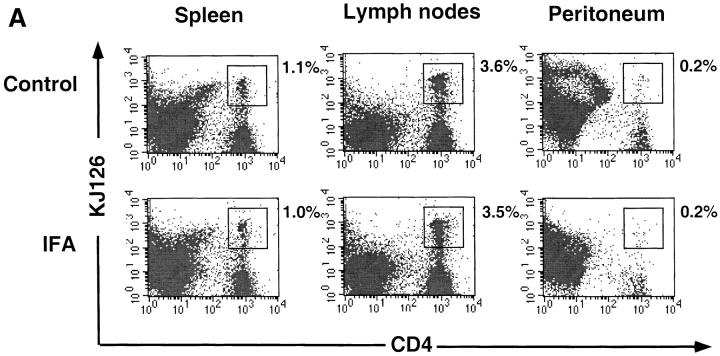
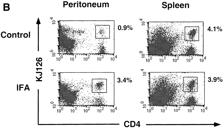
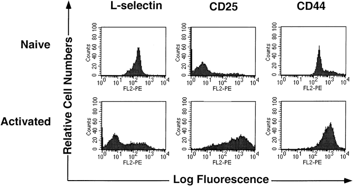
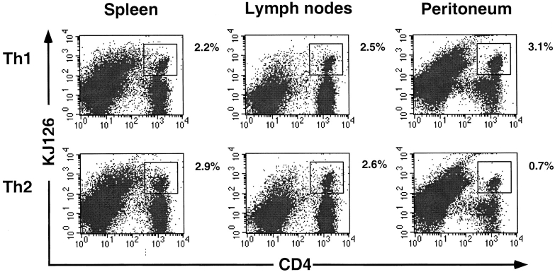
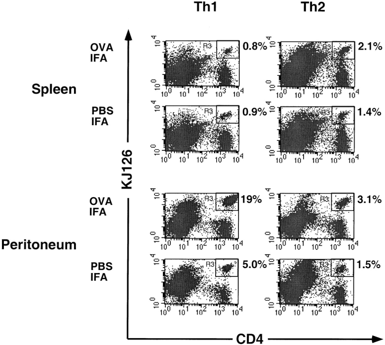
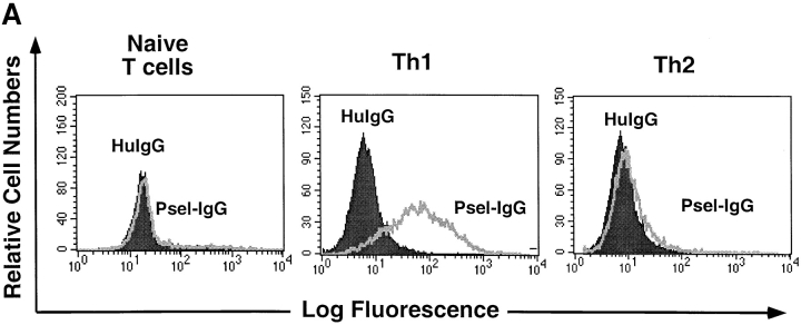
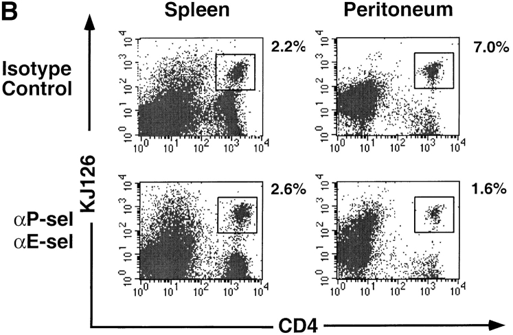
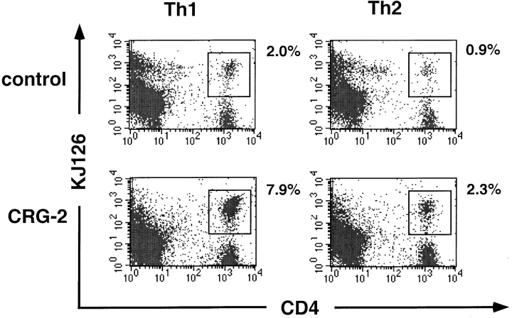
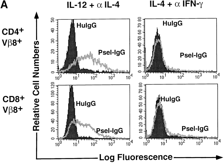
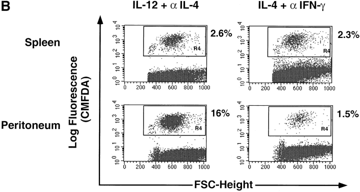
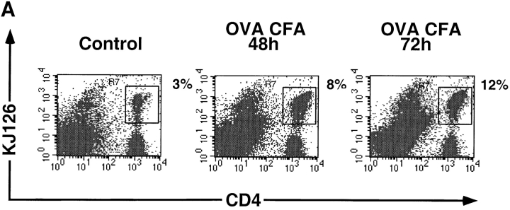
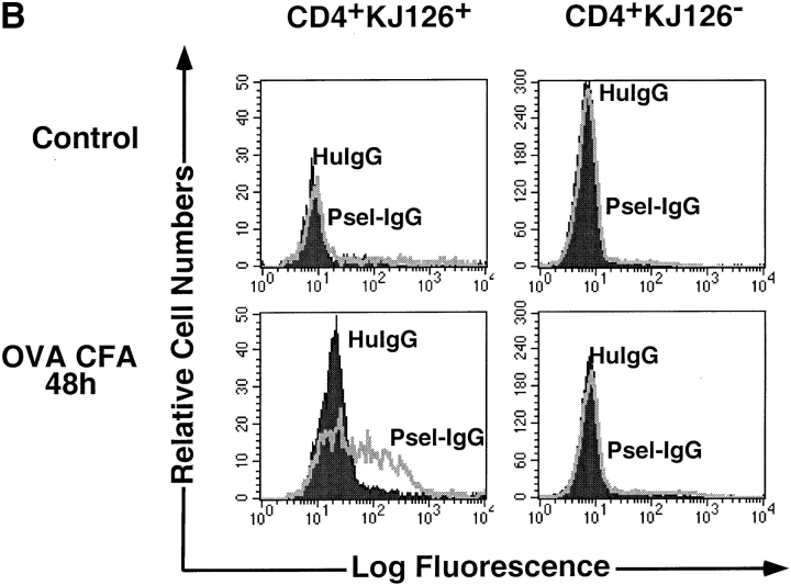
Different T cell subsets exhibit distinct capacities to migrate into peripheral sites of inflammation, and this may in part reflect differential expression of homing receptors and chemokine receptors. Using an adoptive transfer approach, we examined the ability of functionally distinct subsets of T cells to home to a peripheral inflammatory site. The data directly demonstrate the inability of naive T cells and the ability of effector cells to home to inflamed peritoneum. Furthermore, interleukin (IL)-12 directs the differentiation of either CD4(+) or CD8(+) T cells into effector populations that expresses functional E- and P-selectin ligand and that are preferentially recruited into the inflamed peritoneum compared with T cells differentiated in the presence of IL-4. Recruitment can be blocked by anti-E- and -P-selectin antibodies. The presence of antigen in the peritoneum promotes local proliferation of recruited T cells, and significantly amplifies the Th1 polarization of the lymphocytic infiltrate. Preferential recruitment of Th1 cells into the peritoneum is also seen when cytokine response gene 2 (CRG-2)/interferon gamma-inducible protein 10 (IP-10) is used as the sole inflammatory stimulus. We have also found that P-selectin binds only to antigen-specific T cells in draining lymph nodes after immunization, implying that both antigen- and cytokine-mediated signals are required for expression of functional selectin-ligand.


Figures

Figure 1
Figure 1
Recruitment of naive versus effector CD4+ T cells to inflamed peritoneum. 1.5 × 10⁷ naive CD4+KJ126+ T cells (A) or 1.8 × 10⁷ in vitro– activated CD4+ T cells (B) were adoptively transferred into BALB/c mice. After 6–24 h, mice were either left untreated (control) or were given intraperitoneal injections of IFA. 3 d later, spleens, lymph nodes, and peritoneal cells were collected and stained with PE-conjugated anti-CD4 and Cy-Chrome– labeled KJ126. The numbers adjacent to the upper right corner of each panel are the percentage of the total gated cells that are CD4+KJ126+ cells. One of two comparable experiments is shown.
Figure 1
Figure 1
Recruitment of naive versus effector CD4+ T cells to inflamed peritoneum. 1.5 × 10⁷ naive CD4+KJ126+ T cells (A) or 1.8 × 10⁷ in vitro– activated CD4+ T cells (B) were adoptively transferred into BALB/c mice. After 6–24 h, mice were either left untreated (control) or were given intraperitoneal injections of IFA. 3 d later, spleens, lymph nodes, and peritoneal cells were collected and stained with PE-conjugated anti-CD4 and Cy-Chrome– labeled KJ126. The numbers adjacent to the upper right corner of each panel are the percentage of the total gated cells that are CD4+KJ126+ cells. One of two comparable experiments is shown.
Figure 2
Figure 2
Phenotype of naive and activated T cells. Naive cells represented in this figure were CD4+ T cells freshly isolated from DO11 mice, and activated cells represent DO11 T cells stimulated in vitro with OVA peptide and APCs, as described in Materials and Methods. The histograms depict staining for the indicated molecule using PE-labeled secondary antibody after gating on CD4+ (FITC) plus KJ126+ (Cy-Chrome) cells. One of two comparable experiments is shown.
Figure 3
Figure 3
In vivo recruitment of Th1 versus Th2 cells to adjuvant-treated peritoneum. DO11 Th1 or Th2 cells (2 × 10⁷) were adoptively transferred into BALB/c mice. After 24 h, mice were given intraperitoneal injections of PBS-IFA. 3 d later, lymph nodes (inguinal, axillary, and mesenteric), spleen, and peritoneal cells were collected and stained with PE-conjugated anti-CD4 and Cy-Chrome–labeled KJ126. The numbers adjacent to the upper right corner of each panel are the percentage of the total gated cells that are CD4+KJ126+ cells. One of four comparable experiments is shown.
Figure 4
Figure 4
Influence of antigen on recruitment of Th1 versus Th2 cells to peritoneum. DO11 Th1 or Th2 cells (2 × 10⁷) were adoptively transferred into BALB/c mice. After 24 h, mice were given intraperitoneal injections of either OVA-IFA or PBS-IFA. 3 d later, spleen and peritoneal cells were collected and stained with PE-conjugated anti-CD4 and Cy-Chrome–labeled KJ126. The numbers adjacent to the upper right corner of each panel are the percentage of the total gated cells that are CD4+KJ126+ cells. One of four comparable experiments is shown.
Figure 5
Figure 5
Antigen-induced proliferation of adoptively transferred Th1 cells in response to antigen in peritoneum. DO11 Th1 cells (2 × 10⁷) were labeled with green tracker (CMFDA) before adoptive transfer into BALB/c mice. Recipient mice were given intraperitoneal injections of either OVA-IFA (OVA+) or PBS-IFA (OVA−), and 3 d later peritoneal cells were harvested and stained with PE-conjugated anti-CD4 and Cy-Chrome–labeled KJ126. The green tracker visualized in FL-1 shown here is gated on CD4+KJ126+ cells.
Figure 6
Figure 6
P- and E-selectin–mediated recruitment of Th1 cells into an inflammatory site. (A) Naive, Th1, or Th2 cells were stained with P-selectin Ig chimeric protein (Psel-IgG) or with human Ig control (HuIgG) followed by FITC-conjugated anti–human-Ig. Cells were also stained for CD4 (PE) and KJ126 (Cy-Chrome). The histogram shown represents cells gated on the CD4+KJ126+ population. One of three comparable experiments is shown. (B) Isotype control rabbit IgG or anti–P- and E-selectin antibodies were injected via tail vein together with Th1 cells into BALB/c mice. After 24 h, PBS-IFA was injected into the peritoneum of these mice. 3 d later, spleen and peritoneal cells were collected and stained with PE-conjugated anti-CD4 and Cy-Chrome–labeled KJ126. The numbers adjacent to the upper right corner of each panel are the percentage of the total gated cells that are CD4+KJ126+ cells. One of two comparable experiments is shown.
Figure 6
Figure 6
P- and E-selectin–mediated recruitment of Th1 cells into an inflammatory site. (A) Naive, Th1, or Th2 cells were stained with P-selectin Ig chimeric protein (Psel-IgG) or with human Ig control (HuIgG) followed by FITC-conjugated anti–human-Ig. Cells were also stained for CD4 (PE) and KJ126 (Cy-Chrome). The histogram shown represents cells gated on the CD4+KJ126+ population. One of three comparable experiments is shown. (B) Isotype control rabbit IgG or anti–P- and E-selectin antibodies were injected via tail vein together with Th1 cells into BALB/c mice. After 24 h, PBS-IFA was injected into the peritoneum of these mice. 3 d later, spleen and peritoneal cells were collected and stained with PE-conjugated anti-CD4 and Cy-Chrome–labeled KJ126. The numbers adjacent to the upper right corner of each panel are the percentage of the total gated cells that are CD4+KJ126+ cells. One of two comparable experiments is shown.
Figure 7
Figure 7
IP-10/CRG-2–mediated preferential recruitment of T helper cells in vivo. DO11 Th1 and Th2 cells were adoptively transferred into BALB/c mice through tail vein injection. After 24 h, mice either were left untreated or else received an intraperitoneal injection of 0.5 μg of CRG-2. 3 d later, spleen and peritoneal cells were collected and stained with PE-conjugated anti-CD4 and Cy-Chrome–labeled KJ126. The numbers adjacent to the upper right corner of each panel are the percentage of the total gated cells that are CD4+KJ126+ cells.
Figure 8
Figure 8
The P-selectin binding and peritoneal recruitment of SEB-stimulated BALB/c splenic CD4+ and CD8+ T cells. (A) Spleen cells were stimulated in vitro with 2 μg/ml of SEB with either IL-12 plus anti–IL-4 antibody or with IL-4 plus anti–IFN-γ antibody. After 6 d of culture, cells were harvested and stained for CD4, Vβ8, and P-selectin Ig (Psel-IgG) or control human Ig (HuIgG) as described in previous figures. (B) The same cell populations as described in A were labeled with green tracker dye before tail vein injection into BALB/c mice. After 24 h, mice were injected in the peritoneum with PBS-IFA. 3 d later, spleen and peritoneal cells were collected and visualized by flow cytometry. The numbers in the plots represent the percentage of total gated cells that are tracker dye positive (CMFDA). One of four comparable experiments is shown.
Figure 8
Figure 8
The P-selectin binding and peritoneal recruitment of SEB-stimulated BALB/c splenic CD4+ and CD8+ T cells. (A) Spleen cells were stimulated in vitro with 2 μg/ml of SEB with either IL-12 plus anti–IL-4 antibody or with IL-4 plus anti–IFN-γ antibody. After 6 d of culture, cells were harvested and stained for CD4, Vβ8, and P-selectin Ig (Psel-IgG) or control human Ig (HuIgG) as described in previous figures. (B) The same cell populations as described in A were labeled with green tracker dye before tail vein injection into BALB/c mice. After 24 h, mice were injected in the peritoneum with PBS-IFA. 3 d later, spleen and peritoneal cells were collected and visualized by flow cytometry. The numbers in the plots represent the percentage of total gated cells that are tracker dye positive (CMFDA). One of four comparable experiments is shown.
Figure 9
Figure 9
Induction of functional P-selectin ligand expression on antigen-activated T cells in vivo. Naive (1.5 × 10⁷) CD4+KJ126+ T cells were adoptively transferred into BALB/c mice. After 24 h, mice were either left untreated or further immunized with OVA and CFA by footpad injection. Draining lymph nodes were collected at days 1, 2, and 3, and were stained with PE-conjugated anti-CD4 and Cy-Chrome–labeled KJ126 and with P-selectin Ig chimeric protein (Psel-IgG) or with human Ig control (HuIgG) followed by anti–human Ig conjugated with FITC. The number of DO11 T cells in the draining lymph nodes is shown in A. The numbers adjacent to the upper right corner of each panel are the percentage of the total gated cells that are CD4+KJ126+ cells. P-selectin binding by CD4+KJ126+ or CD4+KJ126 populations is shown in B.
Figure 9
Figure 9
Induction of functional P-selectin ligand expression on antigen-activated T cells in vivo. Naive (1.5 × 10⁷) CD4+KJ126+ T cells were adoptively transferred into BALB/c mice. After 24 h, mice were either left untreated or further immunized with OVA and CFA by footpad injection. Draining lymph nodes were collected at days 1, 2, and 3, and were stained with PE-conjugated anti-CD4 and Cy-Chrome–labeled KJ126 and with P-selectin Ig chimeric protein (Psel-IgG) or with human Ig control (HuIgG) followed by anti–human Ig conjugated with FITC. The number of DO11 T cells in the draining lymph nodes is shown in A. The numbers adjacent to the upper right corner of each panel are the percentage of the total gated cells that are CD4+KJ126+ cells. P-selectin binding by CD4+KJ126+ or CD4+KJ126 populations is shown in B.

Similar articles

Cited by

References

    1. Tietz W, Allemand Y, Borges E, von Laer D, Hallmann R, Vestweber D, Hamann A. CD4+T cells migrate into inflamed skin only if they express ligands for E- and P-selectin. J Immunol. 1998;161:963–970. - PubMed
    1. Bradley LM, Watson SR. Lymphocyte migration into tissue: the paradigm derived from CD4 subsets. Curr Opin Immunol. 1996;8:312–320. - PubMed
    1. Mackay CR, Marston W, Dudler L. Altered patterns of T cell migration through lymph nodes and skin following antigen challenge. Eur J Immunol. 1992;22:2205–2210. - PubMed
    1. Mackay CR, Marston WL, Dudler L. Naive and memory T cells show distinct pathways of lymphocyte recirculation. J Exp Med. 1990;171:801–817. - PMC - PubMed
    1. Swain SL, Bradley LM. Helper T cell memory: more questions than answers. Semin Immunol. 1992;4:59–68. - PubMed

Publication types

MeSH terms