. 1999 Jun;10(6):1763-82.
doi: 10.1091/mbc.10.6.1763.

Membrane tubule-mediated reassembly and maintenance of the Golgi complex is disrupted by phospholipase A2 antagonists

Affiliations
Free PMC article

Membrane tubule-mediated reassembly and maintenance of the Golgi complex is disrupted by phospholipase A2 antagonists

P de Figueiredo et al. Mol Biol Cell. 1999 Jun.
Free PMC article

Abstract

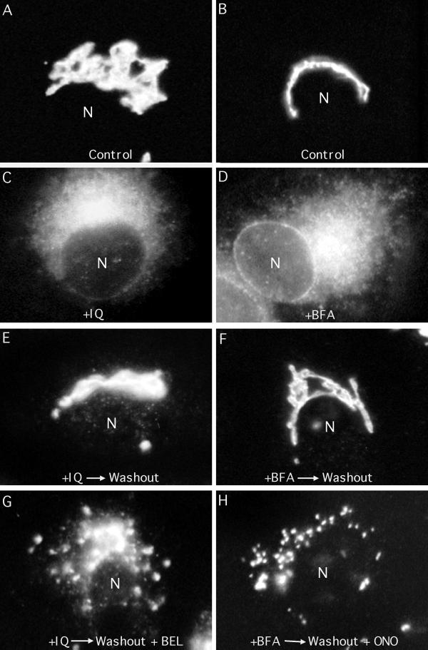
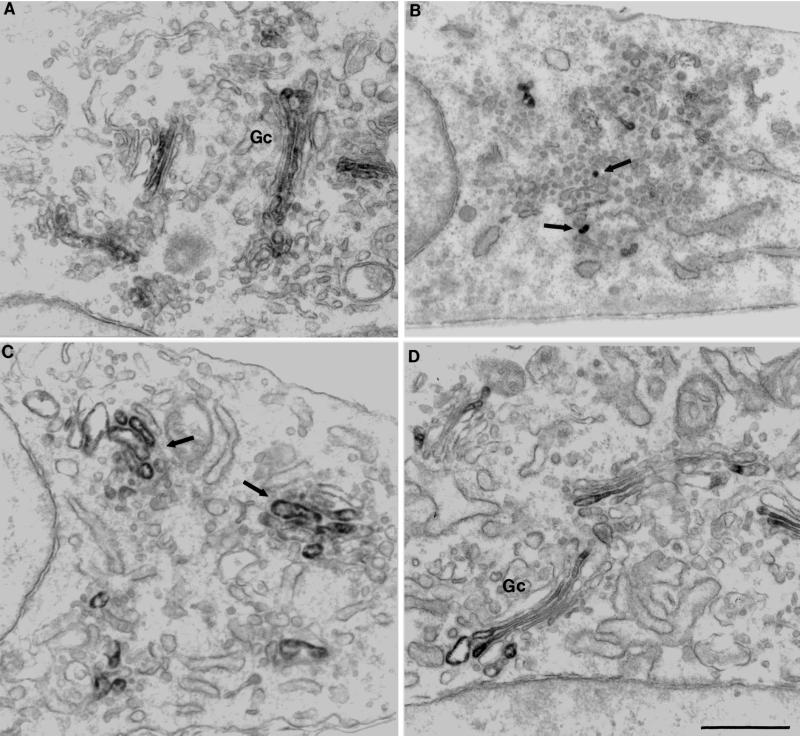
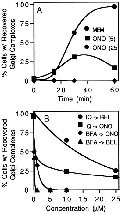
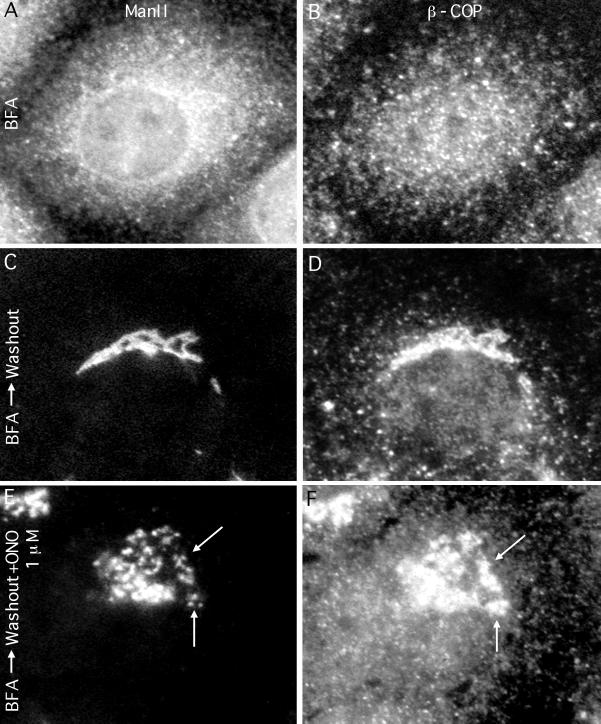
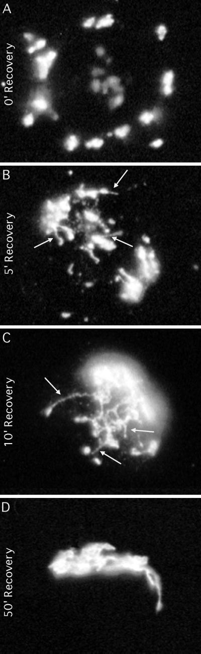
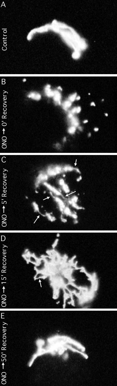
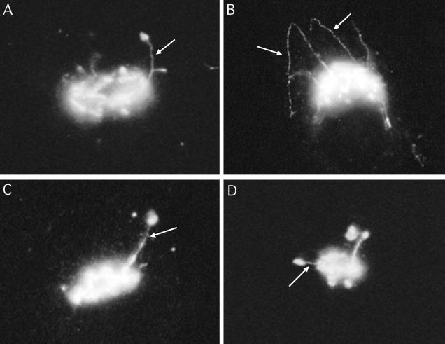
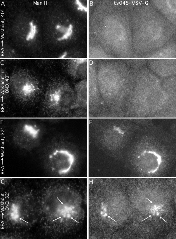
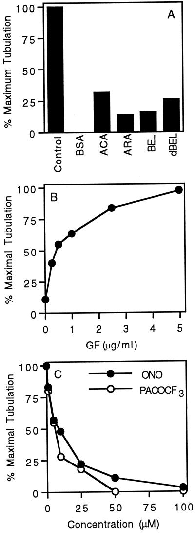
Although membrane tubules can be found extending from, and associated with, the Golgi complex of eukaryotic cells, their physiological function has remained unclear. To gain insight into the biological significance of membrane tubules, we have developed methods for selectively preventing their formation. We show here that a broad range of phospholipase A2 (PLA2) antagonists not only arrest membrane tubule-mediated events that occur late in the assembly of the Golgi complex but also perturb its normal steady-state tubulovesicular architecture by inducing a reversible fragmentation into separate "mini-stacks." In addition, we show that these same compounds prevent the formation of membrane tubules from Golgi stacks in an in vitro reconstitution system. This in vitro assay was further used to demonstrate that the relevant PLA2 activity originates from the cytoplasm. Taken together, these results demonstrate that Golgi membrane tubules, sensitive to potent and selective PLA2 antagonists, mediate both late events in the reassembly of the Golgi complex and the dynamic maintenance of its steady-state architecture. In addition, they implicate a role for cytoplasmic PLA2 enzymes in mediating these membrane trafficking events.


Figures

Figure 1
Figure 1
PLA2 antagonists arrest the reassembly of the Golgi complex during recovery from IQ or BFA treatment as seen by immunofluorescence staining with the medial Golgi enzyme ManII. Control cells display a compact, interconnected Golgi complex in the juxtanuclear region of the cell (A and B). Treatment with either IQ (10 μM for 60 min) or BFA (5 μg/ml for 30 min) resulted in a diffuse Man II staining pattern (D). When IQ- or BFA-treated cells were then washed free of these drugs and incubated in normal media, Golgi complexes were fully reassembled within 1 h (E and F). However, when recovery from IQ or BFA for the same length of time was done in the presence of the PLA2 inhibitors BEL (10 μM; G), or ONO-RS-082 (ONO, 1 μM; H), Golgi complexes reassembled only to the point of forming large, disconnected punctate structures in the juxtanuclear region of the cell. N, nucleus.
Figure 2
Figure 2
Immunoperoxidase localization of ManII during recovery from IQ treatment. (A) Control cells showing the typical stacked Golgi cisternae (Gc) with ManII labeling generally restricted to one or two medial cisternae. (B) Cells treated with IQ (10 μM for 60 min) reveal the formation of clusters of VGMs throughout the cytoplasm; within these clusters a few of the vesicles stain for ManII (arrows). (C) Cells treated with IQ as above and then washed and incubated without IQ but in the presence of the reversible PLA2 inhibitor ONO-RS-082 (1 μM) for 1 h are capable of reassembling stacked Golgi cisternae that are labeled with ManII antibodies (arrows). (D) Cells treated as in C, but then washed free of ONO-RS-082 and incubated for 1 h, demonstrate that the Golgi complex further reassembles into a larger interconnected organelle with ManII staining more restricted to one or two cisternae. All micrographs were printed to the same final magnification. Bar, 0.5 μm.
Figure 3
Figure 3
(A) Time course of Golgi reassembly during recovery from IQ treatment. Cells were first treated with IQ (10 μM for 60 min), washed to remove IQ, and then incubated in the absence or presence of the PLA2 inhibitor ONO-RS-082 (ONO, 5 or 25 μM as indicated). Cells were fixed, processed for ManII immunofluorescence, and then counted to determine the percentage that had fully reassembled Golgi complexes (as illustrated by Figure 1, E and F). (B) Dose response of PLA2 inhibitors on the reassembly of Golgi complexes during recovery from IQ or BFA treatment. Cells were treated with IQ or BFA as in Figure 1, washed free of the drugs, and incubated for 1 h with various concentrations of BEL or ONO-RS-082 as indicated.
Figure 4
Figure 4
ONO-RS-082 inhibits Golgi reassembly but does not prevent the association of β-COP with Golgi membranes during recovery from BFA treatment. Each set of side-by-side panels shows double-immunofluorescent labeling of ManII (left panels) and β-COP (right panels). Cells treated with BFA (5 μg/ml for 15 min) show the typical diffuse immunofluorescence staining pattern of both ManII and β-COP (A and B). Treatment with BFA followed by a washout period of 1 h in drug-free media resulted in the reassembly of the Golgi complex and reassociation of β-COP with these membranes (C and D). Recovery from BFA in the presence of 1 μM ONO-RS-082 (ONO) resulted in the reformation of tightly clustered but unconnected mini-stacks in the juxtanuclear region to which β-COP was localized (E and F).
Figure 5
Figure 5
Membrane tubules form from Golgi mini-stacks after recovery from PLA2 inhibitor arrest of Golgi reassembly. In these experiments, cells were treated with BFA (5 μg/ml for 30 min), washed free of BFA, and incubated with the reversible PLA2 inhibitor ONO-RS-082 (5 μM) for 60 min to allow reassembly up to the point of mini-stack formation (A). Cells were then washed free of the inhibitor and allowed to recover in normal media for 5 min (B), 10 min (C), or 50 min (D). After each experiment, cells were processed for immunofluorescence with anti-ManII antibodies. Cells incubated for 5 min in inhibitor-free media exhibit ManII-stained, thin membrane tubules emanating from many of the mini-stacks (arrows) (B). By 10 min of recovery, nearly all mini-stacks were interconnected by thin membrane tubules (arrows), and by 50 min the Golgi complex had completely reformed (D). Some of the tubules indicated by the arrows appear to be of somewhat different diameters; however, these differences are actually due to out-of-focus light from tubules in different focal planes.
Figure 6
Figure 6
Normal Golgi architecture is reversibly altered by PLA2 antagonists, as shown by immunofluorescence localization of ManII. (A) Control cell. (B) Treatment of cells with ONO-RS-082 (ONO) alone (5 μM for 60 min) produced large fragments that remained in the juxtanuclear region, which were, in fact, also mini-stacks (de Figueiredo et al., 1998). (C–E) Reassembly of intact Golgi complexes during recovery from ONO-RS-082 treatment. Cells were treated as in B and then washed and incubated in normal media for 5 min (C), 15 min (D), and 50 min (E). By 5 min of recovery, thin, ManII-stained membrane tubules can be observed to emanate from, and link together, spatially separate mini-stacks; by 15 min virtually all ManII-stained elements are completely interconnected by thin tubules, and by 50 min the entire Golgi complex has reformed.
Figure 7
Figure 7
Cells recovering from ONO-RS-082 treatment display ManII-stained tubular connections between large centrally located Golgi elements and more peripheral Golgi mini-stacks. (A) Cells were treated with BFA for 30 min, washed free of BFA, incubated with ONO-RS-082 (5 μM) for 60 min to allow reassembly up to the point of mini-stack formation, and then allowed to recover from ONO-RS-082 for 10 min before fixing for immunofluorescence. (B–D) Cells were treated with ONO-RS-082 alone (5 μM for 60 min) to generate mini-stacks and then allowed to recover in drug-free growth medium for 10 min (B and C) or 15 min (D).
Figure 8
Figure 8
Golgi mini-stacks formed in the presence of PLA2 inhibitors during reassembly after BFA washout receive membrane traffic from the ER. Cells were infected with ts045 VSV at 40°C to entrap VSV-G in the ER, treated with BFA (5 μg/ml for 15 min) to recycle the Golgi back to the ER, washed free of BFA, and then incubated under various conditions to allow Golgi reassembly and/or transport of VSV-G out of the ER. Cells were then fixed and processed for double-label immunofluorescence to localize ManII (left panels) and VSV-G (right panels). Conditions during the washout from BFA were as follows. (A and B) Cells incubated with drug-free media at 40°C allow the Golgi complex to completely reform, but VSV-G remains diffusely in the ER. (C and D) Cells incubated in media with ONO-RS-082 (ONO) at 40°C reassemble the Golgi into punctate mini-stacks, but VSV-G still remains in the ER. (E and F) Cells incubated with drug-free media but shifted to the permissive temperature of 32°C reassemble an intact Golgi complex to which VSV-G is now transported, as evidenced by its colocalization with ManII. (G and H) Cells incubated with ONO-RS-082 at 32°C reassemble the Golgi only to the point of forming punctate mini-stacks to which VSV-G is transported (arrows point to double-labeled mini-stacks).
Figure 9
Figure 9
Electron microscopic observations of Golgi membrane tubulation in a cytosol-dependent, in vitro reconstitution system and immunogold localization of ManII on Golgi membrane tubules. Golgi-enriched fractions were incubated in vitro under various conditions and applied as whole-mount preparations onto EM grids for standard negative staining (A–C) or for a modified protocol involving a combination of immunogold labeling of ManII followed by negative staining (D and E). (A) Golgi complex incubated with buffer control. (B) Golgi complex incubated with bovine brain cytosol under conditions that induce membrane tubule formation. Arrows indicate a few of the numerous 60- to 80-nm-diameter membrane tubules that formed. (C) Golgi complex incubated with cytosol that had first been treated with the PLA2 inhibitor BEL (25 μM). (D) Control Golgi complex immunogold labeled to show distribution of ManII. (E) Golgi complexes incubated with cytosol under tubulation conditions and then immunolabeled to show ManII distribution. Arrows indicate several tubules with a nearly uniform distribution of gold particles along the length of each tubule. As shown in E, all of the induced tubules were labeled with anti-ManII antibodies; however, in many other cases, only about half of the Golgi tubules were labeled with ManII antibodies, and in double-labeling experiments that localized ManII and mannose 6-phosphate receptors (located in trans elements), separate tubules were stained. Bar, 0.5 μm.
Figure 10
Figure 10
Quantitation of Golgi membrane tubulation inhibition by PLA2 antagonists. (A) Aliquots of an active fraction of bovine brain cytosol, containing tubulation activity, were incubated in the absence or presence of various PLA2 inhibitors, and then each was mixed with isolated rat liver Golgi complexes in our standard in vitro tubulation assay. Concentrations were BBC (bovine brain cytosol), 1.5 mg/ml; BSA, 1.5 mg/ml; ACA, ARA, and BEL, 5 μM. The bar labeled dBEL (for dialyzed BEL) shows the level of tubulation that resulted when BBC was first incubated with BEL (20 μM for 15 min at 37°C) and then dialyzed to remove excess, unbound BEL. The extent of tubulation was measured as described in MATERIALS AND METHODS, and the data are expressed as the percent maximal tubulation to compare results from different experiments. (B) Addition of the enriched GF fraction restored tubulation activity toBEL-inactivated cytosol. BBC (1.5 mg/ml) was incubated with BEL (as above), dialyzed extensively, incubated with increasing amounts of enriched GF fraction, and then used in the standard in vitro tubulation assay. The data points are averages of duplicate experiments. (C) Typical dose–response experiment showing the loss of cytosolic tubulation activity with increasing amounts of the PLA2 inhibitors ONO-RS-082 and PACOCF3. All data points are the averages of duplicate experiments.

Similar articles

Cited by

References

    1. Acharya U, Jacobs R, Peters JM, Watson N, Farquhar MG, Malhotra V. The formation of Golgi stacks from vesiculated Golgi membranes requires two distinct fusion events. Cell. 1995a;82:895–904. - PubMed
    1. Acharya U, McCaffery JM, Jacobs R, Malhotra V. Reconstitution of vesiculated Golgi membranes into stacks of cisternae: requirement of NSF in stack formation. J Cell Biol. 1995b;129:577–589. - PMC - PubMed
    1. Ackermann EJ, Conde-Frieboes K, Dennis EA. Inhibition of macrophage Ca2+-independent phospholipase A2 by bromoenol lactone and Trifluoromethyl ketones. J Biol Chem. 1995;270:445–450. - PubMed
    1. Alcalde J, Bonay P, Roa A, Vilaro S, Sandoval IV. Assembly and disassembly of the Golgi complex: two processes arranged in a cis-trans direction. J Cell Biol. 1992;116:69–83. - PMC - PubMed
    1. Balboa MA, Balsinde J, Jones SS, Dennis EA. Identity between the Ca2+-independent phospholipase A2 enzymes from P388D1 macrophages and Chinese hamster ovary cells. J Biol Chem. 1997;272:8576–8580. - PubMed

Publication types

MeSH terms

LinkOut - more resources