Comparative Study
. 1999 Jun;10(6):1783-98.
doi: 10.1091/mbc.10.6.1783.

Deciphering the nuclear import pathway for the cytoskeletal red cell protein 4.1R

Affiliations
Free PMC article
Comparative Study

Deciphering the nuclear import pathway for the cytoskeletal red cell protein 4.1R

P Gascard et al. Mol Biol Cell. 1999 Jun.
Free PMC article

Abstract

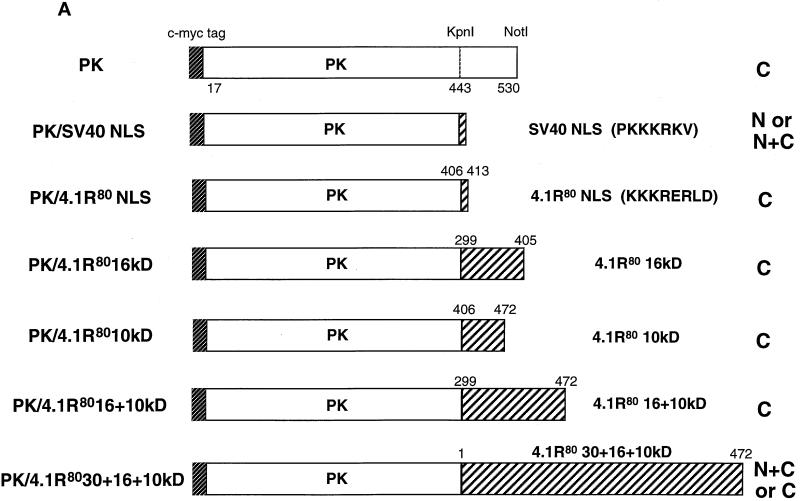
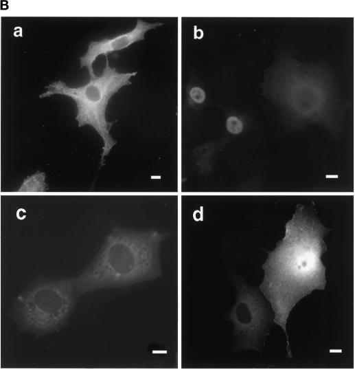
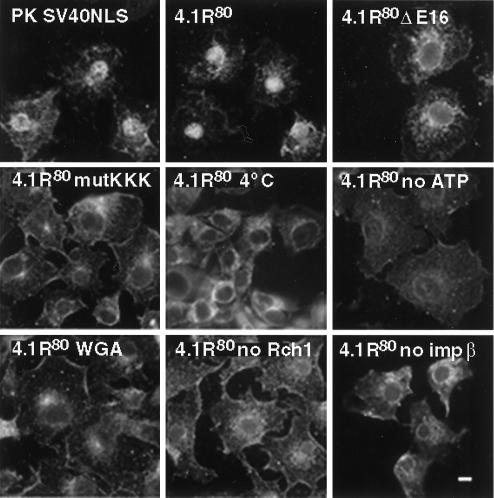
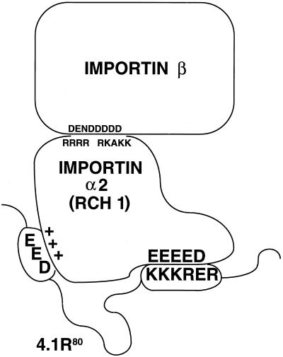
The erythroid membrane cytoskeletal protein 4.1 is the prototypical member of a genetically and topologically complex family that is generated by combinatorial alternative splicing pathways and is localized at diverse intracellular sites including the nucleus. To explore the molecular determinants for nuclear localization, we transfected COS-7 cells with epitope-tagged versions of natural red cell protein 4.1 (4.1R) isoforms as well as mutagenized and truncated derivatives. Two distant topological sorting signals were required for efficient nuclear import of the 4.1R80 isoform: a basic peptide, KKKRER, encoded by alternative exon 16 and acting as a weak core nuclear localization signal (4.1R NLS), and an acidic peptide, EED, encoded by alternative exon 5. 4.1R80 isoforms lacking either of these two exons showed decreased nuclear import. Fusion of various 4.1R80 constructs to the cytoplasmic reporter protein pyruvate kinase confirmed a requirement for both motifs for full NLS function. 4.1R80 was efficiently imported in the nuclei of digitonin-permeabilized COS-7 cells in the presence of recombinant Rch1 (human importin alpha2), importin beta, and GTPase Ran. Quantitative analysis of protein-protein interactions using a resonant mirror detection technique showed that 4.1R80 bound to Rch1 in vitro with high affinity (KD = 30 nM). The affinity decreased at least 7- and 20-fold, respectively, if the EED motif in exon 5 or if 4.1R NLS in exon 16 was lacking or mutated, confirming that both motifs were required for efficient importin-mediated nuclear import of 4.1R80.


Figures

Figure 1
Figure 1
Expression of endogenous protein 4.1R in COS-7 cells and of HA epitope-tagged 4.1R isoforms in transfected COS-7 cells. Nontransfected COS-7 cells (a) or COS-7 cells transfected with empty expression vector (b), HA epitope-tagged 4.1R80 (c), HA epitope-tagged 4.1R80ΔE16 (d), or HA epitope-tagged 4.1R80mutKKK (e) were fixed with 3% paraformaldehyde, permeabilized with 0.5% Triton X-100, and processed for immunofluorescence using, as primary antibody, either the polyclonal protein 4.1R 24-3 antibody (Krauss et al., 1997a) to probe nontransfected cells or a polyclonal anti-HA-tag antibody to probe transfected cells. An anti-rabbit IgG coupled to FITC was used in all samples as secondary antibody. Because of COS-7 cell thickness, the nucleus and cytoplasmic network cannot be in focus in the same field. (a, inset) Focused closeup of nuclear and centrosome staining of the cell displayed in this panel. Bar, 10 μm.
Figure 2
Figure 2
Comparison of cellular distribution of various proteins of the 4.1 protein family in transfected COS-7 cells. COS-7 cells were transfected with HA-tagged 4.1R80, HA-tagged 4.1G, or 4.1N. Cells were fixed with 3% paraformaldehyde, permeabilized with 0.5% Triton X-100, and processed for immunofluorescence using, as primary antibody, either a polyclonal anti-HA tag antibody (4.1R80 and 4.1G) or a polyclonal 4.1N antibody (Walensky et al., unpublished data). An anti-rabbit IgG coupled to FITC was used in all samples as secondary antibody. The 4.1R NLS and the corresponding aligned primary amino acid sequences in 4.1G and 4.1N are displayed. The basic residues are shown in gray. Note that because of poor conservation of the 4.1N sequence within the corresponding region of 4.1R and 4.1G, the 4.1N sequence displayed results from the best alignment achievable (Walensky et al., unpublished data). Bar, 10 μm.
Figure 3
Figure 3
Effects of truncation or point mutation of protein 4.1R domains flanking 4.1R NLS on protein 4.1R nuclear import. (A) Map of protein 4.1R constructs used to investigate nuclear localization. The four chymotryptic fragments of 4.1R80 isoform are shown at the top (30-kDa membrane binding domain, 16-kDa domain, 10-kDa SAB domain [containing the weak core NLS KKKRER], and 24-kDa domain). The protein is 622 amino acids in length. The numbers displayed refer to the amino acids present in each construct. The predominant distribution pattern is displayed for each construct and is based on examination of at least 600 cells per construct: N > C, predominant nuclear staining; C > N, predominant cytoplasmic staining; N = C, equivalent nuclear and cytoplasmic staining. (B) COS-7 cells were transfected with various HA-tagged mutants of 4.1R80. Cells were fixed with 3% paraformaldehyde, permeabilized with 0.5% Triton X-100, and then processed for immunofluorescence using an affinity-purified polyclonal antibody against the HA tag as primary antibody and anti-rabbit IgG coupled to FITC as secondary antibody. Cell imaging was performed on samples analyzed by conventional microscopy. Cells were transfected with the following: (a) 4.1R80ΔC-term; (b) 4.1R80ΔE17; (c) 4.1R80ΔE13; (d) 4.1R80ΔE5 (the protein distribution pattern of cells expressing 4.1R80Δ30-kDa domain isoform is similar to that of 4.1R80ΔE5 isoform); (e) 4.1R80mut KH(X)7KR exon5; (f) 4.1R80mut EED exon 5. Bar, 10 μm.
Figure 3
Figure 3
Effects of truncation or point mutation of protein 4.1R domains flanking 4.1R NLS on protein 4.1R nuclear import. (A) Map of protein 4.1R constructs used to investigate nuclear localization. The four chymotryptic fragments of 4.1R80 isoform are shown at the top (30-kDa membrane binding domain, 16-kDa domain, 10-kDa SAB domain [containing the weak core NLS KKKRER], and 24-kDa domain). The protein is 622 amino acids in length. The numbers displayed refer to the amino acids present in each construct. The predominant distribution pattern is displayed for each construct and is based on examination of at least 600 cells per construct: N > C, predominant nuclear staining; C > N, predominant cytoplasmic staining; N = C, equivalent nuclear and cytoplasmic staining. (B) COS-7 cells were transfected with various HA-tagged mutants of 4.1R80. Cells were fixed with 3% paraformaldehyde, permeabilized with 0.5% Triton X-100, and then processed for immunofluorescence using an affinity-purified polyclonal antibody against the HA tag as primary antibody and anti-rabbit IgG coupled to FITC as secondary antibody. Cell imaging was performed on samples analyzed by conventional microscopy. Cells were transfected with the following: (a) 4.1R80ΔC-term; (b) 4.1R80ΔE17; (c) 4.1R80ΔE13; (d) 4.1R80ΔE5 (the protein distribution pattern of cells expressing 4.1R80Δ30-kDa domain isoform is similar to that of 4.1R80ΔE5 isoform); (e) 4.1R80mut KH(X)7KR exon5; (f) 4.1R80mut EED exon 5. Bar, 10 μm.
Figure 4
Figure 4
Constructs used to identify protein 4.1R domains necessary for targeting cytoplasmic PK to the nucleus. (A) The top construct corresponds to PK lacking its first 16 amino acids, which are replaced by a c-myc epitope tag (Siomi and Dreyfuss, 1995). A construct consisting of PK fused with a KpnI–NotI fragment encoding SV40 NLS (PKKKRKV; Kalderon et al., 1984) was used as a positive control. The PK clone was used to generate fusion proteins with KpnI–NotI fragments encoding various 4.1R domains. Numbers displayed above dashed boxes refer to the 4.1R80 amino acids present in each KpnI–NotI fragment. The predominant distribution pattern is displayed for each construct and is based on examination of at least 600 cells per construct. N, strong nuclear staining with no or weak cytoplasmic staining; C, strong cytoplasmic staining with no nuclear staining; N+C, variable nuclear staining and strong cytoplasmic staining. (B) COS-7 cells transfected with the various c-myc epitope-tagged PK fusion proteins were fixed with 3% paraformaldehyde, permeabilized with 0.5% Triton X-100, and processed for immunofluorescence using a monoclonal anti-c-myc epitope tag antibody as primary antibody and anti-mouse IgG coupled to Texas Red as secondary antibody. Cells were transfected with the following: (a) PK; (b) PK/SV40 NLS; (c) PK/4.1R80KKKRERLD (including amino acids 406–413); (d) PK/4.1R8030 + 16 + 10 kDa (including amino acids 1–472). The image b shows a representative population of the two major patterns observed: strong nuclear and weak cytoplasmic staining or similar nuclear and cytoplasmic staining accounting for 45 and 30% of transfected cells, respectively, 25% of the cells showing no nuclear staining. The image in d shows a representative population of the two major patterns observed: nuclear and cytoplasmic staining or only cytoplasmic staining accounting for 45 and 55% of transfected cells, respectively. An identical pattern was observed if sequences of the 10-kDa domain encoded by exon 17 were deleted (our unpublished results). Nuclear localization was not observed for several fusion constructs containing either the 16-kDa domain (PK/4.1R8016 kDa, including amino acids 299–406) or the NLS with additional flanking sequences (PK/4.1R8010 kDa, including amino acids 406–472; PK/4.1R8016 + 10 kDa, including amino acids 299–472). Bar, 10 μm.
Figure 4
Figure 4
Constructs used to identify protein 4.1R domains necessary for targeting cytoplasmic PK to the nucleus. (A) The top construct corresponds to PK lacking its first 16 amino acids, which are replaced by a c-myc epitope tag (Siomi and Dreyfuss, 1995). A construct consisting of PK fused with a KpnI–NotI fragment encoding SV40 NLS (PKKKRKV; Kalderon et al., 1984) was used as a positive control. The PK clone was used to generate fusion proteins with KpnI–NotI fragments encoding various 4.1R domains. Numbers displayed above dashed boxes refer to the 4.1R80 amino acids present in each KpnI–NotI fragment. The predominant distribution pattern is displayed for each construct and is based on examination of at least 600 cells per construct. N, strong nuclear staining with no or weak cytoplasmic staining; C, strong cytoplasmic staining with no nuclear staining; N+C, variable nuclear staining and strong cytoplasmic staining. (B) COS-7 cells transfected with the various c-myc epitope-tagged PK fusion proteins were fixed with 3% paraformaldehyde, permeabilized with 0.5% Triton X-100, and processed for immunofluorescence using a monoclonal anti-c-myc epitope tag antibody as primary antibody and anti-mouse IgG coupled to Texas Red as secondary antibody. Cells were transfected with the following: (a) PK; (b) PK/SV40 NLS; (c) PK/4.1R80KKKRERLD (including amino acids 406–413); (d) PK/4.1R8030 + 16 + 10 kDa (including amino acids 1–472). The image b shows a representative population of the two major patterns observed: strong nuclear and weak cytoplasmic staining or similar nuclear and cytoplasmic staining accounting for 45 and 30% of transfected cells, respectively, 25% of the cells showing no nuclear staining. The image in d shows a representative population of the two major patterns observed: nuclear and cytoplasmic staining or only cytoplasmic staining accounting for 45 and 55% of transfected cells, respectively. An identical pattern was observed if sequences of the 10-kDa domain encoded by exon 17 were deleted (our unpublished results). Nuclear localization was not observed for several fusion constructs containing either the 16-kDa domain (PK/4.1R8016 kDa, including amino acids 299–406) or the NLS with additional flanking sequences (PK/4.1R8010 kDa, including amino acids 406–472; PK/4.1R8016 + 10 kDa, including amino acids 299–472). Bar, 10 μm.
Figure 5
Figure 5
In vitro nuclear import assay of PK and 4.1R80 in permeabilized COS-7 cells. Subconfluent COS-7 cells grown on coverslips were permeabilized in import buffer containing 50 μg/ml digitonin and washed twice in import buffer. Cells were then incubated with recombinant import substrates together with GTP, an ATP regeneration system, and recombinant importin α2 (Rch1), importin β, and GTPase Ran. Import substrates include PK/SV40NLS, 4.1R80, 4.1R80ΔE16, and 4.1R80mutKKK (first four panels). Control experiments showed that import was blocked by incubation at 4°C, by preincubation with WGA, by omission of GTP and the ATP regeneration system, or by omission of Rch1 or importin β (remaining panels). Cells were fixed in 3% paraformaldehyde and permeabilized in 0.5% Triton X-100. Samples were then processed for immunofluorescence using an affinity-purified polyclonal antibody against the S tag as primary antibody and anti-rabbit IgG coupled to FITC as secondary antibody. Specificity of detection was assessed using samples in which primary S-tag antibody was either omitted or pre-exhausted with control peptide. Cell imaging was performed on samples analyzed by conventional microscopy. The pattern of cells probed with wild-type PK was similar to that of 4.1R80 mutants. Bar, 10 μm.
Figure 6
Figure 6
Schematic representation of the interaction between 4.1R and Rch1. We propose a model similar to that of Xiao et al. (1997) for the interaction of 4.1R with Rch1. In that model, the positively charged 4.1R NLS KKKRER in exon 16 and the negatively charged motif EED in exon 5 of 4.1R interact with clusters of negatively charged and positively charged residues of Rch1, respectively. Amino acids responsible for electrostatic interactions between Rch1 and importin β are also displayed.

References

    1. Adam SA, Sterne Marr R, Gerace L. Nuclear protein import in permeabilized mammalian cells requires soluble cytoplasmic factors. J Cell Biol. 1990;111:807–816. - PMC - PubMed
    1. Amankwah KS, De Boni U. Ultrastructural localization of filamentous actin within neuronal interphase nuclei in situ. Exp Cell Res. 1994;210:315–325. - PubMed
    1. Anderson RA, Correas I, Mazzucco C, Castle JD, Marchesi VT. Tissue-specific analogues of erythrocyte protein 4.1 retain functional domains. J Cell Biochem. 1988;37:269–284. - PubMed
    1. Anderson RA, Lovrien RE. Glycophorin is linked by band 4.1 protein to the human erythrocyte membrane skeleton. Nature. 1984;307:655–658. - PubMed
    1. Ankenbauer T, Kleinschmidt JA, Walsh MJ, Weiner OH, Franke WW. Identification of a widespread nuclear actin binding protein. Nature. 1989;342:822–825. - PubMed

Publication types

MeSH terms

LinkOut - more resources